


1. Background
The emergence of resistance to antimicrobial agents in hospitals and in the community has become an important public health concern (1-4). Each year in the United States, at least 2 million people acquire serious infections with bacteria that are not susceptible to one or more of the antibiotics designed to treat those infections. At least, 23,000 people die each year as a direct result of these antibiotic-resistant infections. Much more also die from other conditions that were complicated by antibiotic-resistant infections (5). The emergence of resistance to an antimicrobial agent is associated with increased morbidity and mortality (6-9). The importance of decreased sensitivity to available antibiotics is a growing concern in hospitals due to increased rates of multi-drug resistant (MDR) pathogens in Iranian healthcare facilities (9-12). Since there are a few data regarding the bacterial susceptibility pattern in Iran, it is essential to prospectively evaluate the distribution of bacterial species isolated and their susceptibility pattern, especially in the case of threatening bacteria.
In the present study, we aimed to identify the antimicrobial susceptibility pattern of Escherichia coli, Pseudomonas aeruginosa, Acinetobacter baumannii, Klebsiella pneumoniae, Staphylococcus aureus, and Enterococcus spp. among hospitalized patients and outpatients to provide a feasible guide for clinicians.
2. Methods
2.1. Study Area and Bacterial Identification
This retrospective cross-sectional study was conducted within six months from October 2015 to April 2016. We surveyed antimicrobial sensitivity of E. coli, P. aeruginosa, Acinetobacter spp., K. pneumoniae, S. aureus, and Enterococcus spp., which were detected from urine and non-urine sites (wound, CSF, blood, sputum, trachea, eye, and nose discharge) in the laboratory of Mofid children’s hospital affiliated to Shahid Beheshti University of Medical Sciences. Inpatients and outpatients were included. Mofid Hospital is a referral, tertiary care center that contains 300 beds and different wards in Tehran, the capital of Iran.
2.2. Antimicrobial Susceptibility Testing
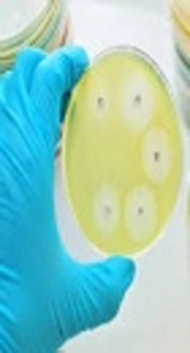
The susceptibility profile was determined by following locally available antibiotics by using the disk (Mast, UK) diffusion method in accordance with the CLSI recommendation (13), and the test was performed on Mueller-Hinton agar (Merck, Germany). The antimicrobial disks were as follows for Gram-positive isolates: penicillin (10 units), ampicillin (10 µg), vancomycin (30 µg), co-trimoxazole (25 µg), chloramphenicol (30 µg), gentamicin (10 µg), ciprofloxacin (5 μg), oxacillin (1 µg), azithromycin (15 ug), linezolid (10 µg), tetracycline (30 µg), doxycycline (30 µg), rifampicin (5 µg), and clindamycin (2 µg) and for Gram-negative isolates: ampicillin (10µg), amikacin (30 µg) tobramycin (10 µg), piperacillin (30 µg), amoxicillin-clavulanic acid (20 – 10 µg), ampicillin-sulbactam (10 - 10µg), cefuroxime (30 µg), cefepime (30 µg), levofloxacin (5 µg), ticarcillin-clavulanic acid (75 - 10μg), meropenem(10 µg), imipenem (10 µg), ceftazidime (30 µg), co-trimoxazole (25 µg), chloramphenicol (30 µg), gentamycin (10 µg), ciprofloxacin (5 µg), tetracycline (30 µg), doxycycline (30 µg), cephazolin (30 µg), and piperacillin-tazobactam (100 – 10 µg). The plates were incubated aerobically at 37°C for 18 hours and the interpretation of the results of the antimicrobial susceptibility was made based on the clinical and laboratory standards institute (CLSI) criteria. In our results, intermediate isolates considered as resistant. S. aureus ATCC 29213, E. coli ATCC25922, and P. aeruginosa ATCC 27853 were used as the standard quality controls. MDR isolates were estimated according to previously described definitions (14).
2.3. Statistical Analysis
The analysis was performed by using SPSSTM software, version 21.0 (IBM Corp., USA). The results are presented as descriptive statistics in terms of relative frequency. Values were expressed as the percentages in every group (for categorical variables).
3. Results
During the six-month period of this study, 867 pathogens were detected; we presented the result based on urine and non-urine site.
3.1. Urine Culture
Among 467 pathogens isolated from urine cultures, 92 (19.7%) Gram-positive bacteria and 375 (80.3%) Gram-negative bacteria were detected.
The most common bacteria were E. coli (293, 62.74%), followed by Enterococcus spp. (73, 15.63%), Klebsiella spp. (63, 13.5%), S. aureus (19, 4.06%), Pseudomonas spp. (17, 3.64%), and Acinetobacter spp. (2, 0.43%). In urine cultures, the majority of samples were gathered from outpatients and the nephrology ward with 60.8% and 13.5%, respectively. Tables 1 and 2 demonstrate the frequency of antimicrobial susceptibility of the important pathogens isolated from urine cultures. For Gram-negative bacteria, the susceptibility pattern of antibiotics showed that imipenem, meropenem, and amikacin were most effective antibiotics and ciprofloxacin and ceftazidime showed an acceptable sensitivity. In addition, for Gram-positive bacteria, vancomycin and chloramphenicol showed 100% sensitivity for staphylococci and 47.9 and 89.7% for enterococci isolates, respectively.
| Gram-Positive Isolates | S. aureus (N = 19) | Enterococcus spp.(N = 73) |
|---|---|---|
| Penicillin | 3 (15.8) | 15 (20.5) |
| Ampicillin | 3 (15.8) | 28 (38.4) |
| Vancomycin | 19 (100) | 35 (47.9) |
| Linezolid | 0 (0) | 0 (0) |
| Tetracycline | 0 (0) | 0 (0) |
| Oxacillin | 5 (26.3) | 0 (0) |
| Azithromycin | 0 (0) | 0 (0) |
| Clindamycin | 0 (0) | 1 (1.4) |
| Trimethoprim-sulfamethoxazole | 6 (31.6) | 2 (2.7) |
| Doxycycline | 0 (0) | 0 (0) |
| Rifampicin | 0 (0) | 0 (0) |
| Gentamicin | 5 (26.3) | 20 (27.4) |
| Ciprofloxacin | 13 (68.4) | 14 (19.2) |
| Chloramphenicol | 19 (100) | 65 (89) |
aValues are presented as No. (%).
| Gram-Negative Isolates | E. coli (N = 293) | Klebsiella spp. (N = 63) | Acinetobacter spp. (N = 2) | Pseudomonas spp. (N = 17) |
|---|---|---|---|---|
| Ampicillin | 20 (6.8) | 6 (9.5) | 0 (0) | 0 (0) |
| Cephazolin | 31 (10.6) | 7 (11.1) | 0 (0) | 0 (0) |
| Gentamicin | 167 (57) | 34 (54) | 0 (0) | 10 (58.8) |
| Tobramycin | 43 (14.7) | 6 (9.5) | 0(0) | 3 (17.6) |
| Piperacillin | 18 (6.1) | 3 (4.8) | 1 (50) | 7 (41.2) |
| Amoxicillin clavulanic acid | 0 (0) | 0 (0) | 0 (0) | 0 (0) |
| Ampicillin sulbactam | 40 (13.7) | 9 (14.3) | 0 (0) | 0 (0) |
| Piperacillin-tazobactam | 0 (0) | 0 (0) | 0 (0) | 0 (0) |
| Cefepime | 11 (3.8) | 4 (6.3) | 0 (0) | 1 (5.9) |
| Cefotaxime | 27 (9.2) | 3 (4.8) | 0 (0) | 0 (0) |
| Cefuroxime | 10 (3.4) | 3 (4.8) | 0 (0) | 1 (5.9) |
| Imipenem | 229 (78.2) | 47 (74.6) | 0 (0) | 12 (70.6) |
| Meropenem | 277 (94.5) | 58 (92.1) | 1 (50) | 15 (88.2) |
| Amikacin | 273 (93.2) | 47 (74.6) | 1 (50) | 14 (82.4) |
| Ciprofloxacin | 186 (63.5) | 42 (66.7) | 0 (0) | 11 (64.7) |
| Trimetoprim-sulfametoxazol | 33 (11.3) | 11 (17.5) | 0 (0) | 0 (0) |
| Ceftazidime | 148 (50.5) | 28 (44.4) | 1 (50) | 16 (94.1) |
| Ticarcillin-clavulanic acid | 0 (0) | 0 (0) | 0 (0) | 0 (0) |
| Tetracycline | 0 (0) | 0 (0) | 0 (0) | 0 (0) |
| Chloramphenicol | 1 (3) | 0 (0) | 0 (0) | 1 (5.9) |
| Levofloxacin | 0 (0) | 0 (0) | 0 (0) | 0 (0) |
| Doxycycline | 0 (0) | 0 (0) | 0 (0) | 0 (0) |
aValues are presented as No. (%).
3.2. Non-Urine Culture
Out of 400 pathogens isolated from non-urine clinical samples, 97 (24.2%) Gram-positive bacteria and 303 (75.8%) Gram-negative bacteria were detected. The predominant isolates were Pseudomonas spp. (169, 42.25%), followed by S. aureus (88, 22%), Klebsiella spp. (65, 16.25%), E. coli (45, 11.25%), Acinetobacter spp. (24, 6%), and Enterococcus spp. (9, 2.25%). The majority of these samples were obtained from pediatric intensive care unit (PICU) and outpatients as 19.5% and 18%, respectively.
The antimicrobial susceptibility pattern of the isolated pathogens from non-urine samples is shown in Tables 3 and 4. Enterococcus spp. were most susceptible to chloramphenicol (88.9%) and vancomycin (33.3%). S. aureus isolates were sensitive to vancomycin and chloramphenicol as 100% and 78.4%, respectively. Most effective antibiotics for Pseudomonas spp. were ciprofloxacin (81.1%) and ceftazidime (70.4%). E. coli and Klebsiella spp. showed high sensitivity rates to meropenem, imipenem, and amikacin. In overall, the most MDR isolates were enterococci and Acinetobacter spp. both with 100% prevalence (Table 5).
| Gram-Positive Isolates | S. aureus (N = 88) | Enterococcus spp. (No = 9) |
|---|---|---|
| Penicillin | 2 (2.3) | 0 (0) |
| Ampicillin | 5 (5.7) | 0 (0) |
| Vancomycin | 88 (100) | 3 (33.3) |
| Linezolid | 0 (0) | 0 (0) |
| Tetracycline | 0 (0) | 0 (0) |
| Oxacillin | 21 (23.9) | 0 (0) |
| Azithromycin | 2 (2.3) | 0 (0) |
| Clindamycin | 1 (1.1) | 0 (0) |
| Trimethoprim-sulfamethoxazole | 20 (22.7) | 0 (0) |
| Doxycycline | 0 (0) | 0 (0) |
| Rifampicin | 3 (3.4) | 0 (0) |
| Gentamicin | 4 (4.5) | 0 (0) |
| Ciprofloxacin | 39 (44.3) | 0 (0) |
| Chloramphenicol | 69 (78.4) | 8 (88.9) |
aValues are presented as No. (%).
| Gram -Negative Isolates | E. coli (N = 45) | Klebsiella spp. (No = 65) | Acinetobacter spp. (N = 24) | Pseudomonas spp. (N = 169) |
|---|---|---|---|---|
| Ampicillin | 3 (6.7) | 2 (3.1) | 0 (0) | 2 (1.2) |
| Cephazolin | 0 (0) | 0 (0) | 0 (0) | 0 (0) |
| Gentamicin | 15 (33.3) | 13 (20) | 1 (4.2) | 36 (21.3) |
| Tobramycin | 2 (4.4) | 2 (3.1) | 4 (16.7) | 27 (16) |
| Piperacillin | 0 (0) | 6 (9.2) | 3 (12.5) | 49 (29) |
| Amoxicillin clavulanic acid | 0 (0) | 0 (0) | 0 (0) | 0 (0) |
| Ampicillin sulbactam | 2 (4.4) | 3 (4.6) | 0 (0) | 0 (0) |
| Piperacillin-tazobactam | 0 (0) | 0 (0) | 0 (0) | 2 (1.2) |
| Cefepime | 11 (24.4) | 8 (12.3) | 0 (0) | 31 (18.3) |
| Cefotaxime | 1 (2.2) | 0 (0) | 0 (0) | 4 (2.4) |
| Cefuroxime | 7 (15.6) | 8 (12.3) | 2 (8.3) | 32 (18.9) |
| Imipenem | 31 (68.9) | 49 (75.4) | 5 (20.8) | 96 (56.8) |
| Meropenem | 43 (95.6) | 56 (86.2) | 5 (20.8) | 113 (66.9) |
| Amikacin | 27 (60) | 28 (43.1) | 8 (33.3) | 89 (52.7) |
| Ciprofloxacin | 25 (55.6) | 49 (75.4) | 13 (54.2) | 137 (81.1) |
| Trimetoprim-sulfametoxazol | 1 (2.2) | 5 (7.7) | 1 (4.2) | 7 (4.1) |
| Ceftazidime | 23 (51.1) | 26 (40) | 6 (25) | 119 (70.4) |
| Ticarcillin-clavulanic acid | 0 (0) | 0 (0) | 0 (0) | 0 (0) |
| Tetracycline | 0 (0) | 0 (0) | 0 (0) | 0 (0) |
| Chloramphenicol | 1 (2.2) | 0 (0) | 2 (8.3) | 8 (4.7) |
| Levofloxacin | 0 (0) | 0 (0) | 0 (0) | 0 (0) |
| Doxycycline | 0 (0) | 0 (0) | 0 (0) | 0 (0) |
aValues are presented as No. (%).
| Isolates | S. aureus | Enterococcus spp. | E. coli | Klebsiella spp. | Acinetobacter spp. | Pseudomonas spp. |
|---|---|---|---|---|---|---|
| Urine culture | 17 (89.4) | 67 (91.8) | 250 (85.3) | 57 (90.5) | 2 (100) | 16 (94.1) |
| Non-urine culture | 83 (94.3) | 9 (100) | 36 (80) | 57 (87.7) | 22 (91.6) | 157 (92.9) |
aValues are presented as No. (%).
4. Discussion
Consistent with our results, the most common nosocomial pathogens in Ott et al. study were E. coli and S. aureus. Moreover, the most common pathogens isolated in Azim et al. study from India were P. aeruginosa and A. baumannii (15). In Aly and Balkhy study from six Arab countries including Saudi Arabia, Qatar, Bahrain, Kuwait, Oman, and United Arab Emirates, the most prevalent determined microorganisms were E. coli, K. pneumoniae, P. aeruginosa, methicillin-resistant Staphylococcus aureus (MRSA), and Acinetobacter spp. (16). In our results, E. coli (62.7%) was the predominant isolated pathogen in urine cultures. The role of E. coli as the predominant causative agent of urinary tract infections (UTIs) was confirmed in most of the previous studies (17-23). In our findings, S. aureus and enterococci were seen in small numbers, but they were recognized as important causes of UTIs (24-26). The general distribution pattern of our study showed the most commonly isolated bacteria from non-urine samples were P. aeruginosa (42.3%). Previously, some other studies showed the same results including Mukherjee et al. as 61, Japoni et al. as 67.7%, and Karlowskyet et al. as 42%, and Ghassemi et al. observed P. aeruginosa as the most common pathogen in their findings (23, 27, 28). However, it could be due to nosocomial infection or contamination of samples with this organism.
The surveillance of bacterial antimicrobial resistance is the most important challenge to understanding the dynamics of decreasing susceptibility to antibiotics in both hospital and community-acquired pathogens (29). The highest antibiotic susceptibility rate of E. coli and Klebsiella isolates obtained from urine and non-urine cultures was to meropenem, followed by amikacin, imipenem, and ciprofloxacin, in sequence. Overall, the most effective antibiotics against Gram-negative bacteria were meropenem, amikacin, and imipenem with approximately 50 – 95% susceptibility.
The rates of imipenem susceptibility in our study were different from those reported in previous studies showing that the isolates of Klebsiella spp. and E. coli were fully susceptible to imipenem (30, 31). The cause of these differences may be due to higher carbapenem prescription for patients in our study hospital and subsequently the emergence of carbapenem-resistant strains.
In our study, the susceptibility rate of P. aeruginosa to imipenem was lower than the susceptibility rate reported by Mohammadi et al. from the west of Iran (32). All of the S. aureus isolates in our study showed 100% susceptibility to vancomycin. This pattern is the same as the findings of other Iranian studies (22, 33-36).
The high rate of sensitivity to vancomycin for Gram-positive cocci was noted in several previous studies from Iran and other parts of the world (18, 21). On the other hand, in our study, meropenem, amikacin, and imipenem are relatively effective drugs for the treatment of the majority of the infections caused by Gram-negative bacterial isolates, especially Enterobacteriaceae. This can be due to that these antibiotics may not be commonly used before and/or they are newly introduced. These findings were in accordance with the previous results from different parts of the world (32, 34, 37), but Armin et al. presented contrary results (38) that could be due to differences in the population study. The emergence of MDR strains is increasing the health concern. In the present study, we found a remarkable rate of MDR isolates. Previously, in accordance with our findings, several reports showed the increasing prevalence of MDR strains in Iranian hospitals (11, 39-42).
4.1. Conclusion
Considerable differences in antimicrobial resistance do exist for every single hospital that may reflect differences in antimicrobial usage pattern, population study, and infection control strategies. In this study, Gram-positive bacteria showed a high rate of susceptibility to chloramphenicol, which recently was rarely prescribed by our physicians. Gram-negative bacteria showed the most susceptibility to amikacin, meropenem, and imipenem, in sequence.
