


1. Background
Idiopathic pulmonary fibrosis (IPF) is a chronic and progressive lung disease, and leads to tissue destruction, decline in lung function, respiratory distress, and finally decreased quality of life that causes death during three to five years (1, 2). Although IPF is unknown, causes disease, and is poorly diagnosed, several factors, such as reduced lung alveolar epithelial cells, environmental factors, excessive sensitivity in the respiratory tract, high body mass index, and high grade on the modified Medical Research Council (MRC) breathlessness scale contribute to pathogenesis of IPF. There is no definite care for IPF therapy and lung transplantation is the only alternative to save a small number of patients from the danger of death (3-6).
Because of changing viscosity of mucus in patients with progressive lung disease, use of broad-spectrum antibiotics and oral-respiratory steroid, the lung tissue is susceptible to colonization of fungal, bacterial, and viral agents, creating chronic infections (7). Previous studies investigated the role of microorganisms in the etiology of IPF pathogenesis (8, 9).
Colonization of Candida species in the respiratory system tract, especially in high risk patients with impaired lung epithelial cells, has been increased, which can improve pathogenecity of Candida and subsequently produce disease (10, 11).
It has been reported that Candida albicans (C. albicans) is the most common fungal agent isolated from the respiratory system of patients, whereas other Candida species are less frequent. The significance of Candida species is their ability to form a rigid network of yeast and hyphal complex as defined by biofilm structure with high resistance to common antifungal agents (12, 13). However, the role of fungi in progression of cyctic fibrosis (CF) is well-known (10, 11, 14), while research on the prevalence of fungal colonization in IPF is limited. Therefore, the discovery of fungal colonization in IPF subjects is important for antifungal prophylaxis and treatment management of patients.
2. Objectives
This study aimed at investigating the identification of fungal agents in the respiratory system of IPF patients and determination of the anti-fungal susceptibility pattern of isolates, which is imperative for administrating a proper treatment in IPF cases.
3. Methods
3.1. Patients Characteristics and Specimens
Forty Iranian IPF patients were enrolled in this study, according to the results of lung NP swab or BAL or CT scans related to referral hospitals of Iran University of Medical Sciences (Tehran, Iran). During 12 months (from 2015 to 2016), 10 nasopharyngeal (NP) swabs and 30 bronchoalveolar lavage (BAL) specimens were collected from patients. The IPF cases had underlying disease, such as liver cirrhosis, pulmonary disease, and heart failure. Some cases took antibiotic and prednisolone for the treatment of IPF. Moreover, IPF cases did not show any clinical sign of fungal infections during the study. Demographic information of subjects, including age, gender, familial history of lung disease, underlying disease, and use of drugs, were recorded. All patients filled out the consent form designed for the study. The study protocol was approved by the Research Ethics Committee of Iran University of Medical Sciences (IUMS) with ethical code number IR.IUMS.REC25488.
3.2. Culture of Samples
Specimens were collected in a sterile container and immediately transported to Medical Mycology Laboratory of Iran University. The samples were smeared on a slide and observed for the presence of fungal elements using 10% potassium hydroxide (KOH), directly under the light microscope. Then, an equal volume of 0.5% pancreatin was added and centrifuged. The precipitate was cultured on Sabouraud Dextrose Agar medium (SDA; Merck, Germany) with chloramphenicol, incubated for two weeks at 37°C and 25°C for yeast and filamentous fungi growth, respectively. Fungal was growth tested every two to three days. Candida species were identified by conventional methods, including Germ tube formation in Calf Fetal serum, Chlamydospore production on corn-meal agar (Difco, USA), color differentiation on CHROMagar Candida (Paris, France) and API 20C AUX (bioMe´rieux Italia S.p.A., Rome, Italy) (15). Finally, the isolates were identified using PCR and sequencing methods.
Indeed, filamentous fungi were recognized using morphological characterization on Czapek Dox Agar (Sigma-Aldrich, USA) and slide culture technique.
3.3. Genomic DNA Extraction from Cadida Species
Fungal genomic DNA was extracted, as described previously. Briefly, genomic DNA was extracted using phenol, chloroform, isoamyl alcohol (25, 24, 1) glass beads, and lysis buffer (100 mM Tris pH 8, 100 mM NaCl, 1% SDS, Triton 2% X-100). The DNA was eluted in 20 µL of Tris-EDTA buffer (5 mM), and stored at -20°C until for analysis. The quality and quantity of DNA was checked by gel electrophoresis and Nanodrop spectrophotometer (Thermo fisher scientific), respectively (16).
3.4. Polymerase Chain Reaction (PCR) and Sequenceing Analysis
Polymerase chain reaction was accomplished to amplify universal primer ITS1-5.8S rDNA-ITS2 region sequence in ribosomal DNA. The sequences of ITS primers were ITS1 5’-TCC GTA GGT GAA CCT GCG G-3’ and ITS4 5’-TCC TCC GCT TAT TGA TAT GC-3’, respectively.
The PCR assay was performed in a 25-μL reaction mixture, including 12.5 µL of Mastermix (Sinaclon, Iran), 2 µL of forward and revers primer (10 pmol), 1 µL of DNA template (25 ng), and sterile distilled water to reach a total volume of 25 µL.
After initial denaturation of DNA at 95°C for five minutes, 30 cycles of amplification were completed in a thermal cycler (PeQlab, UK), including a denaturation step at 94°C for 30 seconds, an annealing step at 56°C for 30 seconds, an extension step at 72°C for one minute and a final extension step at 72°C for seven minutes. Proper positive and negative controls were used in each run. Then, the PCR products were electrophoresed through 1.8% agarose gel and visualized by ethidium bromide staining (17). The PCR products were sequenced by ITS primers from the Bioneer company (Korea).
All positive PCR amplicons were purified using a QIAquick PCR purification kit (Qiagen., UK) and were eluted in Tris-HCl (10 mM, pH 8.5) prior to sequencing, and then were sequenced in the forward direction, employing the ITS primer. The result of sequences was aligned, employing the MegAlign software (DNAStar Inc. Wisconsin, USA), and compared with those stored in the Genbank Data system using the BLAST alignment software tool (http://www.blast.genome.ad.jp/).
3.5. Antifungal Susceptibility Testing
Antifungal suscebtibility was conducted with Candida isolates against fluconazole, amphotricin B, and itraconazole by microdilution broth, according to Clinical Laboratory Standards Institute CLSI (M27-S3) guidelines (18). Amphotericin B, itraconazole, and fluconazole (Sigma-Aldrich, USA) were prepared by the manufacturers standard assay powders. Microdilution trays (96 U-bottom shaped, Germany) containing antifungal dilutions were equipped and incubated at 35°C for 24 hours. In each experiment, proper positive and negative controls were used.
The MICs were determined by visual examination and considered as the lowest concentration of agents that inhibited the growth of yeast. The C. glabrata CBS 138 was used as the reference strain, and all experiments were conducted in duplicates. Species-specific clinical breakpoints (CBPs), recently published by the CLSI (M27-S4), were used for interpretation of results (19).
3.6. Statitical Analysis
The data were analyzed using the SPSS software, version 20 (SPSS, Chicago, IL, USA). The correlation between the presence of fungal species and patient characteristics were examined using either Pearson’s chi-square or Fisher’s exact test. Variations in the fungal agents colonization among underlying diseases were measured using the Mann Whitney U test. A P value of ≤ 0.05 was considered statistically significant.
4. Results
4.1. Study Population
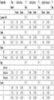
Of 40 IPF cases, 22 (55%) were male and 18 (45%) were female. The male to female ratio was 22:18 patients (1.22). Patients’ ages ranged between 18 and 70 years old (mean age: 60 years). The age of 21 (52.5%) patients was younger than 60, and 19 (45%) patients were older than 60 years old. Twenty-seven (68%) patients were undergoing treatment with antibiotic and prednisolone, whereas 13 (32%) patients only took prednisolone. The IPF patients, did not use any antifungal drugs during the sampling. All of the IPF patients had chronic pneumonia, had been diagnosed by CT scan in the recent years, and did not show any occupational exposure. Seven (17.5%) of 40 IPF patients represented fungal agent, however, no relationship was found between the presence of fungal species with age and gender of IPF patients. Demographic data of the IPF patients are shown in Table 1.
| Patients Data | Total Number | Candida albicans | P Value | Candida glabrata | P Value | Aspergillus fumigatus | P Value | |||
|---|---|---|---|---|---|---|---|---|---|---|
| Presence | Absence | Presence | Absence | Presence | Absence | |||||
| Age (mean = 60) | 0.65 | 0.73 | 0.52 | |||||||
| ≤ 60 | 21 (53) | 2 (9) | 19 (91) | 1 (5) | 20 (95) | 1 (5) | 20 (95) | |||
| > 60 | 19 (47) | 2 (10) | 17 (90) | 0 (0) | 19 (100) | 1 (5) | 18 (95) | |||
| Gender | 0.41 | 0.29 | 0.45 | |||||||
| Female | 22 (55) | 2 (9) | 20 (91) | 0 (0) | 22 (100) | 2 (9) | 20 (91) | |||
| Male | 18 (45) | 2 (11) | 16 (89) | 1 (6) | 17 (94) | 0 (0) | 18 (100) | |||
| Disease | 0.04a | 0.18 | 0. 36 | |||||||
| IPF | 6 (15) | 4 (67) | 2 (33) | 2 (33) | 4 (67) | 1 (17) | 5 (83) | |||
| IPF + other | 34 (85) | 0 (0) | 34 (100) | 0 (0) | 34 (100) | 0 (0) | 34 (100) | |||
| Type of drug + | 0.39 | 0.45 | 0.32 | |||||||
| prednisolone | 27 (68) | 2 (7) | 25 (93) | 0 (0) | 27 (100) | 2 (7) | 25 (93) | |||
| Prednisolone | 13 (32) | 2 (15) | 11 (85) | 1 (8) | 12 (92) | 0 (0) | 13 (100) | |||
| Severity | 0.39 | 0.58 | 0.42 | |||||||
| 0 | 4 (10) | 0 (0) | 4 (100) | 0 (0) | 4 (100) | 0 (0) | 4 (100) | |||
| I | 17 (42) | 1 (6) | 16 (94) | 1 (6) | 16 (94) | 1 (6) | 16 (94) | |||
| II | 19 (48) | 3 (16) | 16 (84) | 0 (0) | 19 (100) | 0 (0) | 19 (100) | |||
aP value is statistically significant.
4.2. Charecterization of Fungi Species by Conventional and PCR Methods
In direct microscopy examination of samples, seven (17.5%) of 40 specimens represented yeast, hypha, and branching of fungal hyphae forms. Indeed, these specimens were positive for fungal growth in SDA media. The distribution of fungi was as follows, Candida albicans four (10%), Candida glabrata two (5%), and (2.5%) Aspergillus fumigatus one (A. fumigatus).
Candida species were identified definitely by PCR and sequencing. By sequencing analysis, the Candida species showed high similarity in gene bank data and identified as C. albicans and C. glabrata. Also, A. fumigatus were identified mainly on morphological criteria.
4.3. Correlation Between Fungal Colonization and Risk Factors in IPF Patients
There was a significant association between the colonization of C. albicans in IPF patients and underlying diseases, including liver cirrhosis, pulmonary and heart disease as well as antibiotics and corticosteroid therapy (P < 0.05). Interstingly, the findings showed no significant relationship between the C. glabrata colonization and risk factors in IPF patients (P = 0.08). Patients with A. fumigatus colonization were older than those with Candida species. However, no significant relationship was found between the presence of A. fumigatus and other charecteristics in IPF patients (P = 0.06). The finding are summarized and presented in Table 1.
4.4. Antifungal Susceptibility Testing
Regarding antifungal susceptibility findings, four (100%) isolates of C. albicans were resistant to itraconazole (MIC ≥1 µg/mL) and it was found that three (75%) isolates of C. albicans were resistant to amphoterecin B (MIC ≥ 1 µg/mL), whereas three (75%) and one (25%) C. albicans isolates acted in a dose dependent manner (MIC ≥ 4 µg/mL) and were resistant to fluconazole (MIC ≥ 8 µg/mL), respectively. Also, 25% of isolates of C. albicans were sensitive (MIC = 0.5 µg/mL) to amphotricin B, while 75% of isolates were resistant to amphotricin B. Furthermore, C. glabrata isolates were resistant to fluconazole (MIC ≥ 64 µg/mL), itraconazole (MIC ≥ 1 µg/mL), and amphotricin B (MIC ≥ 1 µg/mL). The results are shown in Table 2. Breakpoint for fluconazole provided by CLSI M27-S4. As breakpoint for AMB and itraconazole was not provided by CLSI M27-S4, therefore, the researchers interpreted according to the M27-S3 document (18).
| Clinical Isolates | Fluconazole (MIC, µg/mL) | Itraconazole (MIC, µg/mL) | Amphotricin B (MIC, µg/mL) | ||||||
|---|---|---|---|---|---|---|---|---|---|
| S | SDD | R | S | SDD | R | S | SDD | R | |
| C. glabrata (n = 2) | 0 | 0 | 2 | 0 | 0 | 2 | 0 | 0 | 2 |
| C. albicans (n = 4) | 0 | 3 | 1 | 0 | 0 | 4 | 1 | 0 | 3 |
Abbreviations: MIC, minimum inhibitory concentration; S, sensitive; SDD, susceptible-dose dependent; R, resistance.
5. Discussion
The role of fungi in terms of colonization and pathogenesis of IPF are less well described. Notably, previous published data demonsterated the role of bacteria and viral infection in the pathogenesis of IPF (8, 9, 20-25), however, the role of fungal agents as etiology of IPF is less addressed. This reflects the fact that little attention has been paid to the outcomes of fungal agents in IPF patients. For this reason, in the current study, the researchers evaluated the colonization of fungal agents in Iranianin patients with IPF for the first time. Also, antifungal susceptibility of isolates was examined. Assessment of fungal agents in IPF patients needs greater attention because of colonization of fungal agents in IPF subjects with underlying disease, which may ultimately lead to lethal infection by dispersing to the blood stream.
To the best of the author’s knowledge, this study was the first report of fungal colonization in Iranian IPF patients. The findings expressed that C. albicans was the most prevalent species isolated from IPF patients followed by C. glabrata and A. fumigatus.
Moreover, a significant correlation was found between the presence of C. albicans and predisposing factors in IPF patients that result in growthe of Candida species colonization in the respiratory system, which leads to severe infections and morbidity in high risk patients.
The findings are consistent with other related studies that have focused on the pathigenesis of respiratory disease; peltrochelacsahuanga isolated C. albicans and C. dubliniensis with a similar percentage of the respiratory tract in cystic fibrosis (CF) patients (26, 27).
In a similar study performed by Gungor et al. in 2013, Candida albicans was the most common agent isolated from the respiratory tract of Turkish CF patients, although C. parapsilosis, C. dubliniensis, and Aspergillus fumigatus were reported in the second most common fungi. Similarity, the current findings reported that there is no correlation between Candida growth and age and gender of patients (10).
Gammelsroud et al. reported that Children with CF had the highest prevalence of Candida albicans (11). Similarity, Candida albicans was the predominant yeast isolated from turkish patients with CF, followed by C. parapsilosis and C. dubliniensis. Also, Aspergillus fumigatus detected the most common filamentous fungus (11).
Horre et al. in 2004 investigated Wangiella dermatitidis as the black yeast could be colonized and recovered from sputum culture of CF patients (14).
Indeed, in a previous study carried out by Pihet, Aspergillus fumigatus, Scedosporium apiospermum, Aspergillus terreus, and Candida albicans offered the main fungal species in respiratory secretions associated with CF cases, nevertheless, other species are less common (28).
Interestingly, in a case report by Kumar, coexistence of aspergilloma (fungal ball) with Idiopathic pulmonary fibrosis (IPF) was reported in 55-year-old female (29).
Antifungal susceptibility test indicated a high rate of resistance in Candida albicans and C. glabrata isolates. Overall, 100% of the isolates were resistant to itraconazole, 75% of isolates were resistant to amphotricin B, and 25% were resistant to fluconazole. Candida glabrata isolates were resistant to fluconazole, itraconazole, and amphotricin B. It is well known that the resistance of Candida glabrata is developing in clinical isolates, and previous studies supported this data. This resistance may be related to genetic diversity of species and the emergence of resistant genes due to using common antifungal drugs (12, 13, 30).
The main obstacle of non-albicans Candida infection is high intrinsic resistance to several antifungal drugs, especially azoles. Additionally, the progress of antifungal resistance, during prolonged and prophylactic treatment, leads to a decrease in theraputic efficacies (31).
Therefore, management and appropriate antifungal therapy must be mentioned for fungal infection treatment of IPF patients and to decrease the drug resistance rate. Some of the limitations of the current study was the low sample size of IPF patients due to the low rate of IPF incidence, nevertheless, a more dependable finding will be accomplished in a large papulation.
5.1. Conclusions
In conclusion, in IPF patients, fungi may contribute to destruction of lung function and infection. However, the clinical relevance of the fungal airway colonization and its correlation with IPF still needs consideration. Based on the current findings, yeast and filamentous fungi may be responsible for local inflammatory response and leads to infection in susceptible patients.
In addition, the effects of antifungal prophylaxis and cancer therapy medications can be investigated on Candida colonization rate.
