Abstract
Background:
Low back pain (LBP) is a common cause of lost playing time and can be a challenging clinical condition in competitive athletes. LBP in athletes may be associated with joint and ligamentous hypermobility and impairments in activation and coordination of the trunk musculature, however there is limited research in this area.Objectives:
To determine if there is an association between altered lumbar motor control, joint mobility and low back pain (LBP) in a sample of athletes.Materials and Methods:
Fifteen athletes with LBP were matched by age, gender and body mass index (BMI) with controls without LBP. Athletes completed a questionnaire with questions pertaining to demographics, activity level, medical history, need to self-manipulate their spine, pain intensity and location. Flexibility and lumbar motor control were assessed using: active and passive straight leg raise, lumbar range of motion (ROM), hip internal rotation ROM (HIR), Beighton ligamentous laxity scale, prone instability test (PIT), observation of lumbar aberrant movements, double leg lowering and Trendelenburg tests. Descriptive statistics were compiled and the chi square test was used to analyze results.Results:
Descriptive statistics showed that 40% of athletes with LBP exhibited aberrant movements (AM), compared to 6% without LBP. 66% of athletes with LBP reported frequently self-manipulating their spine compared to 40% without LBP. No significant differences in motor control tests were found between groups. Athletes with LBP tended to have less lumbar flexion (63 ± 11°) compared to those without LBP (66 ± 13°). Chi-Square tests revealed that the AM were more likely to be present in athletes with LBP than those without (X2 = 4.66, P = 0.03).Conclusions:
The presence of aberrant movement patterns is a significant clinical finding and associated with LBP in athletes.Keywords
1. Background
Competitive athletes commonly lose playing time due to low back pain and it is one of the most common reasons for missed playing time in professional athletes (1). According to previous studies, rates of low back pain in athletes have ranged from 1 to > 30% (1). Low back pain in athletes has been linked to certain sporting activities that place greater stress on the lumbar spine (2-4). These sports include gymnastics, wrestling, rowing, diving, and football (2-5). A combination of strength and mobility of the lumbar spine is necessary for many athletic activities and sports (1).
Hypermobility is typically benign and may provide an inherent advantage in certain sports; however research has found that it may also lead to increased risk for injury, therefore it appears to be important to identify hypermobility in athletes (6-8). There is some preliminary research indicating an association between ligamentous laxity and LBP in athletes (6) and non-athletes (7), but most of the research in this area has focused on lower extremity injuries (8-10). The Beighton Ligamentous Laxity Scale (BLLS) is used to assess generalized joint hypermobility (11). A number of additional clinical examination measures are useful for assessing flexibility and mobility in this region. They include the passive straight leg raise (PSLR), range of motion (ROM) of the lumbar spine, and hip internal rotation range of motion (HIR). Patients with radiographic instability of the lumbar spine have been found to have greater ligamentous laxity as measured with the Beighton scale and greater lumbar flexion range of motion measurements (12). Deficits in hip internal rotation have been found in patients with LBP, in athletes playing rotation-based sports and specifically in judo players, golfers and tennis players (13-16).
Impairments in activation and coordination of the trunk musculature have also been identified in patients with low back pain (17-19). Clinical tests such as the Active Straight Leg Raise Test (ASLR), Leg Lowering Test (LL), and Trendelenburg are used to measure lumbar motor control. These tests have been found to be reliable for measuring movement control in the lumbar spine (20-23).
Research suggests that classifying patients with low back pain into more homogenous subgroups based on their clinical presentation, and matching interventions accordingly may result in improved clinical outcomes (24). One such sub group includes patient with impairments in lumbopelvic movement control and possible lumbar clinical or functional segmental instability. This clinical or functional instability appears to be related to a loss of segmental stiffness and of mid-range control of spinal segments during motion resulting in aberrant motion (25). A lumbar stabilization program is advocated to address these impairments. Hicks et al. (27) developed a clinical prediction rule to identify patients with low back who would benefit from lumbar stabilization exercises. They found that the following four factors had the greatest predictive value of success with stabilization exercises: a passive straight leg raise of 91 degrees or greater, a positive prone instability test, the presence of aberrant movement of the lumbar spine, and age younger than 40. These clinical measurements, which have been found to be reliable and predictive of success with a stabilization program, were assessed in our study (24).
There has been very little research investigating a combination of all of the previously mentioned clinical tests of flexibility and motor control in athletes. Most research articles have limited their view to a single test such as Kim et al. (7), who only utilized the Beighton Laxity Scale, or Leao Almeida et al. (15) who studied the correlation between hip internal rotation range of motion and lumbar hypermobility associated with low back pain. Roussel et al. (22) investigated the role of lumbopelvic movement control and joint hypermobility in predicting injury in dancers. They found that altered movement control but not hypermobility was associated with development of lumbar or lower extremity injuries in dancers. As with many injuries, there is typically more than one contributing factor.
2. Objectives
The purpose of this study was to investigate whether there is an association between altered lumbar motor control, joint mobility and low back pain (LBP) in a sample of athletes.
3. Materials and Methods
3.1. Participants
A cross sectional analysis was used and fifteen athletes with LBP were conveniently recruited for this study. They were matched by age, gender and BMI with controls without LBP. To be included in the study, participants had to be athletes actively participating in their respective sport at the time of the study. Exclusion criteria included any subjects who were currently or had previously had physical therapy treatment or instruction in motor control exercises for the lumbar spine in the previous 6 months. Athletes who were receiving medical care or diagnostic workup for low back pain or lower extremity injury at the time of the study were also excluded. This study was approved by the Northeastern University Institutional Review Board and written consent was obtained from all participants.
3.2. Procedure
The testing was carried out by four fifth year Physical Therapy Doctoral students and two experienced clinicians. The tests were divided between the examiners and they each performed the same tests on each subject. To ensure test reliability, all testers practiced the testing procedures prior to the experiment. The testers were not aware to if the athletes had back pain or not while performing the tests, making the procedures blinded.
All athletes signed a written informed consent form and a completed a questionnaire regarding demographic information, medical history, questions pertaining to the number of hours a week participating in their sport and if they frequently needed to self-manipulate their spine. If the athletes checked “yes” to having low back pain within the past 7 days, on the initial questionnaire, they were asked to describe the location of pain, complete the Numerical Pain Rating Score (NPRS), to assess lumbar pain severity, the Oswestry Disability Index (ODI) to assess levels of functional impairment and Fear Avoidance Beliefs Questionnaire Score (FABQ) (Physical Activity Subscale) to assess fear of pain and avoidance of movement. Next, the athletes were taken to the testing room in groups of four and randomly assigned to one of the four testing stations. The order of tests within each station was randomized. After each test was performed on each subject, the subject was then taken into a second room where their height and weight was recorded and their data was inputted into an excel spreadsheet which was later analyzed.
3.3. Outcome Measures
Beighton Ligamentous Laxity Scale (continuous variable): This test consisted of four bilateral tests: passive hyperextension of the elbow > 10°; passive hyperextension of the fifth finger > 90°; passive abduction of the thumb to contact the forearm; and passive hyperextension of the knees > 10°. The last test consisted of placing both hands flat on the floor while bending forward without flexing the knees. A point was given for each test the subject could perform (11). The scores ranged from 0 to 9. With a test result of ≥ 4, hypermobility is generally considered to be present.
Hip Internal Range of Motion (continuous variable): These measurements were taken with the patient prone, with one leg slightly abducted and the measured side with the knee flexed to 90 degrees. We measured hip internal rotation by first passively observing the subject’s internal rotation, followed by an inclinometer measurement of their range of motion as described by Van Dillen et al. (14) Manual stabilization of the pelvis was utilized as opposed to stabilization with a belt in order to prevent or detect any compensatory motion.
Range of Motion of the Lumbar Spine (continuous variable): Inclinometers were placed at the T12 and S1 levels. Lumbar and sacral flexion and extension were tested in this way with the difference between the two values representing true flexion and extension. Side bending was also evaluated with the inclinometer placed at the T12 level (26).
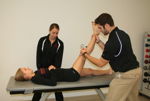
Passive Straight Leg Raise Range of Motion (continuous variable) Figure 1: This test was performed according to Hicks et al. (27), with an inclinometer placed on the tibial tuberosity, while next raising the leg passively with the non-measured leg flat on the table. The lumbar spine was monitored and palpated for compensatory movement.
Passive Straight Leg Raise Range of Motion
3.4. Motor Control Tests and Instrumentation
For the clinical tests, a pressure biofeedback unit (PBU) (Stabilizer,TM Chattanooga Group, Hixson, TN), was utilized as well as a goniometer and an inclinometer. The PBU measures pressure changes in mmHg when placed under the lumbar spine. The PBU has been proven to be a reliable source of detection of even the slightest changes in pressure during movement (28). It was used to assess the subjects’ ability to control movement of the lumbopelvic region while performing lower extremity movements. The pressure change from baseline for each test was recorded. A lower score indicated a better performance on the tests using the PBU.
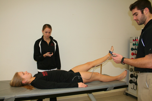
Active Straight Leg Raise Test (continuous variable) Figure 2 : This test was performed following the procedure described by Roussel et al. (21). The subject was instructed to pull their lower abdomen towards their spine, with the PBU under their lumbar spine and inflated to a baseline pressure of 40 mmHg. The subject was instructed to raise their leg off table table 20 inches to a clearly marked yardstick (phase 1) and hold for 20 seconds (phase 2). Pressure changes were recorded during both phases to determine overall motor control (29). Each subject was given one trial while looking at the PBU, followed by a trial without looking at the PBU.
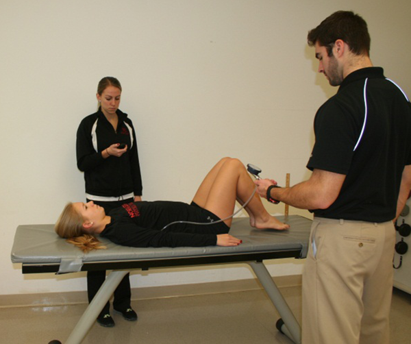
Leg Lowering Test (continuous variable) Figure 3 : This test was performed with a PBU under the subject’s lumbar spine to detect small changes in pressure, which correlated to movement of the lumbar spine. The procedure was performed similar to the procedure described by Enoch et al. (23). The athlete started with both hips flexed to ninety degrees and was asked to lower both legs toward the table while maintaining a neutral spine position. The athlete had two chances to have visual feedback from the PBU while performing the maneuver. The athlete then performed three repetitions in which visual feedback was not given. The pressure deviation on the PBU was recorded when the feet were as close to the table as possible, and averaged for the three trials. All athletes performed the test without shoes.
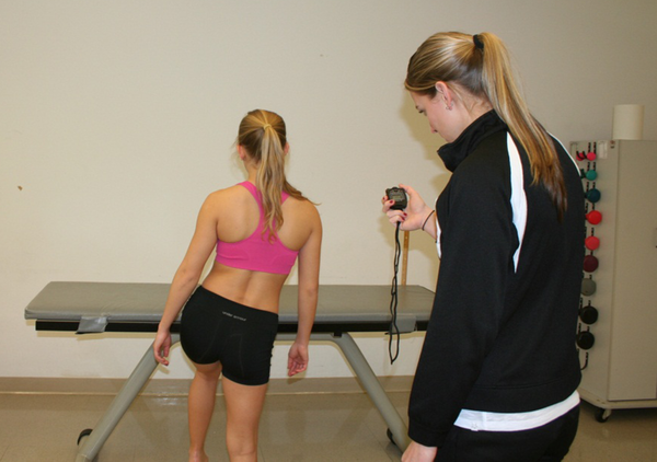
Trendelenburg Test (categorical variable: Yes and No) Figure 4 : The test was performed with the patient standing as described by Hardcastle et al. (30) and Roussel et al. (21). The athlete was asked to stand on one leg, and flex the other leg to 30 degrees, trying to keep the non-stance pelvis elevated compared to the stance leg for 30 seconds. Perceived Exertion with Trendelenburg test: As described by Roussel (21), a 6-point Likert scale was used to evaluate the athletes' perceived effort. The scale was from 0-5 with the perceived effort being not difficult at all, minimally difficult, somewhat difficult, fairly difficult, very difficult or unable to perform.
Aberrant Movement Patterns (categorical variable: Yes or No)-Aberrant movement patterns of the lumbar spine include five observational elements that can help a clinician determine whether clinical instability of the spine is present (31). To perform the test the subject was asked to flex the trunk forward as far as possible then extend back to anatomical position. If any of the following aberrant movements were observed then the test was considered positive: painful arc in flexion, painful arc on return, Gower's sign, instability catch, or reversal of lumbopelvic rhythm. Two examiners, one of whom was an experienced physical therapist, simultaneously observed for the presence of aberrant movement patterns.
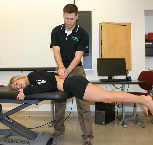
Prone Instability Test (categorical variable: Yes or No) Figure 5 : This test is based on the hypothesis that if pain is present during passive provocation testing of the lumbar spine, but disappears when the patient activates the spinal extensor muscles, the muscle activity is stabilizing the segment, indicating the presence of lumbar segmental instability (24). The first part of the test was performed with the patient prone with their legs over the edge of the table and feet resting on the floor. With the patient in this position, the examiner applied a posterior to anterior pressure to the lumbar spine. The patient was asked to report any provocation of pain. If a painful segment was identified, the second part of the test was performed. For this, the patient lifted the legs off the floor and posterior compression was applied again to the lumbar spine at the level of the painful segment. If pain was present in the resting position but subsided in the second position, the test was considered positive (24).
Active Straight Leg Raise Test
Leg Lowering Test
Trendelenburg Test
Prone Instability Test
3.5. Data Analysis
Descriptive statistics were first conducted to understand the general data distribution. Independent t-tests were used to determine whether the continuous outcome measures varied as a function of low back pain (with or without pain). For categorical outcome measures, Chi-Square tests were used. All statistical analyses were carried out using SPSS v21 (Chicago IL).
4. Results
4.1. Participants
Participants’ characteristics in each group are shown in Table 1. The average NPRS score of athletes with LBP was 3.2 ± 2.2; ODI score 14.7 ± 2.2 and FABQ score 8.3 ± 5.2. Although the low back pain group was on average older and had a slightly higher BMI, the groups were very similar in these characteristics. The low back pain group included 5 lacrosse players, 2 runners, 2 cheerleaders, 2 baseball players, 1 cyclist, 1 softball player, 1 wrestler, and 1 ice hockey player. The control group included 3 runners, 3 cheerleaders, 2 lacrosse players, 2 ice hockey players, 1 cyclist, 1 baseball player, 1 soccer player, 1 water polo player, and 1 ultimate Frisbee player.
Results were summarized in Table 2. Notably, athletes with LBP had less lumbar flexion, and greater lumbar extension compared to those without LBP. In addition, those with LBP demonstrated a lower PSLR bilaterally than those without LBP. Surprising, those with LBP had a similar performance in the Leg Lowering and Active Straight Leg Raise Tests compared to those without LBP Approximately 40% of athletes with low back pain (6 out of 15) exhibited AM, compared to 6% without LBP (1 out of 15). Sixty-six percent of athletes with LBP (9 out of 15) reported frequently self-manipulating their spine compared to 40% without LBP (6 out of 15).
Chi-Square tests revealed that the AM was significantly different between the LBP and non LBP groups (X2 = 4.66, P = 0.03). For other outcome measures, no significant results were detected by independent t-test or Chi-Square analyses (P > 0.05).
Patient Characteristics
| Parameter | Low Back Pain Group | Control Group |
|---|---|---|
| Age, y | 21.26 (2.05) | 20.87 (1.3) |
| Body mass index | 23.75 (2.16) | 23.64 (1.93) |
| Height, in | 66.43 (3.17) | 67.03 (2.98) |
| Weight, lbs. | 149.09 (19.15) | 151.15 (17.94) |
Patient Outcome Measures
| Low Back Pain Group | Control Group | Mean Difference 95% Confidence Interval | |
|---|---|---|---|
| Beighton Ligamentous Laxity Scale | 2.47 (2.67) | 2.07 (2.19) | -0.4 (-2.23, 1.43) |
| Perceived Exertion of Trendelenburg R | 1.00 (1.00) | 1.07 (0.59) | 0.07 (-0.55, 0.68) |
| Perceived Exertion of Trendelenburg L | 1.00 (1.07) | 1.27 (0.7) | 0.27 (-0.41, 0.94) |
| Hip IR L, deg | 46.87 (9.06) | 43.53 (9.08) | -0.33 (-10.12, 3.45) |
| Hip IR R, deg | 44 (10.07) | 46.5 (8.69) | 2.53 (-4.51, 9.57) |
| Hip IR Difference, deg | 5.67 (4.40) | 4.87 (3.70) | -0.8 (-3.85, 2.25) |
| Leg Lowering Test, mmHg | 14.14 (12.35) | 13.49 (10.63) | -0.65 (-9.27, 7.97) |
| Lumbar Flexion, deg | 63 (10.97) | 66.4 (12.94) | 3.4 (-5.58, 12.38) |
| Lumbar Extension, deg | 28.27 (15.07) | 24.07 (11.44) | -4.2 (-14.21, 5.81) |
| Lumbar Side Bending R, deg | 35.67 (6.58) | 33.13 (4.69) | -2.53 (-6.81, 1.74) |
| Lumbar Side Bending L, deg | 35.13 (6.8) | 32.73 (4.67) | -2.4 (-6.76, 1.96) |
| Passive Straight Leg Raise R, deg | 61.8 (12.9) | 62.87 (10.89) | 1.07 (-7.87, 10.007) |
| Passive Straight Leg Raise L, deg | 62.27 (15.618) | 64.27 (11.16) | 2.0 (-8.15, 12.15) |
| Leg Lowering Test, mmHg | 14.14 (12.35) | 13.49 (10.63) | -0.65 (-9.27, 7.97) |
| Active Straight Leg Raise R difference in pressure Phase 1, mmHg | 3.3 (2.87) | 3.6 (4.91) | 0.27 (-2.74, 3.28) |
| Active Straight Leg Raise R difference in pressure-Phase 2 (mmHg) | 4.2 (2.27) | 3.33 (3.59) | -0.87 (-3.12, 1.39) |
| Active Straight Leg Raise L difference in pressure-Phase 1, mmHg | 3 (3.14) | 4.33 (4.47) | 1.33 (-1.55, 4.22) |
| Active Straight Leg Raise L difference in pressure-Phase 2, mmHg | 5.2 (4.28) | 4.86 (2.59) | -0.33 (-2.98, 2.31) |
| Trendelenburg R | N = 1 | N = 1 | N/A |
| Trendelenburg L | N = 0 | N = 0 | N/A |
| Prone Instability Test | N = 1 | N = 0 | N/A |
| Aberrant Movements a | N = 6 (+) | N = 1(+) | N/A |
| Self-Manipulation | N = 10 (+) | N = 6 (+) | N/A |
5. Discussion
Our results indicate that aberrant movement patterns were more commonly found in individuals with LBP. This may suggest that athletes with low back pain may be more likely to have poor motor control or a loss of segmental stiffness in the lumbar spine. It has been suggested that the presence of aberrant movements may also be useful in identifying individuals at risk of injury (32). Patients with LBP who exhibit aberrant lumbar movement have been found to benefit from a lumbar stabilization program (27, 33). Teyhen et al. (34) also suggest that individuals with mid-range aberrant motion without signs of hypermobility are likely to benefit from a stabilization exercise program. More research would be beneficial to determine whether a lumbar stabilization program would decrease low back pain in athletes with lumbar segmental instability.
A higher proportion of athletes also reported frequent self-manipulation of the spine. Self-manipulation has been identified by experienced physical therapists as an identifier for patients with clinical lumbar instability (35). It is thought to be a pain control mechanism associated with clinical instability however more research is needed to investigate this mechanism.
Previous research has indicated that limits in hip internal rotation range of motion have been found to correlate with low back pain (36). It is theorized that limited hip internal rotation is compensated by hypermobility of the lumbar spine, generating overload with repetitive compensatory movements (14). Vad et al. (16) theorized that decreased hip internal rotation correlated with increased forces being transmitted to the lumbar spine, resulting in low back pain in professional golfers. Inconsistent with previous studies, we did not find a statistical relationship between hip internal rotation and low back pain. These previous studies investigated rotation based sports and our study also included athletes who participated in non-rotation sports. Further research is needed to investigate the relationship between hip internal rotation and low back pain in athletes participating in non-rotation sports. This research should also include assessment of hip motion in all planes.
Fenety et al. (37) found that female hockey players had less lumbar extension than pain-free controls. Our results were not consistent with these findings as our descriptive statistics showed that athletes with LBP had more lumbar extension that those without LBP. We found that athletes with LBP tended to have less lumbar flexion, unlike Ashmen et al. (38) who did not find any impairment in lumbar flexion in athletes with chronic low back pain. This may be due to athletes in our study participating in a wider variety of sports with differing physical demands on the lumbar spine.
A surprising finding in our study was that there was no correlation between performance on the clinical tests of the motor control tests and LBP, and in the case of the LLT the patients with LBP performed slightly better on that test. The motor control tests performed in our study may not have been sensitive enough to detect abnormalities in motor control in the LBP group.
Some limitations of the study include a limited sample size of 15 athletes with LBP, which may have had a negative impact on the power of the statistical analysis. Athletes participated in different sports with different physical demands. The athletes with LBP had relatively low NPRS, ODI and FABQ scores indicating milder levels of disability. Results may not be applicable to athletes with more severe or chronic LBP. Further study of the interaction between joint mobility and motor control in athletes with LBP is needed. A larger scale study is ongoing to test the relationship between low back pain and aberrant movements, self-manipulation, hip and lumbar range of motion. In this larger scale study we will also determine the effects of different sports.
The purpose of this study was to examine the association between altered lumbar motor control, flexibility and low back pain (LBP) in a sample of athletes. Our results suggest that aberrant movement patterns may be a predictor for LBP in athletes. The clinical implication of this is that many athletes with LBP exhibit clinical findings of aberrant movement patterns indicating that they will benefit from a lumbar stabilization program.
Acknowledgements
References
-
1.
Bono CM. Low-back pain in athletes. J Bone Joint Surg Am. 2004;86-A(2):382-96. [PubMed ID: 14960688].
-
2.
Foss IS, Holme I, Bahr R. The prevalence of low back pain among former elite cross-country skiers, rowers, orienteerers, and nonathletes: a 10-year cohort study. Am J Sports Med. 2012;40(11):2610-6. [PubMed ID: 22972850]. https://doi.org/10.1177/0363546512458413.
-
3.
Teitz CC, O'Kane J, Lind BK, Hannafin JA. Back pain in intercollegiate rowers. Am J Sports Med. 2002;30(5):674-9. [PubMed ID: 12239000].
-
4.
Sward L, Hellstrom M, Jacobsson B, Nyman R, Peterson L. Disc degeneration and associated abnormalities of the spine in elite gymnasts. A magnetic resonance imaging study. Spine (Phila Pa 1976). 1991;16(4):437-43. [PubMed ID: 1828629].
-
5.
Fritz JM, Clifford SN. Low back pain in adolescents: a comparison of clinical outcomes in sports participants and nonparticipants. J Athl Train. 2010;45(1):61-6. [PubMed ID: 20064050]. https://doi.org/10.4085/1062-6050-45.1.61.
-
6.
Nadler SF, Wu KD, Galski T, Feinberg JH. Low back pain in college athletes. A prospective study correlating lower extremity overuse or acquired ligamentous laxity with low back pain. Spine (Phila Pa 1976). 1998;23(7):828-33. [PubMed ID: 9563115].
-
7.
Kim HJ, Yeom JS, Lee DB, Kang KT, Chang BS, Lee CK. Association of benign joint hypermobility with spinal segmental motion and its clinical implication in active young males. Spine (Phila Pa 1976). 2013;38(16):E1013-9. [PubMed ID: 23846448]. https://doi.org/10.1097/BRS.0b013e31828ffa15.
-
8.
Baeza-Velasco C, Gely-Nargeot MC, Pailhez G, Vilarrasa AB. Joint hypermobility and sport: a review of advantages and disadvantages. Curr Sports Med Rep. 2013;12(5):291-5. [PubMed ID: 24030301]. https://doi.org/10.1249/JSR.0b013e3182a4b933.
-
9.
Konopinski MD, Jones GJ, Johnson MI. The effect of hypermobility on the incidence of injuries in elite-level professional soccer players: a cohort study. Am J Sports Med. 2012;40(4):763-9. [PubMed ID: 22178581]. https://doi.org/10.1177/0363546511430198.
-
10.
Stewart DR, Burden SB. Does generalised ligamentous laxity increase seasonal incidence of injuries in male first division club rugby players? Br J Sports Med. 2004;38(4):457-60. [PubMed ID: 15273185]. https://doi.org/10.1136/bjsm.2003.004861.
-
11.
Smits-Engelsman B, Klerks M, Kirby A. Beighton score: a valid measure for generalized hypermobility in children. J Pediatr. 2011;158(1):119-23. [PubMed ID: 20850761]. https://doi.org/10.1016/j.jpeds.2010.07.021.
-
12.
Fritz JM, Piva SR, Childs JD. Accuracy of the clinical examination to predict radiographic instability of the lumbar spine. Eur Spine J. 2005;14(8):743-50. [PubMed ID: 16047211]. https://doi.org/10.1007/s00586-004-0803-4.
-
13.
Ellison JB, Rose SJ, Sahrmann SA. Patterns of hip rotation range of motion: a comparison between healthy subjects and patients with low back pain. Phys Ther. 1990;70(9):537-41. [PubMed ID: 2144050].
-
14.
Van Dillen LR, Bloom NJ, Gombatto SP, Susco TM. Hip rotation range of motion in people with and without low back pain who participate in rotation-related sports. Phys Ther Sport. 2008;9(2):72-81. [PubMed ID: 19081817]. https://doi.org/10.1016/j.ptsp.2008.01.002.
-
15.
Almeida GP, de Souza VL, Sano SS, Saccol MF, Cohen M. Comparison of hip rotation range of motion in judo athletes with and without history of low back pain. Man Ther. 2012;17(3):231-5. [PubMed ID: 22281524]. https://doi.org/10.1016/j.math.2012.01.004.
-
16.
Vad VB, Bhat AL, Basrai D, Gebeh A, Aspergren DD, Andrews JR. Low back pain in professional golfers: the role of associated hip and low back range-of-motion deficits. Am J Sports Med. 2004;32(2):494-7. [PubMed ID: 14977679].
-
17.
Hodges PW, Richardson CA. Altered trunk muscle recruitment in people with low back pain with upper limb movement at different speeds. Arch Phys Med Rehabil. 1999;80(9):1005-12. [PubMed ID: 10489000].
-
18.
Hides J, Gilmore C, Stanton W, Bohlscheid E. Multifidus size and symmetry among chronic LBP and healthy asymptomatic subjects. Man Ther. 2008;13(1):43-9. [PubMed ID: 17070721]. https://doi.org/10.1016/j.math.2006.07.017.
-
19.
Teyhen DS, Bluemle LN, Dolbeer JA, Baker SE, Molloy JM, Whittaker J, et al. Changes in lateral abdominal muscle thickness during the abdominal drawing-in maneuver in those with lumbopelvic pain. J Orthop Sports Phys Ther. 2009;39(11):791-8. [PubMed ID: 19881003]. https://doi.org/10.2519/jospt.2009.3128.
-
20.
Luomajoki H, Kool J, de Bruin ED, Airaksinen O. Reliability of movement control tests in the lumbar spine. BMC Musculoskelet Disord. 2007;8:90. [PubMed ID: 17850669]. https://doi.org/10.1186/1471-2474-8-90.
-
21.
Roussel NA, Nijs J, Truijen S, Smeuninx L, Stassijns G. Low back pain: clinimetric properties of the Trendelenburg test, active straight leg raise test, and breathing pattern during active straight leg raising. J Manipulative Physiol Ther. 2007;30(4):270-8. [PubMed ID: 17509436]. https://doi.org/10.1016/j.jmpt.2007.03.001.
-
22.
Roussel NA, Nijs J, Mottram S, Van Moorsel A, Truijen S, Stassijns G. Altered lumbopelvic movement control but not generalized joint hypermobility is associated with increased injury in dancers. A prospective study. Man Ther. 2009;14(6):630-5. [PubMed ID: 19179101]. https://doi.org/10.1016/j.math.2008.12.004.
-
23.
Enoch F, Kjaer P, Elkjaer A, Remvig L, Juul-Kristensen B. Inter-examiner reproducibility of tests for lumbar motor control. BMC Musculoskelet Disord. 2011;12:114. [PubMed ID: 21612650]. https://doi.org/10.1186/1471-2474-12-114.
-
24.
Hicks GE, Fritz JM, Delitto A, Mishock J. Interrater reliability of clinical examination measures for identification of lumbar segmental instability. Arch Phys Med Rehabil. 2003;84(12):1858-64. [PubMed ID: 14669195].
-
25.
Beazell JR, Mullins M, Grindstaff TL. Lumbar instability: an evolving and challenging concept. J Man Manip Ther. 2010;18(1):9-14. [PubMed ID: 21655418]. https://doi.org/10.1179/106698110X12595770849443.
-
26.
Alaranta H, Hurri H, Heliovaara M, Soukka A, Harju R. Flexibility of the spine: normative values of goniometric and tape measurements. Scand J Rehabil Med. 1994;26(3):147-54. [PubMed ID: 7801064].
-
27.
Hicks GE, Fritz JM, Delitto A, McGill SM. Preliminary development of a clinical prediction rule for determining which patients with low back pain will respond to a stabilization exercise program. Arch Phys Med Rehabil. 2005;86(9):1753-62. [PubMed ID: 16181938]. https://doi.org/10.1016/j.apmr.2005.03.033.
-
28.
Falla DL, Campbell CD, Fagan AE, Thompson DC, Jull GA. Relationship between cranio-cervical flexion range of motion and pressure change during the cranio-cervical flexion test. Man Ther. 2003;8(2):92-6. [PubMed ID: 12890436].
-
29.
Richardson C, Jull G, Toppenberg R, Comerford M. Techniques for active lumbar stabilisation for spinal protection: A pilot study. Aust J Physiother. 1992;38(2):105-12. [PubMed ID: 25025642]. https://doi.org/10.1016/S0004-9514(14)60555-9.
-
30.
Hardcastle P, Nade S. The significance of the Trendelenburg test. J Bone Joint Surg Br. 1985;67(5):741-6. [PubMed ID: 4055873].
-
31.
Fritz JM, Erhard RE, Hagen BF. Segmental instability of the lumbar spine. Phys Ther. 1998;78(8):889-96. [PubMed ID: 9711212].
-
32.
Biely SA, Silfies SP, Smith SS, Hicks GE. Clinical observation of standing trunk movements: what do the aberrant movement patterns tell us? J Orthop Sports Phys Ther. 2014;44(4):262-72. [PubMed ID: 24450372]. https://doi.org/10.2519/jospt.2014.4988.
-
33.
Rabin A, Shashua A, Pizem K, Dickstein R, Dar G. A clinical prediction rule to identify patients with low back pain who are likely to experience short-term success following lumbar stabilization exercises: a randomized controlled validation study. J Orthop Sports Phys Ther. 2014;44(1):6-B13. [PubMed ID: 24261926]. https://doi.org/10.2519/jospt.2014.4888.
-
34.
Teyhen DS, Flynn TW, Childs JD, Abraham LD. Arthrokinematics in a subgroup of patients likely to benefit from a lumbar stabilization exercise program. Phys Ther. 2007;87(3):313-25. [PubMed ID: 17311885]. https://doi.org/10.2522/ptj.20060253.
-
35.
Cook C, Brismee JM, Sizer PS, Jr. Subjective and objective descriptors of clinical lumbar spine instability: a Delphi study. Man Ther. 2006;11(1):11-21. [PubMed ID: 15996889]. https://doi.org/10.1016/j.math.2005.01.002.
-
36.
Hoffman SL, Johnson MB, Zou D, Van Dillen LR. Sex differences in lumbopelvic movement patterns during hip medial rotation in people with chronic low back pain. Arch Phys Med Rehabil. 2011;92(7):1053-9. [PubMed ID: 21704784]. https://doi.org/10.1016/j.apmr.2011.02.015.
-
37.
Fenety A, Kumar S. Isokinetic trunk strength and lumbosacral range of motion in elite female field hockey players reporting low back pain. J Orthop Sports Phys Ther. 1992;16(3):129-35. [PubMed ID: 18796763]. https://doi.org/10.2519/jospt.1992.16.3.129.
-
38.
Ashmen K, Swanik CB, Lephart S. Strength and flexibility characteristics of athletes with chronic low-back pain. J Sport Rehabil. 1996;9:275-86.