Abstract
Background:
Early determination of fetal gender during pregnancy is essential for the early detection of gender-linked diseases in the fetus. Thus, the purpose of this study was to evaluate the sensitivity and specificity of ultrasonography in determining fetal gender in pregnant mothers at 11 to 14 weeks of gestation.Methods:
The study included 227 pregnant mothers at 11 to 14 weeks of gestational age. Ultrasonography results were recorded for fetal gender determination based on gestational age and body mass index (BMI).Results:
The sensitivity and specificity of ultrasonography for male gender determination were 91.73% and 99.05%, respectively. This value for female gender determination was 99.05% and 91.73%, respectively.Conclusions:
The results of our study showed that ultrasonography at 11 to 14 weeks of gestation had high sensitivity and specificity in detecting gender, and its sensitivity in female gender determination was higher.Keywords
Gender Determination Ultrasonography Sensitivity Specificity
1. Background
With the advancement of technology in health and pregnancy care, the use of medical science is increasing in the field of pregnancy, and routine prenatal ultrasonography is a case in point (1). Trying to determine fetal gender during pregnancy has long been done in communities with different cultures in various forms. Regardless of humankind curiosity, fetal gender screening provides important information in varied clinical contexts (1-3), facilitates the detection of specific anomalies, such as Turner in female or posterior urethral valve in male (1, 4), Zygosity in heterosexual twin pregnancies (3-5), and hermaphroditism in the lack of coordination between sonographic and amniocentesis results (6, 7), and decision-making on invasive tests (8, 9).
The clinical value of fetal gender determination by ultrasonography in decision-making for invasive prenatal testing is gender-linked, because invasive testing is essential in male fetuses. Previous research suggests that the final decision to perform invasive tests should be made after 11 weeks of gestation (1).
In this way, the risk of miscarriage or damage will be reduced (8, 10, 11). The accuracy of fetal gender determination by ultrasonography may be reduced by biological factors such as small size and incomplete differentiation of external genitalia, fetal hyperactivity, the umbilical cord between the thighs, poor posture of the fetus, or by technical factors, including difficulty in obtaining the mid-sagittal section or achieving the direct goal such as genital tubercle (10, 12).
In terms of embryology, there are no morphological differences between male and female genitalia up to 7 weeks of pregnancy. Fetal gender determination can be done by ultrasonography by observing the sagittal sign and genital tubercle with a percentage of errors (2, 9). Although fetal sex can be detected early at 13 to 14 weeks of gestation, but most experts believe that the diagnostic power of ultrasonography increases at more than 18 weeks of gestation (3, 5).
Tahmasebi performed a study entitled "the accuracy of ultrasonography for fetal gender determination at 16 to 40 weeks of gestation", in which the diagnostic accuracy of ultrasonography in male gender was 99.6% and in female was 100%, and the overall accuracy was 99.8% (1, 13). In 2010, a study entitled "the accuracy of ultrasonography in fetal gender determination at the gestational age of less than 16 weeks by Igbinedion" was conducted. In that study, the sensitivity and value of prenatal gender determination in female fetuses (100%) was higher than in the male fetuses (98.2%) (8). In 2009, a study by Youssef et al. entitled "the accuracy of fetal gender determination in the first trimester of pregnancy using ultrasonography" was conducted among 85 pregnant women with an average age of 31.0 years old, the mean body mass index of (BMI)1 22.3 ± 4.6, and the average gestational age of 87.7 days (12 weeks and 5 days). In that study, it was found that the accuracy of fetal gender determination by ultrasonography may be reduced by some factors including biological factors such as small size and incomplete differentiation of external genitalia, fetus hyperactivity, umbilical cord between the thighs, and poor posture of the fetus, or by technical factors, including difficulty in obtaining the mid-sagittal section or achieving the direct goal such as genital tubercle (6).
A study entitled "fetal gender determination in the first trimester of pregnancy" was performed in Fetal Health Research Centre in London by Efart et al. among 172 pregnant women at 11 to 14 weeks of pregnancy. Before sampling, villous has been used to evaluate the karyotypes. In that study, the accuracy of fetal gender determination was enhanced by increasing the gestational age from 70.3% at 11 weeks to 98.7% at 12 weeks and to 100% at 13 weeks. The rate of male fetuses that were incorrectly reported a female was 56% in fetuses aged 11 weeks, 3% in fetuses aged 12 weeks, and 0% in fetuses aged 13 weeks. However, only 5% of female fetuses were reported incorrectly, and this false positive result was reported 0% at 12 and 13 weeks of gestation (12). In another study, the rate of correct diagnosis based on age of pregnancy increased from 46% to 75%, 79%, and 90% at 11, 12, 13, and 14 weeks of gestation (2). Sifrash investigated the accuracy of fetal gender determination by ultrasonography during routine obstetric ultrasound scans. The study included mothers with gestational age of less than 16 weeks. Ultrasonography results over the age of 14 weeks had 100% accuracy. The group success in the first trimester of pregnancy (11 to 14 weeks) was 75%. The results had less accuracy for fetuses less than 12 weeks of gestation. However, the success rate was 54%. Male fetuses aged less than 13 weeks of gestation did not have the necessary conditions for a definitive determination (5).
2. Objectives
The purpose of this study was to evaluate the sensitivity and specificity of ultrasonography in determining fetal gender in pregnant mothers at 11 to 14 weeks of gestation.
3. Methods
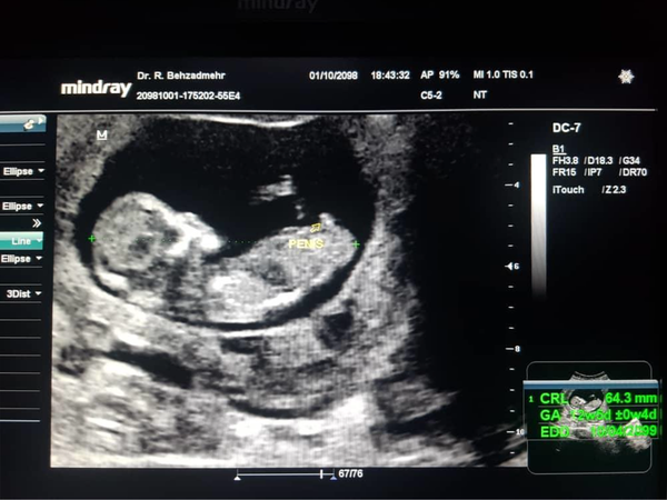
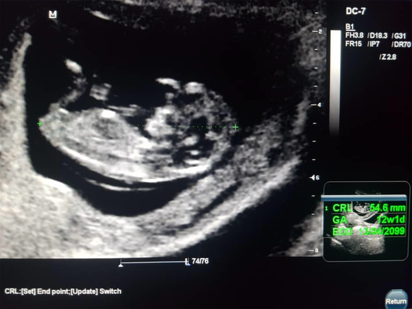
This was a prospective cross-sectional study performed among patients admitted to the radiology clinic of Amir al-Momenin Hospital in Zabol City in the first semester of 2015. In this study, 225 pregnant women with the gestational age of 11 to 14 weeks who were admitted for obstetric ultrasonography were examined with personal consent. The study procedure was explained to the participants, and the information sheet was completed by the operator. Prenatal gender determination was performed for the participants by ultrasonography and the mothers were informed of the result. Information such as phone number, gestational age estimated by ultrasonography, the estimated date of delivery, notification number of each patient, and other related information were recorded in the information sheet. Follow up was done through phone call after the end of pregnancy, the definitive fetus recording of gender and the fetal gender was compared with the ultrasonography result. Mindray DC7 ultrasound device was used. Ultrasonography scans were obtained by a radiologist. Detection of the vulva, clitoris, and labia represents females (Figure 1). Observing scrotum, testicles and penis were allocated to males (Figure 2). Data was recorded in the information sheet after scanning and informing the fetal gender to the pregnant women. Other information and experts' opinion were recorded in the information sheet. In the following, the sensitivity of ultrasonography was calculated by dividing the correct determinations by the sum of the correct determinations and the wrong determinations.
Ultrasound of female fetus at 12 weeks and 3 days

Ultrasound of male fetus at 13 weeks

Ultrasonography specificity was calculated by dividing the correct determinations of the opposite gender by the sum of the correct determinations of the opposite gender and the wrong assignments the positive predictive value was calculated by dividing the correct identification of each gender divided by the sum of the correct and wrong determinations of the desired gender. The negative predictive value was calculated by dividing the correct determinations of the opposite gender divided by the sum of the correct and wrong determinations of the opposite gender. Finally, the overall accuracy was calculated by dividing the correct fetal gender assignments and the correct determinations of the opposite gender by the sum of the correct and wrong identifications and the correct and wrong determinations of the opposite gender. After collecting the data, data analysis was performed by SPSS version 18, and the results were analyzed through descriptive statistics (frequency tables and diagrams). The logistic regression models and ROC curve were used at the significance level of 5% to the significance level of 5% determination accuracy.
4. Results
The ultrasonography results showed that 49.3% of the fetuses were boys, and 50.7% were girls. However, the final result showed that 53.7% of the fetuses were boys, and 46.3% were girls. It was also found that most mothers were at 12 to 13 weeks of gestational age (44.5%), and most of the mothers had BMI 18 to 25 kg/m2 (68.3%; tables 1 & 2).
Sensitivity, Specificity, Positive and Negative Predictive Values of Ultrasonography
| Sensitivity | Specificity | Positive Predictive Value | Negative Predictive Value | Overall Accuracy | |
|---|---|---|---|---|---|
| Ultrasonography | |||||
| Boy | 91.73% | 99.05% | 99.1% | 91.3% | 95.15% |
| Girl | 99.05% | 91.73% | 91.3% | 99.1% | 95.15% |
| Ultrasonography 11 - 12 | |||||
| Boy | 78.26% | 94.73% | 94.73% | 78.26% | 85.71% |
| Girl | 94.73% | 78.26% | 78.26% | 94.73% | 85.71% |
| Ultrasonography 12 - 13 | |||||
| Boy | 91.22% | 100% | 100% | 89.79% | 95.04% |
| Girl | 100% | 91.22% | 89.79% | 100% | 95.04% |
| Ultrasonography 13 - 14 | |||||
| Boy | 100% | 100% | 100% | 100% | 100% |
| Girl | 100 % | 100 % | 100 % | 100 % | 100 % |
Sensitivity, Specificity, Positive and Negative Predictive Value of Ultrasonography in Women by Body Mass Index
| Sensitivity | Specificity | Positive Predictive Value | Negative Predictive Value | Overall Accuracy | |
|---|---|---|---|---|---|
| Ultrasonography less than 18 | |||||
| Boy | 88.88% | 100% | 100% | 87.5% | 93.75% |
| Girl | 100%% | 88.88% | 87.5% | 100% | 93.75% |
| Ultrasonography 18 - 25 | |||||
| Boy | 98.64% | 100% | 100% | 98.78% | 99.35% |
| Girl | 100% | 98.64% | 98.78% | 100% | 99.35% |
| Ultrasonography 25 - 30 | |||||
| Boy | 84.84% | 93.3% | 96.55% | 73.68% | 87.5% |
| Girl | 93.3% | 84.84% | 73.68% | 96.55% | 87.5% |
| Ultrasonography more than 30 | |||||
| Boy | 40% | 100% | 100% | 50% | 62.5% |
| Girl | 100% | 40% | 50% | 100% | 62.5% |
The sensitivity and specificity of ultrasonography for determining male gender were 91.73% and 99.5%, respectively. In addition, the positive and negative predictive values of this method were also equal to 99.1% and 91.3%, respectively, and the overall accuracy of the test was 95.15%. The sensitivity and specificity of ultrasonography for determining the female gender were 99.05% and 91.76%, respectively. In addition, the positive and negative predictive values of this method were equal to 91.3% and 99.1%, respectively, and the overall accuracy of the test was 95.15%.
The sensitivity and specificity of ultrasonography for determining male gender at 11 to 12 weeks were 78.26% and 94.73%, respectively. In addition, the positive and negative predictive values of this method were equal to 94.73% and 78.26%, respectively, and the overall accuracy of the test was 85.71%. The sensitivity and specificity of ultrasonography for determining female gender at 11 to 12 weeks were 94.73% and 78.26%, respectively. Moreover, the positive and negative predictive values of this method were equal to 4.73% and 78.26%, respectively, and the overall accuracy of the test was 85.71%.
The sensitivity and specificity of ultrasonography for determining male gender at 12 to 13 weeks were 91.22% and 100%, respectively. In addition, the positive and negative predictive values of this method were equal to 100% and 89.79%, respectively, and the overall accuracy of the test was 95.04%. The sensitivity and specificity of ultrasonography for determining female gender at 12 to 13 weeks were 91.22% and 100%, respectively. In addition, the positive and negative predictive values of this method were equal to 100% and 89.79%, respectively, and the overall accuracy of the test was 95.04%. The sensitivity and specificity of ultrasonography for determining male gender at 13 to 14 weeks were 100% and 100%, respectively. In addition, the positive and negative predictive values of this method were respectively equal to 100% and 100%, and the overall accuracy of the test was 100%.
The sensitivity and specificity of ultrasonography for determining female gender at 13 to 14 weeks were 100% and 100%, respectively. In addition, the positive and negative predictive values of this method were respectively equal to 100% and 100% and 89.79%, and the overall accuracy of the test was 100%.
As can be observed, the sensitivity and specificity of ultrasonography for determining male gender at less than 18 weeks were 88.88% and 100%, respectively. In addition, the positive and negative predictive values of this method were equal to 100% and 87.5%, respectively, and the overall accuracy of the test was 93.75%.
The sensitivity and specificity of ultrasonography for determining female gender at less than 18 weeks were 88.88% and 100%, respectively. In addition, the positive and negative predictive values of this method were equal to 100% and 87.5%, respectively, and the overall accuracy of the test was 93.75%.
The sensitivity and specificity of ultrasonography for determining male gender at 18 to 25 weeks were 98.64% and 100%, respectively. In addition, the positive and negative predictive values of this method were equal to 100% and 98.78%, respectively, and the overall accuracy of the test was 99.35%.
The sensitivity and specificity of ultrasonography for determining female gender at 18 to 25 weeks were 98.64% and 100%, respectively. In addition, the positive and negative predictive values of this method were respectively equal to 100% and 98.78%, and the overall accuracy of the test was 99.35%.
The sensitivity and specificity of ultrasonography for determining male gender at 25 to 30 weeks were 84.84% and 93.3%, respectively. In addition, the positive and negative predictive values of this method were equal to 96.55% and 73.68%, respectively, and the overall accuracy of the test was 87.5%. The sensitivity and specificity of ultrasonography for determining female gender at 25 to 30 weeks were 84.84% and 93.3%, respectively. In addition, the positive and negative predictive values of this method were equal to 96.55% and 73.68%, respectively, and the overall accuracy of the test was 87.5%.
The sensitivity and specificity of ultrasonography for determining male gender at more than 30 weeks were 40% and 100%, respectively. In addition, the positive and negative predictive values of this method were equal to 100% and 50%, respectively, and the overall accuracy of the test was 62.5%. The sensitivity and specificity of ultrasonography for determining female gender at more than 30 weeks were 40% and 100%, respectively. In addition, the positive and negative predictive values of this method were equal to 100% and 50%, respectively, and the overall accuracy of the test was 62.5%.
5. Discussion
This study aimed to evaluate the ultrasonography sensitivity for determining fetal gender in pregnant women at 11 to 14 weeks of pregnancy in Amir-al-Momenin Hospital, Zabol, Iran, in 2015. In this study, 227 pregnant women were enrolled whose average age was 25.05 ± 5.4 years. The lowest age was 16 years old, and the highest age was 41 years old. The possible outcome of ultrasonography for gender determination showed that 49.3% of the fetuses were boys, and 50.7% were girls. However, the final results showed that 53.7% of the fetuses were boys, and 46.3% were girls. In other words, the consistency of the possible outcomes of ultrasonography for male and female fetal gender determination was 73.91% and 99.05%, respectively.
Tahmasebi conducted a study entitled "the accuracy of ultrasonography for fetal gender determination at 16 to 40 weeks of pregnancy". According to this study, the accuracy of ultrasonography for male and female fetuses was 99.6% and 99.8%, respectively (1). The result of this study is consistent with our findings. Igbinedion (2010) performed a study entitled "the accuracy of ultrasonography for fetal gender determination at the gestational age of less than 16 weeks". The results showed that the sensitivity and value of prenatal gender determination in females (100%) was higher than in males (98.2%) (8). The results of the study are entirely in line with the findings of our study.
Efart et al. conducted a study entitled "fetal gender determination in the first trimester of pregnancy in a Fetal Health Research Centre in London" among 172 pregnant women at 11 to 14 weeks of pregnancy. The rate of male fetuses that were incorrectly reported female was 56% in fetuses aged 11 weeks, 3% in fetuses aged 12 weeks, and 0% in fetuses aged 13 weeks. Accordingly, only 5% of female fetuses were reported incorrectly, and this false positive value was reported 0% at 12 and 13 weeks of gestation (12).
Since ultrasonography is entirely operator-dependent, the high level of accuracy in our study may be due to the high operator experience. Sifrash included mothers with gestational age of less than 16 weeks. Gender identifications over the age of 14 weeks had 100% accuracy. The group success in the first trimester of pregnancy (11 to 14 weeks) was 75%. The results had less accuracy for fetuses less than 12 weeks of gestation. However, the success rate was 54%. Male fetuses less than 13 weeks of gestation did not have the necessary conditions for a definitive determination (5).
In our study, the gender determination accuracy for male fetuses was 85.71% at 11 to 12 weeks, 85.71% at 12 to 13 weeks, and 100% at 13 to 14 weeks. The resulting difference may be due to operator experience and the differences of the studied population in the two studies. Our results showed that ultrasonography at 11 to 14 weeks had high sensitivity and specificity in gender determination, and this sensitivity was higher in girls. In addition, this sensitivity is enhanced with increasing gestational age in both genders. It was also found that the highest sensitivity and specificity for determining both genders were in women with normal BMI 18 to 25 kg/m2, followed by in women with BMI less than 18 kg/m2, in those with BMI 25-30 kg/m2, and finally, in women with a BMI greater than 30 kg/m2. With decreasing or increasing BMI, the gender identification power reduced.
The results of Gharekhanloo showed that in 150 women, the gender was identified as female in 32 (21.3%) cases, male in 65 (43.3%), and not specified in 53 (35.3%); overall, gender identification was performed in 64.6% of the cases. A total of 57 male fetuses were correctly identified as boys, and 8 female fetuses were wrongly identified as boys. The positive predictive value for the ultrasound imaging gender identification was 87.6% for the male fetuses and 96.8% for female fetuses (14).
The results obtained by Awad showed that 13 (76%) of the responses indicated that fetal gender determination by ultrasound has a high precision in the second trimester (13 - 27 weeks of gestational age), while the remaining 4 (24%) found that gender can be identified in the first trimester. Of the 52 women with ultrasound scans, 50 (96%) knew about the fetal gender during pregnancy, while only 2 (4%) answered no. Finally, in 46 (92%) of the women, the fetal gender was the same as what was determined by ultrasound, while only in 4 (8%) of the women the ultrasound result was wrong. The overall accuracy to correctly determine fetal gender was 92% (15).
5.1. Conclusion
The present study is the first step toward more comprehensive studies. In the case of approving the present results and the findings of other respected medical communities, gender-linked disorders will be detected earlier by adding ultrasonography at 11 to 14 weeks of pregnancy.
References
-
1.
Morteza T, Nasim N, Negar A, Mohammad B, Armaghan MA, Mohammad RJ, et al. Accuracy of ultrasonography in fetal gender determination at 16 to 40 weeks of pregnancy. Sci Med J. 2008;7(3).
-
2.
Whitlow BJ, Lazanakis MS, Economides DL. The sonographic identification of fetal gender from 11 to 14 weeks of gestation. Ultrasound Obstet Gynecol. 2000;15(3):262-3. [PubMed ID: 10380291]. https://doi.org/10.1046/j.1469-0705.1999.13050301.x.
-
3.
Ekele BA, Maaji SM, Bello SO, Morhason-Bello IO. Profile of women seeking fetal gender at ultrasound in a nigerian obstetric population. Ultrasound. 2008;16(4):199-202. https://doi.org/10.1179/174313408x353837.
-
4.
Mubuuke AG, Kiguli-Malwadde E, Byanyima R, Businge F. Evaluation of community based education and service courses for undergraduate radiography students at Makeree University, Uganda. Rural Remote Health. 2008;8(4):976. [PubMed ID: 19063589].
-
5.
Gelaw SM, Bisrat H. The role of ultrasound in determining fetal sex. Ethiop J Health Dev. 2016;25(3).
-
6.
Youssef A, Arcangeli T, Radico D, Contro E, Guasina F, Bellussi F, et al. Accuracy of fetal gender determination in the first trimester using three-dimensional ultrasound. Ultrasound Obstet Gynecol. 2011;37(5):557-61. [PubMed ID: 20814877]. https://doi.org/10.1002/uog.8812.
-
7.
Pajkrt E, Chitty LS. Prenatal gender determination and the diagnosis of genital anomalies. BJU Int. 2004;93 Suppl 3:12-9. [PubMed ID: 15086437]. https://doi.org/10.1111/j.1464-410X.2004.04704.x.
-
8.
Igbinedion BO, Akhigbe TO. The accuracy of 2D ultrasound prenatal sex determination. Niger Med J. 2012;53(2):71-5. [PubMed ID: 23271849]. [PubMed Central ID: PMC3530251]. https://doi.org/10.4103/0300-1652.103545.
-
9.
Hackett LK, Tarsa M, Wolfson TJ, Kaplan G, Vaux KK, Pretorius DH. Use of multiplanar 3-dimensional ultrasonography for prenatal sex identification. J Ultrasound Med. 2010;29(2):195-202. [PubMed ID: 20103789]. https://doi.org/10.7863/jum.2010.29.2.195.
-
10.
Hsiao CH, Wang HC, Hsieh CF, Hsu JJ. Fetal gender screening by ultrasound at 11 to 13(+6) weeks. Acta Obstet Gynecol Scand. 2008;87(1):8-13. [PubMed ID: 17851807]. https://doi.org/10.1080/00016340701571905.
-
11.
Begum A, Saha M, Milah SR, Hosian G. Ultrasonographic determination of fetal sex -a study on 630 cases in bangladesh. J Nepal Med Assoc. 2003;40(139):128-33. https://doi.org/10.31729/jnma.831.
-
12.
Efrat Z, Perri T, Ramati E, Tugendreich D, Meizner I. Fetal gender assignment by first-trimester ultrasound. Ultrasound Obstet Gynecol. 2006;27(6):619-21. [PubMed ID: 16493625]. https://doi.org/10.1002/uog.2674.
-
13.
Cunningham F, Levevo K, Bloom L, Hauth C, Rouse D, Spong Y. Williams obstetrics. 24th ed. new york city: Mcgraw-Hill; 2014.
-
14.
Gharekhanloo F. The ultrasound identification of fetal gender at the gestational age of 11-12 weeks. J Family Med Prim Care. 2018;7(1):210-2. [PubMed ID: 29915761]. [PubMed Central ID: PMC5958571]. https://doi.org/10.4103/jfmpc.jfmpc_180_17.
-
15.
Awad IA, AI-Safwani ZM. The accuracy of sonographic determination of fetal gender. Int J Health Sci Res. 2016;6(7).