Abstract
Background:
None of plantar arch characteristics such as arch height, heel varus and arch flexibility can affect balance indices in different manners.Objectives:
The current study aimed to evaluate the relation between these structural characteristics and balance indices.Patients and Methods:
The study population was 100 male and female students from Semnan University of Medical Sciences, Semnan, Iran. To evaluate plantar arch, indices such as heel height, arch angle index, rear angle, navicular drop, longitudinal arch angle and footprint angle were recorded and Staheli and Denis methods were used. Static and dynamic data were recorded using the Biodex Balance System.Results:
There was a significant correlation between arch height, and static and dynamic balance indices (maximum correlation coefficient = 0.46, moderate correlation coefficient); also, there was a direct significant correlation between rear and footprint angles and some other balance indices (correlation coefficient = 0.2, weak correlation). There was a significant inverse association between navicular drop and balance indices (maximum correlation coefficient = 0.3, moderate correlation coefficient). Evaluating arch and longitudinal arch angle indices by the Staheli and Denis correlation methods showed insignificant association between these variables and balance indices (P > 0.05).Conclusions:
It seems that plantar changes have insignificant effect on static and dynamic indices evaluated by the Biodex Balance System.Keywords
1. Background
Foot performance significantly depends on its shape. Biomechanical foot changes affect its dynamic stability (1). Foot is the last part of lowest extremity, and this small supporting surface provides balance of the entire body. It seems that any biomechanical changes in this supporting surface may affect body posture. To evaluate plantar structure, different static and dynamic clinical methods have been introduced, but none of them provide an specific index to evaluate arch changes (1). Foot injuries may change walking mechanism (2). Foot performance is greatly influenced by its shape (3, 4). Studies conducted in this field indicated that there is no method to categorize the plantar arch (5). In clinical evaluations, plantar arch measuring methods are usually based on the foot structure. Different methods have been provided in this regard (6). Considering the fact that foot arch plays an important role in distribution of body weight on legs, neutralizing weight impacts and conforming feet to different levels of land during walking, any changes in the structure of plantar foot may disrupt the relation between line of gravity and supportive surface (7). The situation of gravity line with supportive surface and detailed mechanical axis is one of the factors affecting body balance (8-10). Almost 80% of people are dealing with foot disorders, especially flat feet (2), and different age groups spend great expenses over insoles and surgeries (5). Plantar arch structure can predict resulting problems. On the other hand, plantar arch changes may affect body balance and walking (6). In the study conducted by Khramtsov et al. the level of stability in 112 children with normal arch and flat feet was evaluated. The level of vertical stability in cases with flat feet was lower than those with normal arch (11). Cote et al. evaluated the effect of increased and decreased plantar arch on dynamic and static balance indices. They categorized cases in three groups of 16 each based on navicular drop. Results showed that the level of stability in pronated feet was higher than supinated feet. There was no significant difference between these two groups and those with normal plantar arch (12). Differences in dynamic and static stabilities were studied by Dicherry through evaluating talonavicular dynamic stability in walking. Level of navicular drop in 72 healthy athletes was evaluated using two static methods. There was no significant difference in navicular drop between different types of feet, during walking and running. Small differences were reported only in running between those with low mobility and high mobility foot (13). Franettovich et al. evaluated the ability to predict dynamic foot position, using static measurements. According to their results, arch height and the relation in two feet were highly associated with static and dynamic measurements (14). Cobb et al. studied the effects of forefoot heel varus on single leg upright standing postural stability. According to their results, postural stability was measured in the two groups of 20 each (first group with varus ≥ 70°, the second group with varus ≤ 70°). Anterior-posterior instability in the first group was significantly higher than the second one (15). Totally, few studies were conducted on biomechanical effect of feet on body balance (16); since it is not obvious that which clinical method is preferred to evaluate plantar arch, it seems that none of these methods can singly categorize plantar arch to normal, reduced and increased groups (2). Therefore, in the current study, the following points can be mentioned: first, plantar arch characteristics dealing with balance disorders could be identified; second, considering the defects of conventional methods, cases under study were not randomly categorized into decreased and increased plantar arch groups. Nonetheless, their plantar arch characteristics, which singly affect walking and balance disorders and apply abnormal forces on the joints were evaluated and the effect of each one of these characteristics on balance indices was studied. According to the conducted studies, increase and decrease in plantar arch may interrupt body balance, but the point is still unclear ignoring the amount of increasing or decreasing the amount of arch height, which characteristic is more associated with balance disorder.
2. Objectives
The current study aimed to evaluate the relation between these structural characteristics and balance indices.
3. Patients and Methods
This cross-sectional study was designed to examine the association of some clinical measurements of foot arch and static and dynamic stability indicators in Neuromuscular Rehabilitation Research Center (Semnan University of Medical Sciences, Iran). The studied participants were volunteer male and female students of Semnan University of Medical Sciences, Semnan, Iran. The sampling method was simple and none-probable. Based on previous studies, the sample size of 100 patients (feet) was determined. Personal and demographic information of participants were recorded using a questionnaire (Table 1). Participants with pain or other problems (surgeries in the past three months, history of dislocation or semi-dislocation of the ankle, any other significant joint injuries in the lower extremity), use of corticosteroids, fractures/breaks in lower extremity, addiction (cigarettes, drugs and alcohol), any significant deformities of the lower extremity (extreme genu varum and genu valgum), any who worked out drastically, and those with body mass index of less than 18.5 or over 24.9 were excluded from the study. Participants were studied in specific times of the day only after obtaining informed consent and becoming familiar with different stages of investigation. The study was approved by the Ethical Committee of Semnan University of Medical Sciences and all participants completed the consent form. For clinical evaluation of foot arch, different measuring methods of footprints and direct foot measuring methods were used. The indicators related to the evaluation of arch of foot were measured in 50% weight baring position of the subjects. Both lower extremities were placed on two equally heighted scales. Subjects were asked to vertically place their weight on the scales without shifting it to either side; measurements were performed under these conditions. To study footprint indicators, participants put their feet in a plate of ink and placed them on a sheet of paper placed on the scales. This was performed once for the right foot and once for the left one. Clinical indicators of longitudinal arch and footprint measurements were performed similarly as follows. Arch length: measures the distance between the highest points of the medial longitudinal arch to the ground using a ruler. Longitudinal arch angle: Using a rule m set-square between the longitudinal lines that connect the medial malleolus to the medial tubercle navicular and the first metatarsal head was determined. Rearangle: Using a m rule, the angel between the longitudinal line of the heel bone where divided into two parts with the line that divides one third of distal leg line into two halves was determined.
Mean and Standard Deviation of Demographic Variables
| Variable | Mean | Standard Deviation |
|---|---|---|
| Age, y | 20.73 | 1.248 |
| Weight, kg | 63.88 | 11.72 |
| Height, cm | 168.3 | 9.08 |
Navicular drop: first, the distance of tubercle navicular from the floor was determined in a way that the foot was on the floor without bearing weight. Then subjects were asked to put 50% of his or her weight on the foot. Tubercle displacement in the sagittal plane was assessed with a ruler. Arch length indicator: The ratio of the length of the inner boundary line between the most medial point of the metatarsal heads and heel over the enclosed arch length between these points, which were determined with a ruler. Arch (footprint) angel: is the angle between the inner edge of the footprint and the line that connects the most medial point of the metatarsal and the highest point of the heel. The Stahly method: The width of the arch of the footprint is divided by the heel width. The obtained ratio is called arch indicator.
Denis method: according to this method, the footprint is divided in three ways:
Grade 1: the width of the supporting surface of central region of the leg is a half of the supporting surface of the metatarsal, Grade 2: the width of the supporting surface of central region of the leg is equal to the supporting surface of the metatarsal, Grade 3: the width of the supporting surface of central region of the leg is larger than the supporting surface of the metatarsal, Grades 1 and 2 are considered flatfoot, The information of balance indicators was obtained using the Biodex Balance system manufactured in the Biodex Company of the United States of America. Participants were standing barefooted on the device, and the test performed as follows; first, subjects stood handles in a predefined condition on the balance disk in a way that his or her center point of gravity would overlap the center point of coordinate on the balance disk; and the balance disk was completely placed horizontally. Device monitor was adjusted according to the height of subjects. Then subjects stayed possible to determine the position of the heel and the angle of the toes. This was used so all the measurements were recorded in the same position. After announcing readiness and pressing the start button, subjects maintained the referred position for 20 seconds handles or changing position of their hands. Each test was performed three times, and an average of these three replications was recorded as the score of participants for sensory-motor indicators in test. Then both postural stability in the stationary state (standing on one foot) and a dynamic test in two conditions of open and closed eyes were performed. In static balance test, the balance disk under the participant's foot was fixed, and during the whole test and with minimal fluctuation, he or shetried to maintain the center of pressure and the center of a circle displayed on the screen by repositioning the body. This test was performed three times with 10 minutes break intervals. In dynamic balance test, the balance disk under the participant foot swayed, and during the whole test and with minimal fluctuation, he or she tried to maintain the center of pressure and center of a circle displayed on the screen by repositioning the body. The test was performed with two different levels of stability index of biodex system: leval 3 and 6. The rest was similar to the static balance test. At the end of each test, data including average scores of three replications from each test, general balance; anterior-posterior and internal-external indicators were collected. The order of recording the balance conditions was randomized, and for each participant these orders were drawn randomly. According to the normal distribution of data confirmed by Kolmogorov-Smirnov test, Pearson's correlation coefficient was used to determine the association of each test results with obtained results from the Biodex balance indicators.
4. Results
Normal distribution of data was confirmed using Kolmogorov-Smirnov test. The correlation between balance indicators and clinical arch indicators was determined using Pearson correlation test.
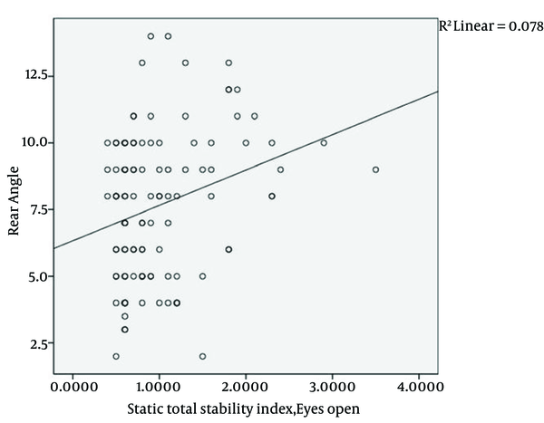
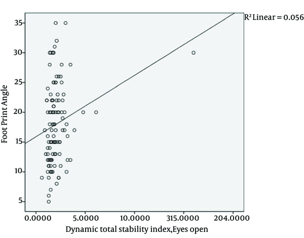
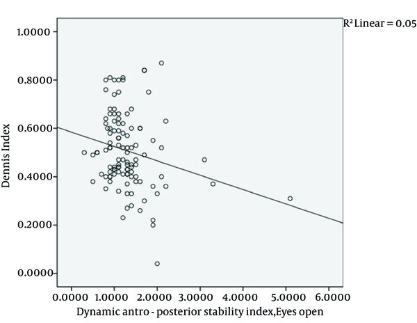
Our results showed a correlation between the length of arch and almost all of the static and dynamic balance indicators. Maximum correlation coefficient in these indicators was 0.46, which showed a medium correlation (Figure 1). Determining the correlation between rear angel with the indicators of balance showed a positive correlation in three general conditions of opened eyes, anterior-posterior opened eyes, and interior-exterior opened eyes, and an interior-exterior dynamic balance with opened eyes; however, the correlation coefficient in these indicators was 0.2, which showed a weak correlation (Figure 2). Determining the correlation between footprint and indicators of balance had a meaningful correlation only with two states of dynamic balance in overall conditions of opened eyes and anterior-posterior opened eyes. The correlation coefficient was less than 0.2, which showed a weak correlation (Figure 3). There was no association between the Denis method and balance indicators (P > 0.05) (Figure 4).
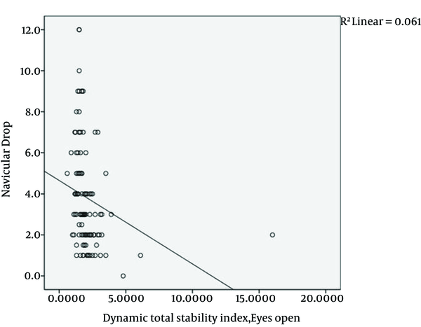
With most balance indicators including both static and dynamic, a negative correlation was shown when investigating the correlation of navicular drop with balance indicators. The maximum correlation coefficient among these indicators was 0.3, which showed a medium correlation (Figure 5). There were no meaningful associations between Stahly methods, arch index and longitudinal archangel with balance indicators (P > 0.05).
The Association Between Arch Height and Static Total Stability Index, Eyes Open

The Association Between Rear Angle and Static Total Stability Index, Eyes Open

The Association Between Foot Print Angle and Dynamic Total Stability Index, Eyes Open

The Association Between Dennis Index and Dynamic Antro-Posterior Stability Index, Eyes Open

The Association Between Navicular Drop and Dynamic Total Stability Index, Eyes Open

5. Discussion
There was a meaningful correlation between static and dynamic indicators of balance and clinical indicators of arch length, navicular drop, and rear angel; however, there was no meaningful association between balance indicators and longitudinal index, arch index, footprint angel, Denis method and Stahly method. Current study was the only study investigating the associations of foot structure and balance indicators. In this research, we focused more on characteristics of shape of foot and its effects on balance indicators, rather than considering the faults of traditional methods of classifying people into stereotypical groups with decreased or increased foot arches. In evaluating balance systems, various factors such as proprioceptive, vestibular system and visual perception were involved. The proprioceptive is a feedback system in the body, which allows us to perceive the position of head and body in space. Studies showed that the feedback from the proprioceptive system in movement, depends not only on the sensory information receptors, but also on the information provided by mechanical, cutaneous, articular and muscular receptors (17). Most of people activity is performed in dynamic balance areas, which are the opposite of static balance (18). Therefore, in this research it was decided to evaluate static balance, in addition to dynamic balance by itself which requires more muscle strength, neuromuscular control and more accurate proprioception on the lower extremity joints, which allows a person to maintain his or her dynamic balance to support the surface of the Biodex device (19).Afferent feedbacks from the feet area are considered as proprioceptive, which affects balance. Since joints, skin and muscles are the main sources of proprioception, foot shape characteristics can affect the angle of skin, joint and muscle tension and therefore can affect afferent feedback for postural control and balance of the body (17). Therefore, in this research, the association between some foot characteristics such as arch height, navicular drop, rear angle, longitudinal arch angle, arch index, Denis method, Stahly methods, footprint angle and static and dynamic balance were investigated. However, this study did not show any strong association between these indices. For example, in evaluating navicular drop, which basically indicates the degree of arch flexibility, it was shown that in patients with flexible flat foot, less balance disturbances has been reported. These patients have normal foot arches or even increased arch when there is no weight bearing, but upon connection with the supporting surface and putting pressure and weight on the lower extremity, the arch decreases and foot flattens. These patients had a better balance compared to others. This can be attributed to increased contact points in foot during weight bearing, which results in increased stimulation of plantar cutaneous receptors among others. On the other hand, greater plasticity in feet makes better adaptation to different levels. This case is consistent with a study performed by Dicharry (13). In studying the arch height, it was discovered that when the arch height is less, the balance is better. These results are consistent with the study of Lin CH, et al. probably due to increased connection points of the foot with the ground, which in turn improves proprioception and balance (16). In this study, it was shown that with increasing rear angle, which increases the angle of heel valgus, balance disruption was more. It seems that each change in the alignment of the heels, which is the junction of the muscles and ligaments of the foot could change the muscle stretch angles and inactive elements around joints leading to incorrect and inaccurate messages from the foot to the central nervous system, which can in turn affect the balance. Moreover, development of heel valgus results in limited contact of the heel with the ground surface; therefore, fewer sensory receptors participate in sending necessary information to maintain balance. In this regard, a similar study was performed by Cobb et al. (15). Although three arch length, rear angel, and navicular drop indicators showed somewhat strong correlation with static and dynamic indicators of balance, this correlation coefficient was low and insignificant statistically, and not very important clinically. More investigations are needed to find an accurate answer to this question. On the other hand, examining indicators in this study showed no significant association with balance indicators. In this regard, Lin et al. performed a similar study on 64 children. They measured some other common parameters such as print, height, length, width and arch angle. In this study, balance and foot arch indicators were evaluated using a force plate, a light source and a digital camera with a reflecting mirror. The ability to maintain a state by means of analyzing swing area, proprioceptive and visual perception conditions was determined for each participant in different situations. The association between arch height of the foot and the swing area showed a mild correlation only in children with closed eyes standing on foam, and this correlation was not observed in other situations. In addition, children with lower arch height had better balance. However, the correlations obtained in this study were very weak and moderate (16). Since the vestibular system and vision perception can be involved in balance as the proprioceptive sense, impaired proprioceptive information, which comes only from the joints, skin and muscles of the foot, cannot have a close association with monitoring situations. Although with elimination of visual perception in this study, there was no increase in the association of posture control and proprioceptive, which could reflect the importance of other joint proprioceptive or vestibular systems (17). In this study, the role of vestibular system was not removed, especially when plays a major role in creating a balance on moving surfaces. Even though turbulence intensity levels in this study were not too high, it seems that information from vestibular system could compensate deficient information received from arch of the foot and/or visual sensors (20). Whether clinically or foot printing, investigations on indicators of foot shape and its arch may not be a full assessment of the arch and unable to show the arch and distribution of forces exerted on the soles of the feet. While evaluations performed with analyzing system of walking or force plate gait assess the arch more from dynamic views (21). The association between foot structure and balance can be separated from neuromuscular and musculoskeletal characteristics. From neuromuscular point of view, any changes in foot could affect very well strategized muscles through stimulating afferent peripheral by changing contact or secondarily changes of the angles of joints. From an anatomical perspective, leg is the lowest part of the lower extremity, which is a small part in maintaining the body position especially in standing on one foot. From biomechanical aspects, little different foot alignment can affect balance strategies. For maintaininwg balance, coordination between postural muscles of the lower extremities and trunk is essential and controlling body position requires a motion analysis system for knees, hip, spine and muscle activities (16).
It seems that foot structure changes do not have much effect on the indicators of static and dynamic balance evaluated by the Biodex balance system. More studies using other body positioning analysis systems and dynamic tools are required to analyze foot biomechanics.
5.1. Suggestions
1- Performing this study on all age groups.
2- Investigating other clinical indicators and footprints not studied in this research for their relationship with balance indicators.
3- Using all other parameters involved in the assessment of balance in addition to the kinematic parameters; the kinetic parameters should also be evaluated.
4- It is proposed to use Electromyography to examine active role of muscles in patients with different foot arches.
Acknowledgements
References
-
1.
Alcalay J, Lederman N, Kornbrot B. The diagnosis of pes planus and pes cavus in soldiers by the foot-ground pressure pattern. Mil Med. 1985;150(4):215-7. [PubMed ID: 3925378].
-
2.
Hogan MT, Staheli LT. Arch height and lower limb pain: an adult civilian study. Foot Ankle Int. 2002;23(1):43-7. [PubMed ID: 11822691].
-
3.
Saltzman CL, Nawoczenski DA, Talbot KD. Measurement of the medial longitudinal arch. Arch Phys Med Rehabil. 1995;76(1):45-9. https://doi.org/10.1016/S0003-9993(95)80041-7.
-
4.
Rose GK, Welton EA, Marshall T. The diagnosis of flat foot in the child. Bone Joint J. 1985;67(1):71-8.
-
5.
Nawoczenski DA, Saltzman CL, Cook TM. The effect of foot structure on the three-dimensional kinematic coupling behavior of the leg and rear foot. Phys Ther. 1998;78(4):404-16. [PubMed ID: 9555923].
-
6.
Razeghi M, Batt ME. Foot type classification: a critical review of current methods. Gait Posture. 2002;15(3):282-91. [PubMed ID: 11983503].
-
7.
Levangie P, Norkin C. Joint Structure and Function. 4th ed. F.A. Davis Company; 2005. p. 465-661.
-
8.
Sarafraz Z. Basics of kinesiology and biomehanics of motion system. University of Social Welfare and Rehabilitation; 2006. p. 50-4.
-
9.
Stewart SF. Human gait and the human foot: an ethnological study of flatfoot. II. Clin Orthop Relat Res. 1970;70:124-32. [PubMed ID: 5463031].
-
10.
Volpon JB. Footprint analysis during the growth period. J Pediatr Orthop. 1994;14(1):83-5. [PubMed ID: 8113378].
-
11.
Khramtsov PI, Kurganskii AM. [Functional stability of the vertical posture in children depending on foot arch condition]. Vestn Ross Akad Med Nauk. 2009;(5):41-4. [PubMed ID: 19507353].
-
12.
Cote KP, Brunet ME, Gansneder BM, Shultz SJ. Effects of Pronated and Supinated Foot Postures on Static and Dynamic Postural Stability. J Athl Train. 2005;40(1):41-6. [PubMed ID: 15902323].
-
13.
Dicharry JM, Franz JR, Della Croce U, Wilder RP, Riley PO, Kerrigan DC. Differences in static and dynamic measures in evaluation of talonavicular mobility in gait. J Orthop Sports Phys Ther. 2009;39(8):628-34. [PubMed ID: 19648718]. https://doi.org/10.2519/jospt.2009.2968.
-
14.
Franettovich MM, McPoil TG, Russell T, Skardoon G, Vicenzino B. The ability to predict dynamic foot posture from static measurements. J Am Podiatr Med Assoc. 2007;97(2):115-20. [PubMed ID: 17369317].
-
15.
Cobb SC, Tis LL, Johnson BF, Higbie EJ. The effect of forefoot varus on postural stability. J Orthop Sports Phys Ther. 2004;34(2):79-85. [PubMed ID: 15043000]. https://doi.org/10.2519/jospt.2004.34.2.79.
-
16.
Lin CH, Lee HY, Chen JJ, Lee HM, Kuo MD. Development of a quantitative assessment system for correlation analysis of footprint parameters to postural control in children. Physiol Meas. 2006;27(2):119-30. [PubMed ID: 16400199]. https://doi.org/10.1088/0967-3334/27/2/003.
-
17.
Riemann BL, Lephart SM. The Sensorimotor System, Part II: The Role of Proprioception in Motor Control and Functional Joint Stability. J Athl Train. 2002;37(1):80-4. [PubMed ID: 16558671].
-
18.
Bernier JN, Perrin DH. Effect of coordination training on proprioception of the functionally unstable ankle. J Orthop Sports Phys Ther. 1998;27(4):264-75. [PubMed ID: 9549710]. https://doi.org/10.2519/jospt.1998.27.4.264.
-
19.
Olmsted LC, Carcia CR, Hertel J, Shultz SJ. Efficacy of the Star Excursion Balance Tests in Detecting Reach Deficits in Subjects With Chronic Ankle Instability. J Athl Train. 2002;37(4):501-6. [PubMed ID: 12937574].
-
20.
Shumway Cook A, Woollacott MH. Motor Control. 3th ed. Washington: Lippincott Williams & Wilkins; 2007. p. 121-5.
-
21.
Van Schie CHM, Boulton AJM. The effect of arch height and body mass on plantar pressure. Health Manag Publication Inc. 2000;12(4):88-95.
.png)