Abstract
Introduction:
Pituitary adenoma producing symptomatic carotid compression of the internal carotid artery without any apoplexy sign would be extremely rare and there was only one report regarding to this condition.Case Presentation:
In this case report we have described a 57-year-old woman with a nonfunctional pituitary macro adenoma which has resulted to symptomatic internal carotid occlusion. Magnetic resonance imaging (MRI) revealed a large pituitary adenoma caused tight stenosis of right internal carotid. The patient has also experienced the transient ischemic attack which has confirmed to be the cause of internal carotid artery occlusion by this macro adenoma tumor. There was not any sign of apoplexy at the time of admission and the patient has not shown a history of pituitary adenoma. The patient then has undergone an endonasal transsphenoidal resection because of this nonfunctional pituitary adenoma.Conclusions:
Pituitary macro adenoma producing symptomatic internal carotid occlusion might develop to several serious conditions including transient ischemic attack. Urgent surgical procedure might be the best approach to prevent further severe complications in such patients.Keywords
1. Introduction
Pituitary adenoma (PA), a lesion arising from adenohypophysial cells, would be one of the most prevalent neuroendocrine intracranial tumors that accounted for 10% of all primary intracranial t111umors (1). Pituitary adenomas were usually benign lesions and less than 10 millimeters (mm). If they have grown to larger sizes they might result in compression with surrounding structures including cavernous sinus (2, 3). It has demonstrated that invasion of the cavernous sinus by tumors might lead to the compression and occlusion of the internal carotid artery (ICA) (4). The consequence of this event would be the presence of various clinical manifestations (4). On the other hand, the prevalence of pituitary adenomas has increased in the last few years (5). Some studies have also indicated the genetic predisposition of pituitary adenoma occurrence in individuals (6, 7). Despite huge pituitary tumors have reported as producer of stenosis or occlusion of the ICA, pituitary adenomas causing carotid compression of the ICA were rarely symptomatic (8-10). In addition, the incidence was extremely rare when there was no evidence of apoplectic event. To the best of our knowledge there was only one case report that has presented a patient with pituitary adenoma producing symptomatic carotid compression of the ICA without any apoplectic signs. In this case report we have described a 57 year old woman presenting with transient ischemic attacks (TIA) and a huge nonfunctional pituitary adenoma which produced symptomatic internal carotid occlusion. Patient has signed an informed consent for publishing data regarding her disease without her name divulged.
2. Case Presentation
A 57-year-old woman has referred to the neurology department of the Loghman Hakim hospital with complaints of sudden left hemiparesis involving her face and left extremities. She had an episode of transient hemiparesis one week before admission that lasts 12 hours. The patient has also suffered from disequilibrium, dizziness and vertigo. Physical examinations have revealed severe left hemiparesis [1/5]; mild left central facial paresis and bitemporal hemianopsis. Other neurologic examinations were normal. Patient's medical history was negative for cerebrovascular or cardiovascular diseases or other risk factors of stroke. She has declared no craniofacial trauma.
2.1. Diagnosis
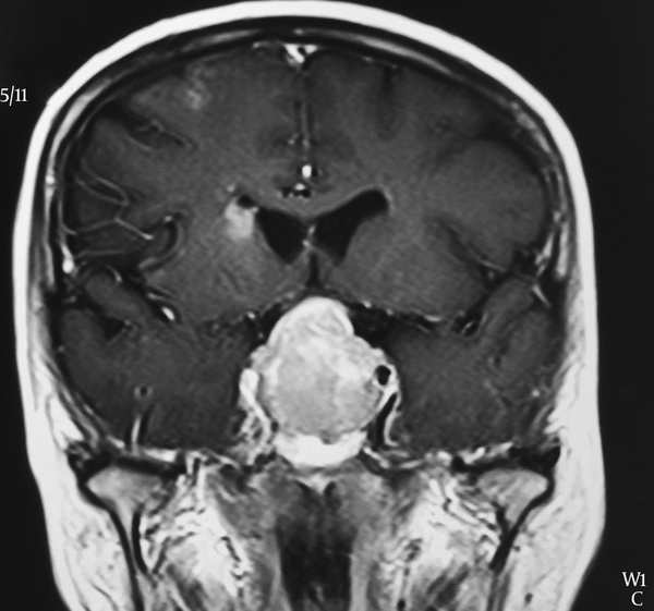
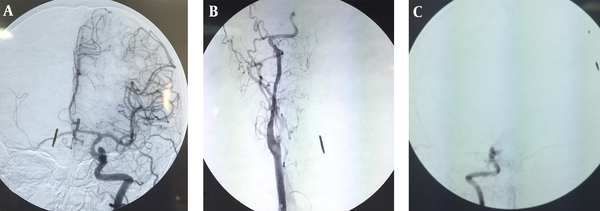
Magnetic resonance imaging (MRI) has revealed two distinct findings: high signal abnormality indicating ischemic injury in periventricular white matter and a huge sellar-suprasellar mass that has encased both of internal carotid arteries. After Gadolinium injection the mass has become heterogeneously enhanced and the right internal carotid artery has appeared to be occluded (Figure 1). Color doppler ultrasound sonography (CDUS) of carotid arteries and vertebral arteries has shown high resistance monophasic wave form with no retrograde flow in diastole in proximal part of right ICA. There was no significant plaque formation. These findings have suggested the presence of either occlusion or severe stenosis of internal carotid artery in distal region of right ICA. Spiral computed tomography (CT) scan of brain has revealed an intra sellar mass 31 × 21 mm. CT scan findings of the other areas were normal. Electrocardiogram and echocardiography have demonstrated no arrhythmia, valvular abnormality or vegetation. Digital subtraction angiography (DSA) has confirmed the occlusion in right internal carotid artery (Figure 2). She had high level of prolactin (threefold of normal limit) in laboratory investigation and other hormones were within normal limits.
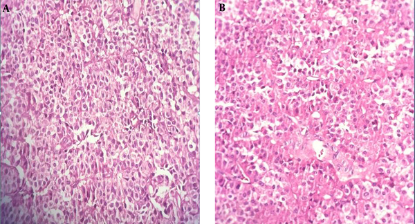
Histopathology: Nonfunctional Pituitary Adenoma Without Atypical Invasive Features, Hemorrhage or Infarction

MRI Has Revealed a Large Pituitary Adenoma Resulted in Tight Stenosis of Right Internal Carotid

Angiography Has Revealed Complete Occlusion of the Right Internal Carotid Artery

2.2. Surgery
The patient has referred to our neurosurgery ward. She has undergone endonasal trans-sphenoidal surgery and the mass has resected. Sella has reconstructed as stage I according to Jalessi et al.’s (11) classification. Histopathologic examination has shown sheets of monomorphic polygona epithelial cells with amphophilic cytoplasms and round nuclei mostly in diffuse pattern with rare mitotic figures consistent with non-functional pituitary adenoma. No evidence of necrosis or hemorrhage has observed (Figure 1 A and B).
2.3. Post Operation
She has shown an uncomplicated post operation course and has discharged 6 days after surgery. Hemiparesis has improved with physiotherapy and other rehabilitation techniques during six month follow up.
3. Discussion
Pituitary adenoma was the third most common intracranial neoplasms (12). Approximately 40 percent of all pituitary adenomas were macroadenomas which their growth might invade suprasellar area, cavernous sinus and sphenoid sinus. Internal carotid occlusion in cavernous sinus region has numerously reported in the setting of pituitary apoplexy (13, 14). In addition, several studies have indicated that pituitary adenoma is an important precursor of pituitary apoplexy (15-18). Although cases of carotid arteries occlusion due to other intracranial lesions such as tumors (19) and huge aneurysms (20) have reported, compression and occlusion of ICA with pituitary adenoma were very rare especially in cases without apoplectic evidence. Molitch et al. (8) has evaluated the rate of internal carotid artery occlusion caused by pituitary adenomas. In their study 141 patients with cavernous sinus invasion have caused by different types of tumor have investigated. Eighty three patients had carotid artery encasement with 58 cases of them having pituitary adenoma. They have found that only one out of 58 cases (1.7%) has developed compression of the artery. This result has shown that pituitary adenoma producing internal carotid artery occlusion was extremely rare. In literature review we have found five case reports describing pituitary tumors producing carotid artery occlusion without the presence of apoplectic events (10, 21-24). Table 1 has summarized the details of these reports.
Cases of Pituitary Adenomas Causing Vascular Occlusion Without Apoplectic Signs
| Authors | Age/Gender | Clinical Manifestation | Tumor Size | Vascular Occlusion | Anatomical site of the Tumor | Treatment | Type of Adenoma |
|---|---|---|---|---|---|---|---|
| Yaghmai et al. 1996 (24) | 47/M | progressive loss of vision + severe headaches | 3 × 2.5-cm | supraclinoid portion of the right ICA | Area of the sella. and extended laterally toward both cavernous sinuses, | transsphenoidal resection | Non-functional |
| Spallone 1981 (10) | 55/F | TIA + left hemiplagia | Not mentioned | Right supracavernous ICA | Not mentioned | None | Not mentioned |
| Cavalcanti et al. 1997 (25) | 54/ F | Visual loss + right hemiparesis | Not mentioned | Narrowing of right supraclinoid ICA | sellar-suprasellar lesion | Craniotomy | Non-functional |
| Alentorn et al. 2011 (22) | 65/M | Acute aphasia Right hemiparesis | 4.2 × 3.3 × 4.2-cm | left internal carotid artery in the left cavernous sinus | macro adenoma in the pituitary region | endoscopic transsphenoidal subtotal resection | Non-functional |
| Rey-Dios et al. 2014 (23) | 48/M | TIA | Not mentioned | left ICA and severe stenosis of the right ICA at the level of the clinoid process | large pituitary adenoma encasing and narrowing both ICA | endonasal transsphenoidal resection | Not mentioned |
| This case | 57/F | TIA + left hemiparesis | 31 × 21 mm | internal carotid artery in distal region of right ICA | huge sellar-suprasellar mass that encased both of internal carotid arteries | endoscopic transsphenoidal subtotal resection | Non-functional |
The microscopic or endoscopic transsphenoidal surgery has emerged as the appropriate approach in cases with pituitary adenoma. In our case report the patient has presented with TIA without the evidence of apoplexy. Histopathologic findings has indicated that the tumor was a nonfunctional pituitary adenoma with no intra tumoral hemorrhage and no necrosis which was in consistent with lack of the apoplectic signs. Angiography in this case has confirmed the occlusion in right internal carotid artery that appears to be due to direct compression effect of the mass. Rey-Dios et al. (23) has reported a 48-year-old patient with right-hand weakness, left hemiparesis and blurred speech. He has experienced TIA which led to stroke. Further evaluation and MRI have revealed a huge pituitary adenoma causing direct compression and occlusion of the left ICA and excessive stenosis of the right ICA. Similar to our result there was no sign of apoplexy in this case. In the study by Spallone (10) three cases with internal carotid artery occlusion by intracranial tumors have reported. Two had meningioma and the other one had pituitary adenoma. The occlusion has confirmed with carotid angiography in all cases.
3.1. Conclusion
Pituitary adenomas causing symptomatic carotid compression of the ICA without any apoplexy event would be extremely rare. However, it might cause several clinical manifestations including TIA or stroke. If other causes for ischemic event have ruled out, urgent surgical procedure would be the best approach to prevent further complication in such patients.
Acknowledgements
References
-
1.
Surawicz TS, McCarthy BJ, Kupelian V, Jukich PJ, Bruner JM, Davis FG. Descriptive epidemiology of primary brain and CNS tumors: results from the Central Brain Tumor Registry of the United States, 1990-1994. Neuro Oncol. 1999;1(1):14-25. [PubMed ID: 11554386].
-
2.
Scheithauer BW, Kovacs KT, Laws ER, Randall RV. Pathology of invasive pituitary tumors with special reference to functional classification. J Neurosurg. 1986;65(6):733-44. [PubMed ID: 3095506]. https://doi.org/10.3171/jns.1986.65.6.0733.
-
3.
Buchfelder M, Fahlbusch R, Adams EF, Kiesewetter F, Thierauf P. Proliferation parameters for pituitary adenomas. Acta Neurochir Suppl. 1996;65:18-21. [PubMed ID: 8738487].
-
4.
Lecoq AL, Kamenicky P, Guiochon-Mantel A, Chanson P. Genetic mutations in sporadic pituitary adenomas--what to screen for? Nat Rev Endocrinol. 2015;11(1):43-54. [PubMed ID: 25350067]. https://doi.org/10.1038/nrendo.2014.181.
-
5.
Aflorei ED, Korbonits M. Epidemiology and etiopathogenesis of pituitary adenomas. J Neurooncol. 2014;117(3):379-94. [PubMed ID: 24481996]. https://doi.org/10.1007/s11060-013-1354-5.
-
6.
Ye Z, Li Z, Wang Y, Mao Y, Shen M, Zhang Q, et al. Common variants at 10p12.31, 10q21.1 and 13q12.13 are associated with sporadic pituitary adenoma. Nat Genet. 2015;47(7):793-7. [PubMed ID: 26029870].
-
7.
Elston MS, McDonald KL, Clifton-Bligh RJ, Robinson BG. Familial pituitary tumor syndromes. Nat Rev Endocrinol. 2009;5(8):453-61. [PubMed ID: 19564887]. https://doi.org/10.1038/nrendo.2009.126.
-
8.
Molitch ME, Cowen L, Stadiem R, Uihlein A, Naidich M, Russell E. Tumors invading the cavernous sinus that cause internal carotid artery compression are rarely pituitary adenomas. Pituitary. 2012;15(4):598-600. [PubMed ID: 22307822]. https://doi.org/10.1007/s11102-012-0375-y.
-
9.
Launay M, Fredy D, Merland JJ, Bories J. Narrowing and occlusion of arteries by intracranial tumors. Review of the literature and report of 25 cases. Neuroradiology. 1977;14(3):117-26. [PubMed ID: 563534].
-
10.
Spallone A. Occlusion of the internal carotid artery by intracranial tumors. Surg Neurol. 1981;15(1):51-7. [PubMed ID: 7256527].
-
11.
Jalessi M, Sharifi G, Mirfallah Layalestani MR, Amintehran E, Yazdanifard P, Rezaee Mirghaed O, et al. Sellar reconstruction algorithm in endoscopic transsphenoidal pituitary surgery: experience with 240 cases. Med J Islam Repub Iran. 2013;27(4):186-94. [PubMed ID: 24926179].
-
12.
Scheithauer BW, Gaffey TA, Lloyd RV, Sebo TJ, Kovacs KT, Horvath E, et al. Pathobiology of pituitary adenomas and carcinomas. Neurosurgery. 2006;59(2):341-53. [PubMed ID: 16883174]. https://doi.org/10.1227/01.NEU.0000223437.51435.6E.
-
13.
Chokyu I, Tsuyuguchi N, Goto T, Chokyu K, Chokyu M, Ohata K. Pituitary apoplexy causing internal carotid artery occlusion--case report. Neurol Med Chir (Tokyo). 2011;51(1):48-51. [PubMed ID: 21273745].
-
14.
Yang SH, Lee KS, Lee KY, Lee SW, Hong YK. Pituitary apoplexy producing internal carotid artery compression: a case report. J Korean Med Sci. 2008;23(6):1113-7. [PubMed ID: 19119461]. https://doi.org/10.3346/jkms.2008.23.6.1113.
-
15.
Dogan S, Kocaeli H, Abas F, Korfali E. Pituitary apoplexy as a cause of internal carotid artery occlusion. J Clin Neurosci. 2008;15(4):480-3. [PubMed ID: 18262423]. https://doi.org/10.1016/j.jocn.2006.08.020.
-
16.
El-Zammar D, Akagami R. ICA Occlusion by an ACTH-secreting pituitary adenoma post-TSS and irradiation. Mcgill J Med. 2011;13(1):31. [PubMed ID: 22399870].
-
17.
Bills DC, Meyer FB, Laws ER, Davis DH, Ebersold MJ, Scheithauer BW, et al. A retrospective analysis of pituitary apoplexy. Neurosurgery. 1993;33(4):602-8. [PubMed ID: 8232799].
-
18.
Elsasser Imboden PN, De Tribolet N, Lobrinus A, Gaillard RC, Portmann L, Pralong F, et al. Apoplexy in pituitary macroadenoma: eight patients presenting in 12 months. Medicine (Baltimore). 2005;84(3):188-96. [PubMed ID: 15879908].
-
19.
Matsuo R, Kamouchi M, Inoue T, Okada Y, Ibayashi S. Cerebral infarction due to carotid occlusion caused by cervical vagal neurilemmoma: report of a case. Stroke. 2002;33(5):1428-31. [PubMed ID: 11988627]. https://doi.org/10.1161/01.STR.0000015241.40071.99.
-
20.
Samadian M, Alavi E, Bakhtevari MH, Rezaei O. Surgical Management of Giant Basilar Tip Aneurysm Associated with Moyamoya Disease: A Case Report and Literature Review. World Neurosurg. 2015;84(3):865 e7-11. [PubMed ID: 25862110]. https://doi.org/10.1016/j.wneu.2015.03.059.
-
21.
Kim JP, Park BJ, Kim SB, Lim YJ. Pituitary Apoplexy due to Pituitary Adenoma Infarction. J Korean Neurosurg Soc. 2008;43(5):246-9. [PubMed ID: 19096606]. https://doi.org/10.3340/jkns.2008.43.5.246.
-
22.
Alentorn A, Bruna J, Acebes JJ, Velasco R. Stroke and carotid occlusion by giant non-hemorrhagic pituitary adenoma. Acta Neurochir (Wien). 2011;153(12):2457-9. [PubMed ID: 21913065]. https://doi.org/10.1007/s00701-011-1159-2.
-
23.
Rey-Dios R, Payner TD, Cohen-Gadol AA. Pituitary macroadenoma causing symptomatic internal carotid artery compression: surgical treatment through transsphenoidal tumor resection. J Clin Neurosci. 2014;21(4):541-6. [PubMed ID: 24211140]. https://doi.org/10.1016/j.jocn.2013.08.002.
-
24.
Yaghmai R, Olan WJ, O'Malley S, Bank WO. Nonhemorrhagic pituitary macroadenoma producing reversible internal carotid artery occlusion: case report. Neurosurgery. 1996;38(6):1245-8. [PubMed ID: 8848074].
-
25.
Cavalcanti CE, de Castro Junior AN. [Non-hemorrhagic pituitary macroadenoma producing reversible internal carotid artery occlusion: case report]. Arq Neuropsiquiatr. 1997;55(3B):659-60. [PubMed ID: 9629423].