Exogenous Lipoid Pneumonia: Dramatic Clinical and Radiological Improvement After Multiple Segmental Bronchoalveolar Lavages

Authors

Mohammadreza Modaresi1, Minoo Dadkhah2,*, Sayed Javad Sayedi1
1Division of Pediatrics Pulmonary Disease, Children’s Medical Center, Pediatrics Center of Excellence, Tehran University of Medical Sciences, Tehran, IR Iran
2Department of Pediatrics, Children’s Medical Center, Tehran University of Medical Sciences, Tehran, IR Iran
*Corresponding Author: Corresponding author: Minoo Dadkhah, Department of Pediatrics, Children’s Medical Center, Tehran University of Medical Sciences, Tehran, IR Iran. Tel: +98-9124990059, E-mail: Email: [email protected]

Innovative Journal of Pediatrics:Vol. 25, issue 6; e3172
Published online:Dec 23, 2015
Article type:Letter
Received:Jul 12, 2015
Accepted:Aug 12, 2015
How to Cite:Modaresi M, Dadkhah M, Sayedi SJ. Exogenous Lipoid Pneumonia: Dramatic Clinical and Radiological Improvement After Multiple Segmental Bronchoalveolar Lavages. Inn J Pediatr. 2015;25(6):e3172. doi: https://doi.org/10.5812/ijp.3172

Dear Editor,
Hydrocarbon compounds such as petroleum jelly and mineral oil can cause lower respiratory disease named exogenous lipoid pneumonia because of high viscosity and surface tension (1).
Acute exogenous lipoid pneumonia occurs after exposure to large amounts of mineral oils. The acute form in children is mostly a consequence of accidental poisoning. (2). Consolidation, nodular lesions, reticular and alveolar-interstitial patterns are the most common findings in the chest X-rays. Lower lobes or right middle lobe commonly get involved, but multifocal and bilateral lesions are shown too (3). The diagnosis of exogenous lipid pneumonia is made by a positive history of mineral oil exposure and radiologic findings in favor of the disease and revealing lipid-laden macrophages on bronchoalveolar lavages (BALs) or sputum specimen. Multiple BALs can remove Lipid-laden macrophages as a leading cause of fibrosis in the alveoli and interstitium (3, 4).
We present a rare case of exogenous lipoid pneumonia in a 2.5-year old boy with a history of mineral oil aspiration who admitted to the pediatric intensive care unit because of cyanosis and severe respiratory distress. On physical examination he presented with tachycardia (190 min), tachypnea (80 min), O2 saturation 50%, numerous crackles on auscultation of both lungs. Laboratory findings were leukocytosis with a left shift and increased CRP. A chest X-ray showed diffuse opacities with an alveolar pattern in both lungs (Figure 1). CT scans of the chest revealed bilateral diffuse severe pneumonitis with ground glass and alveolar appearance and crazy paving (Figure 2). Bronchoscopy was done. Mucopurulent secretions aspirated then rinsed with saline. After staining the BAL fluid with Sudan III many lipid laden macrophages (grade IV/IV) were seen. Multiple segmental BAL was done weekly, after that BAL fluid became nearly clear and cell counts reached near the normal range values. CT scan which was done after the forth therapeutic BAL, showed dramatic improvement in both lungs (Figure 3). The patient discharged after one month hospitalization and recommended to return to follow up.
The best way to remove the oil is multiple BALs especially in the most severely involved segments (3). According to several surveys, multiple BALs is an effective therapeutic strategy with few risks, and better improvement in the clinical and radiological signs (5). The present study indicates that therapeutic multiple segmental BAL is a successful method in removing intra-alveolar mineral oil with significant resolution of clinical, radiological and laboratory findings.
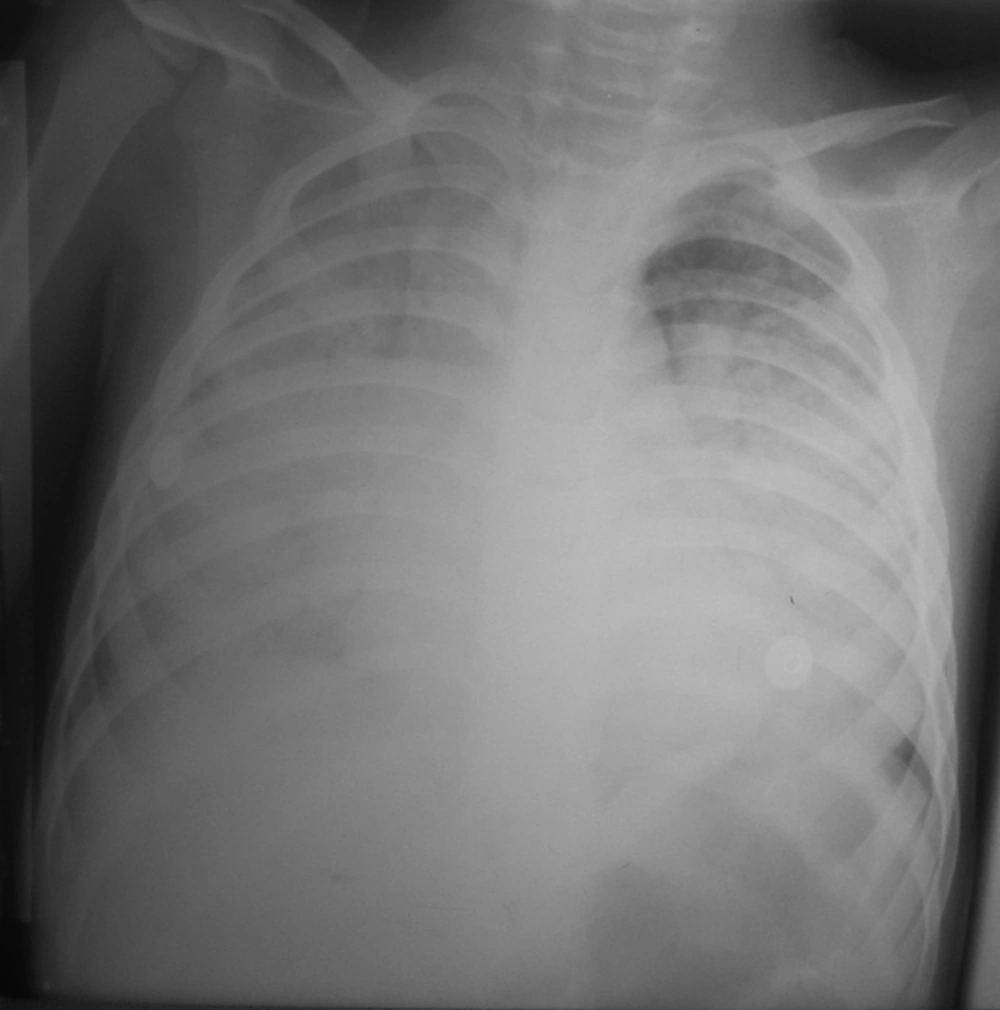
Simple Anteroposterior Chest X-Ray Demonstrating Bilateral Opacities, Mostly in the Lower Lobes
Figure 1.
Simple Anteroposterior Chest X-Ray Demonstrating Bilateral Opacities, Mostly in the Lower Lobes

High-Resolution Computed Tomography Scan of the Chest Demonstrating Alveolar Infiltrate, Mostly in the Lung Bases
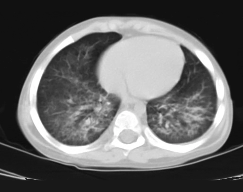
Figure 2.
High-Resolution Computed Tomography Scan of the Chest Demonstrating Alveolar Infiltrate, Mostly in the Lung Bases

High-Resolution Computed Tomography Scan of the Chest Showing Dramatic Improvement in Both Lungs After Multiple Segmental BAL
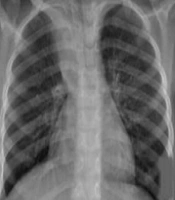
Figure 3.
High-Resolution Computed Tomography Scan of the Chest Showing Dramatic Improvement in Both Lungs After Multiple Segmental BAL


References

  • 1.
    Mylonaki E, Voutsas V, Antoniou D, Papakosta D, Kontakiotis T, Skordalaki A, et al. Hydrocarbon pneumonitis following liquid paraffin aspiration during a fire-eating performance: a case report. J Med Case Rep. 2008;2:214. [PubMed ID: 18565234]. https://doi.org/10.1186/1752-1947-2-214.
  • 2.
    Pielaszkiewicz-Wydra M, Homola-Piekarska B, Szczesniak E, Ciolek-Zdun M, Fall A. Exogenous lipoid pneumonia - a case report of a fire-eater. Pol J Radiol. 2012;77(4):60-4. [PubMed ID: 23269939].
  • 3.
    Sias SM, Daltro PA, Marchiori E, Ferreira AS, Caetano RL, Silva CS, et al. Clinic and radiological improvement of lipoid pneumonia with multiple bronchoalveolar lavages. Pediatr Pulmonol. 2009;44(4):309-15. [PubMed ID: 19283836]. https://doi.org/10.1002/ppul.20918.
  • 4.
    Marchiori E, Zanetti G, Mano CM, Hochhegger B. Exogenous lipoid pneumonia. Clinical and radiological manifestations. Respir Med. 2011;105(5):659-66. [PubMed ID: 21185165]. https://doi.org/10.1016/j.rmed.2010.12.001.
  • 5.
    Simoes MC, Toro IF, Ribeiro JD, Toro AA. Lipoid pneumonia in a 40-day-old infant. J Bras Pneumol. 2012;38(4):535-7. [PubMed ID: 22964941].

Copyright

Copyright © 2015, Growth & Development Research Center.This is an open-access article distributed under the terms of the Creative Commons Attribution-NonCommercial 4.0 International License (http://creativecommons.org/licenses/by-nc/4.0/) which permits copy and redistribute the material just in noncommercial usages, provided the original work is properly cited.

Similar Articles

30
Aug
2007

Clinical and Imaging Findings in 18 Patients with Eosinophilic Pneumonia  

Mehrdad Bakhshayeshkaram

Bakhshayeshkaram M. Clinical and Imaging Findings in 18 Patients with Eosinophilic Pneumonia  . I J Radiol. 2007;4(3):e79114. doi:

13
Dec
2012

Whole Lung Lavage of Nine Children with Pulmonary Alveolar Proteinosis: Experience in a Tertiary Lung Center

Badiozaman Radpay,
Tahereh Parsa,
Shideh Dabir,
Mohammadreza Boloursaz,
Ahmadreza Arbab,
Seyyedahmad Tabatabaei

Radpay B, Parsa T, Dabir S, Boloursaz M, Arbab A, et al. Whole Lung Lavage of Nine Children with Pulmonary Alveolar Proteinosis: Experience in a Tertiary Lung Center. Inn J Pediatr. 2013;23(1):. doi:

8
Oct
2019
Cast Bronchitis in a Cystic Fibrosis Child: Experience with Direct Instillation of Dornase Alfa as an Adjunct Treatment Agent

Cast Bronchitis in a Cystic Fibrosis Child: Experience with Direct Instillation of Dornase Alfa as an Adjunct Treatment Agent

Sedigheh Yousefzadegan,
Rohollah Shirzadi,
Safura Navaie,
Alireza Takzare,
Mahdieh Ghaempanah,
Mohammadreza Modaresi

Yousefzadegan S, Shirzadi R, Navaie S, Takzare A, Ghaempanah M, et al. Cast Bronchitis in a Cystic Fibrosis Child: Experience with Direct Instillation of Dornase Alfa as an Adjunct Treatment Agent. Inn J Pediatr. 2019;29(5):e89703. doi: https://doi.org/10.5812/ijp.89703

23
Dec
2015

Management of Alveolar Proteinosis by Bronchopulmonary lavage under Extra Corporeal Membrane Oxygenation (ECMO)

Tahereh Parsa,
Ardeshir Tajbakhsh,
Zargham Hossein Ahmadi,
Behrooz Farzanegan,
Alireza Jahangirifard

Parsa T, Tajbakhsh A, Hossein Ahmadi Z, Farzanegan B, Jahangirifard A. Management of Alveolar Proteinosis by Bronchopulmonary lavage under Extra Corporeal Membrane Oxygenation (ECMO). J Cell Mol Anesth. 2016;1(1):e150241. doi: https://doi.org/10.22037/jcma.v1i1.10604

21
Aug
2013

Role of Interferon-Gamma in Bronchoalveolar Lavage Fluid on Distinguishing Active Pulmonary Tuberculosis from other Pulmonary Diseases

Nezar Ali Moulaei,
Abbas Ali Niazi,
Ali Saeidpour-Parizi,
Samira Shahba

Ali Moulaei N, Ali Niazi A, Saeidpour-Parizi A, Shahba S. Role of Interferon-Gamma in Bronchoalveolar Lavage Fluid on Distinguishing Active Pulmonary Tuberculosis from other Pulmonary Diseases. Zahedan J Res Med Sci. 2014;16(11):e23371. doi:

More by these authors

Mohammadreza ModaresiPubMedScholar
Minoo DadkhahPubMedScholar
Sayed Javad SayediPubMedScholar
Share
Cited by
Metrics
