Abstract
Background:
Artemisia absinthium L. (known as wormwood) is used as an antihelminthical, antimalarial, antiseptic, anti-inflammatory agent, and also in the treatment of gastric pain, in traditional medicine.Objectives:
In the present study, we have evaluated the effects of the ethanolic extract of A. absinthium L. on the maturation and function of dendritic cells (DCs).Materials and Methods:
The immunomodulatory effects of A. absinthiumL. extract on DCs phenotypic maturation were determined by flow cytometry. The ability of the treated DCs to stimulate allogenic T cells proliferation and cytokines secretion was examined by mixed lymphocyte reaction (MLR) and enzyme-linked immuosorbent assay (ELISA), respectively.Results:
A. absinthium L. extract showed that it could promote DCs phenotypic maturation by increasing the level of surface expression of CD40 as an important costimulatory marker on the DCs, compared to the control. Extract with concentration below 100 µg/mL significantly increased the production of interleukin (IL)-12 cytokine by DCs. The same 100 µg/mL concentration A. absinthium L. inhibited the proliferation of allogenic T cells and also significantly increased the level of IL-10 (P < 0.001). Moreover, the extract with concentrations below 100 µg/mL significantly increased production of interferon-γ (IFN-γ). Nevertheless, these changes were not significant for IL-4 production.Conclusions:
Artemisia absinthium L. extract, at concentrations below 100 µg/mL, can modulate the immune response toward a Th1 pattern, by induction of CD40 expression on DCs and cytokine production. Also, it can inhibit DCs T cell stimulating activity, at high concentrations. Therefore, the traditional use of this plant possibly modulates immune-mediated disorders. However, future studies are needed to confirm these results.Keywords
Artemisia absinthium Immunomodulation Dendritic Cells Chemokines
1. Background
In traditional medicine, worldwide, herbal drugs are used to treat immune disorders, such as inflammatory and autoimmune diseases. Several studies showed that species of herbal medicine have immunomodulatory effects (1, 2). Artemisia (Asteraceae) is commonly used in the treatment of various diseases in traditional medicine. Artemisia absinthium L., known as wormwood, is an antihelminthic, antimalarial and antiseptic agent. This substance has also anti-inflammatory characteristics and is used in the treatment of gastric pain. In addition, it stimulates appetite and facilitates the digestion process (3-5). Previous studies showed that this species represents a source of various biologically active compounds, including essential oils, polysaccharides, polyphenolic compounds, flavonoids, terpenoids, phenolic acids, alkaloids, lignans, saponins, stilbenes and sterols (6-10). In the immune system, dendritic cells (DCs) are potential antigen presenting cells for naive T cells and act as a link between the acquired and innate immunity for the initiation of the protective immune response, or the induction of immune tolerance (11). Factors like maturation status, origin and phenotype are affected by the function of these cells (12). The nature of the cytokines produced by DCs in response to various ligands eventually modulates and determines the type of T helper (Th) cell response. The DCs have the exclusive ability to stimulate naive T cells to transform in either Th1 or Th2 cells, and also effectively down-regulate T-cell responses through the generation of T regulatory cells (13, 14). Although it has been shown that immature DCs can effectively present antigen to naive T cells, because of the low expression of co-stimulatory molecules, such as CD40, CD86 and MHC II, they cannot suitably stimulate the immune system, finally leading to the inhibition of T cell activation and proliferation (12). In this regards, maturation of DCs converts them to the cells that can stimulate immune system.
2. Objectives
Using of dendritic cells as therapeutic targets by pharmacological compounds such as medicinal plant is a valuable strategy to modulate immune responses. In this study, we have evaluated the effects of A. absinthium L. extract, as modulator of the immune response, on the function and maturation of DCs.
3. Materials and Methods
3.1. Animals
Male BALB/c and C57BL/6 mice, 6‒8-week old were purchased from the Razi Institute (Karaj, Iran) and were kept under optimal conditions of hygiene and received standard mouse chow and water, ad libitum. In this study, all experimental procedures on handling the animals were approved by the Ethical Committee of Qazvin University of Medical Sciences.
3.2. Preparation of Dendritic Cells
To purify DCs from spleen, according to the manufacturer’s protocol, the mice CD11c+ isolation kit (Miltenyi Biotec, Bergisch Gladbach, Germany), was used, as previously described (15). Briefly, the mice spleens were isolated and minced to very small pieces and then suspended in cold (4°C) phosphate buffered saline (PBS) containing 1.2 mg/mL collagenase D (Roche, Grenzach-Wyhlen, Germany). The mononuclear cells (MNC) were separated by Nycodenz (Axis-Shield PoC, Oslo, Norway), a material used in density gradient cell separation techniques. According to the manufacturer’s protocol, anti-CD11c magnetic beads (Miltenyi Biotec, Bergisch Gladbach, Germany) were added to the cells and incubated at 4°C for 30 minutes and the non-bonded antibodies were washed out by using of the MiniMACS separator (MS) cell columns into the magnetic field (MS separation unit, Miltenyi Biotec, San Diego, CA, USA). The isolated DCs were collected by disconnecting the column from the magnetic field and injecting it with 1 mL PBS-EDTA under pressure. Purity of the CD11c DCs was determined to more than 94% by flow cytometry.
3.3. Plant Material and Preparation of the Ethanolic Extract
The A. absinthium L. samples were purchased from the herbal market and authenticated by a botanist. Voucher specimens were preserved in the central herbarium of medicinal plants (ACECR). The aerial parts of plant were air-dried at room temperature and then ground into powder. The aerial part of plant (50 g) was extracted, using percolation method, by ethanol (80%) at room temperature. The solvent was completely removed by drying under reduced pressure at 40°C in a rotary evaporator. The samples were stored at 4°C until use (3 g, 14% yield).
3.4. Determination of Dendritic Cells Viability
To study of cytotoxic concentration of the extract on DCs, 3 - (4, 5 -dimethylthiazol- 2 -yl) - 2, 5 -diphenyltetrazolium bromide (MTT) (Sigma-Aldrich, St. Louis, MO, USA) colorimetric assay was used, as described previously (13). Briefly, dendritic cells (105 cells/well) were treated with A. absinthium L. extract at 10 - 200 μg/mL concentrations in the cell culture plates for 24 hours, and then 10 μL MTT (5 mg/mL) were added to each well and cells were incubated for an additional 4 hours at 37°C. Dimethyl sulphoxide (DMSO 0.1%), as vehicle, and lipopolysaccharides (LPS 1 µg/mL), as DCs maturation inducing agent, were used as negative and positive controls, respectively. Finally, the optical density (OD) of each well was measured at 570 nm wavelength, with reference at 630 nm on an enzyme-linked immuosorbent assay (ELISA) plate reader and the viability was determined as following: (OD of extract-treated cells/OD of DMSO-treated cells) × 100.
3.5. Analysis of Markers Expression on Dendritic Cells by Flow Cytometry
In order to evaluate the effect of A. absinthium L. on the expression of co-stimulatory molecules, DCs were treated with the extract for 18 hours and were then analyzed with a flow cytometer (FACSCalibur, BD Biosciences, San Jose, CA, USA). The cells were stained with phycoerithrin (PE)-conjugated anti-CD11c, fluorescein isothiocyanate (FITC)-conjugated anti-CD86, FITC-conjugated anti-CD40, and FITC-conjugated anti-major histocompatibility complex (MHC) II antibody and with appropriate conjugated isotypes, all obtained from BD Biosciences (San Diego, CA, USA). Finally, the percentage of markers expression on extract-treated DCs and DMSO-treated DCs were calculated and also, the mean florescent intensity (MFI) was analyzed using Win MDI software (Scripps, La Jolla, CA, USA).
3.6. Mixed Lymphocyte Reaction Assay
To evaluate of the proliferative effect of A. absinthium L. extract-treated DCs on T lymphocytes, allogeneic mixed lymphocyte reaction (MLR) assay was used. Briefly, T cells were purified from lymph nodes of C57BL/6 mice, using nylon wool. Then, A. absinthium L. treated DCs, from BALB/c mice, were inactivated with mitomycin C (0.5 mg/mL) for 20 minutes, and then cells were washed with PBS for three times and resuspended in culture medium containing 10% fetal calf serum (FCS). Mitomycin-treated DCs (104 cells/well), as stimulator cells, co-cultured with 105 allogenic T cells, as responder cells, were kept in a 96-well culture plate (Nunc A/S, Roskilde, Denmark) in triplicates for 48 hours. As negative control, triplicate wells containing DMSO-treated DCs, plus allogenic T cells were used. Finally, T cell proliferation was measured by a 5-Bromo-20-deoxy-uridine (BrdU) cell proliferation assay kit (Roche, Grenzach-Wyhlen, Germany), according to the manufacturer’s instructions. The result was concluded as following: (OD of extract-treated culture/OD of DMSO-treated culture) × 100.
3.7. Determination of the Effect of the Extract on the Cytokines Production by Enzyme-Linked Immunosorbent Assay
In order to evaluate of immunomodulatory effect of A. absinthium L. extract on the cytokines production, the ELISA method was used. Briefly, the supernatant of extract-treated DCs and MLR cultures were collected and used to measure interferon-γ (IFN-γ), interleukin (IL)- 4, IL- 12 and IL-10 by ELISA kits, according to the manufacturer’s protocol (eBioscience, San Diego, CA, USA), respectively.
3.8. Statistics Analysis
All data were representative of at least three independent experiments performed in triplicate and presented as Mean ± standard deviation (SD). Mann-Whitney test and one-way analysis of variance (ANOVA) were used for the evaluation of statistical differences between the results and P values < 0.05 were considered significant.
4. Results
4.1. Viability of Dendritic Cells After Exposure With Artemisia absinthium L. Extract
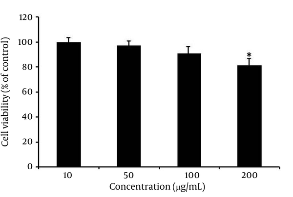
Dendritic cell viability was determined by MTT assay. These cells were treated with different concentrations of the plant extract for 24 hours. Our results indicated that this extract, at concentrations of 10, 50 and 100 μg/mL, had no cytotoxic effect on DCs (Figure 1). However, at 200 μg/mL concentration, the extract had a significant cytotoxic effect on DCs (P < 0.05). Therefore, these concentrations (10 to 100 μg/mL) were used as a safe dose of A. absinthium L. extract for the next experiments on DCs.
Dendritic Cells Viability After Treatment With Artemisia absinthium L. Extract

4.2. Effect of Artemisia. absinthium L. Extract on Maturation Phenotype of Dendritic Cells
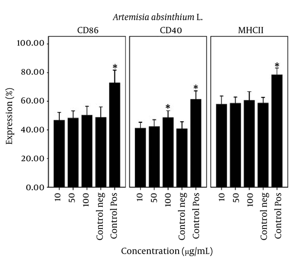
The percentage expression and fluorescent intensity of CD86, CD40 and MHC II co-stimulatory molecules on A. absinthiumL. treated DCs were evaluated by flow cytometry. Our results showed that the extract modulates the percentage expression and fluorescent intensity of CD86, CD40 and MHC II molecules on DCs. As shown in Figure 2, this extract significantly (P = 0.002) increased the percentage of expression of CD40 molecules at 100 μg/mL, compared with negative control. Although, A. absinthiumL. extract increased the percentage of expression and fluorescent intensity of CD86 and MHC-II on DCs, this effect, however, was not significant when comparing with control. Our results showed that A. absinthiumL. extract can modulate the maturation phenotype markers on DCs.
Effect of Artemisia absinthium L. Extract on Maturation Phenotype of Dendritic Cells

4.3. Effect of Artemisia absinthium L. Extract on Allogenic Mixed Lymphocyte Reaction
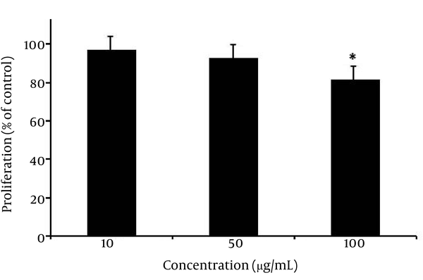
To study the effects of the A. absinthiumL. extract on allogenic immune response and DCs function, allogenic T cells from C57BL/6 were co-cultured with DCs isolated from BALB/c mice. For this, DCs were treated with concentrations of 10 - 100 μg/mL of the extract for 18 hours, and then cells were co-cultured with allogenic T cells in MLR assay. The proliferation of T lymphocytes was evaluated using BrdU incorporation assay. As shown in Figure 3A. absinthiumL. extract significantly decreased the proliferation of T cells at 100 μg/mL (P < 0.05). Our findings indicated that the proliferation of these cells decreased to 78.2 ± 3.6 % of control, when DCs had been treated with 100 μg/mL of the extract.
The Effect of Artemisia absinthium L. Extract on Allogenic Mixed Lymphocyte Reaction

4.4. Effect of Artemisia absinthium L. on the Production of Cytokines
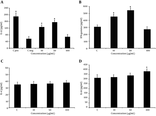
The level of IL-12 cytokine in the supernatant of the extract-treated DCs was measured. As shown in Figure 4 A, treatment of DCs with this extract at lower concentrations (10 - 50 μg/mL) significantly increased production of IL-12 by these cells, compared with the negative control (P < 0.05). However, the extract reduced this production, at 100 μg/mL concentration. Moreover, we have measured the effect of the extract on IFN-γ, IL-4 and IL-10 production, in MLR test. The extract, at lower concentrations (10 - 50 μg/mL), significantly increased production of IFN-γ and slightly increased IL-4 cytokines (P < 0.05). However, these changes were not significant (Figure 4B, 4C). Moreover, as shown in Figure 4 D, our findings indicated that the A. absinthiumL. extract, at 100 µg/mL concentration, significantly increased IL-10 cytokine production (P < 0.05).
Effect of Artemisia absinthium L. Extract on Cytokine Production

5. Discussion
Previous studies have showed that natural agents, such as plants, can modulate DCs activity (16-18). It has been demonstrated that A. absinthium L. represents a reservoir for multiple biologically active compounds, including essential oils, polysaccharides, polyphenolic compounds, flavonoids, terpenoids, phenolic acids, alkaloids, lignans, saponins, stilbenes, sterols (6-9). Anterior studies showed that polysaccharides isolated from A. absinthium have immunomodulatory effects by induction of a Th1 response and stimulation of nitric oxide (NO) production (19). In addition, in vivo studies indicated that A. absinthium suppressed tumor necrosis factor alpha (TNF-α) and accelerated healing in patients with Crohn’s disease and also it has hepatoprotective properties (20, 21).
In the present study, we examined the effect of the A. absinthiumL. extract on the phenotypic maturation and activity of DCs. The expression of CD86, CD40 and MHC-ІІ molecules is important co-stimulatory and maturation markers on DCs and has a critical role in antigen presentation and T cell activation. Our results showed that the extract modulates the percentage expression and fluorescent intensity of CD86, CD40 and MHC II molecules on DCs. Previous studies indicated that several herbal extracts can modulate the immune response by the immunomodulatory effect on the CD40 expression, on the DCs (22, 23). Our results showed that the extract significantly increased the percentage expression of CD40 molecules, at 100 μg/mL concentration, compared with the negative control. Although, A. absinthiumL. extract increased the percentage expression and fluorescent intensity of CD86 and MHC II on DCs, this effect, however, was not significant, when comparing with control. Our finding indicated that A. absinthiumL. extract can modulate the maturation phenotype markers on DCs.
Moreover, we evaluated the immunomodulatory effects of A. absinthiumL. extract on the DCs functions by evaluating the release of cytokines and MLR assay. The IL-12 is a cytokine released by DCs, which can induce differentiation of T cells into Th1 and also induces cellular immunity. By contrast, IL-10 is a Th1 inhibitory cytokine and also, IL-4 and IFN-γ cytokines are landmarks of deviation to Th1 or Th2 response (24). Our results indicated that the treatment of DCs with this extract, at lower concentrations (10 - 50 μg/mL), significantly increased production of IL-12 by these cells, when comparing with control. However, the extract reduced this production at a concentration of 100 μg/mL. In addition, at concentrations of 10 - 50 μg/mL, the extract significantly increased the production of IFN-γ. However, these changes were not significant with regard to IL-4 production. Consistent to our findings, Danilets et al. reported that polysaccharides isolated from A. absinthium possess immunomodulatory effects by inducing the Th1 response (19). We assumed that the increased IL-12 production by DCs, when they were treated with 10 - 50 μg/mL concentrations of the extract, along with production of more IFN-γ by T cells in MLR, suggest the ability of A. absinthiumL. extract to deviate the cytokine pattern of T cells toward a Th1 response. Moreover, the difference between the effects of the extract at higher concentrations may be attributed to the presence of various constituents in the extract, with different modes of actions. Therefore, to confirm Th1/Th2 polarization in co-culture of T cells with A. absinthiumL. treated-DCs, studying of the T cell signaling activity of DCs and the expression of T-bet and GATA3, as related transcription factors for Th1 and Th2 differentiation. is recommended (25).
Nevertheless, our results indicated that the A. absinthiumL. extract, at 100 µg/mL concentration, significantly increased IL-10 cytokine production. Also, this extract, at 100 μg/mL concentration, has increased IL-10 secretion in MLR, and also reduced the proliferation of T cells in allogenic response, which indicated the inhibitory effect of the extract at higher concentration on the immune response. Future research is needed for identifying the main bioactive compound of this plant and other immunomodulatory mechanisms.
Acknowledgements
References
-
1.
Su CA, Xu XY, Liu DY, Wu M, Zeng FQ, Zeng MY, et al. Isolation and characterization of exopolysaccharide with immunomodulatory activity from fermentation broth of Morchella conica. Daru. 2013;21(1):5. [PubMed ID: 23351529]. https://doi.org/10.1186/2008-2231-21-5.
-
2.
Azadmehr A, Hajiaghaee R, Zohal MA, Maliji G. Protective effects of Scrophularia striata in Ovalbumin-induced mice asthma model. Daru. 2013;21(1):56. [PubMed ID: 23837463]. https://doi.org/10.1186/2008-2231-21-56.
-
3.
Setzer WN, Vogler B, Schmidt JM, Leahy JG, Rives R. Antimicrobial activity of Artemisia douglasiana leaf essential oil. Fitoterapia. 2004;75(2):192-200. [PubMed ID: 15030924]. https://doi.org/10.1016/j.fitote.2003.12.019.
-
4.
Bora KS, Sharma A. Neuroprotective effect of Artemisia absinthium L. on focal ischemia and reperfusion-induced cerebral injury. J Ethnopharmacol. 2010;129(3):403-9. [PubMed ID: 20435123]. https://doi.org/10.1016/j.jep.2010.04.030.
-
5.
Nibret E, Wink M. Volatile components of four Ethiopian Artemisia species extracts and their in vitro antitrypanosomal and cytotoxic activities. Phytomedicine. 2010;17(5):369-74. [PubMed ID: 19683909]. https://doi.org/10.1016/j.phymed.2009.07.016.
-
6.
Poiata A, Tuchilus C, Ivanescu B, Ionescu A, Lazar MI. Antibacterial activity of some Artemisia species extract. Rev Med Chir Soc Med Nat Iasi. 2009;113(3):911-4. [PubMed ID: 20191854].
-
7.
Ferracane R, Graziani G, Gallo M, Fogliano V, Ritieni A. Metabolic profile of the bioactive compounds of burdock (Arctium lappa) seeds, roots and leaves. J Pharm Biomed Anal. 2010;51(2):399-404. [PubMed ID: 19375261]. https://doi.org/10.1016/j.jpba.2009.03.018.
-
8.
Zidorn C, Grass S, Ellmerer EP, Ongania KH, Stuppner H. Stilbenoids from Tragopogon orientalis. Phytochemistry. 2006;67(19):2182-8. [PubMed ID: 16890254]. https://doi.org/10.1016/j.phytochem.2006.06.031.
-
9.
Sarker SD, Laird A, Nahar L, Kumarasamy Y, Jaspars M. Indole alkaloids from the seeds of Centaurea cyanus (Asteraceae). Phytochemistry. 2001;57(8):1273-6. [PubMed ID: 11454358].
-
10.
Asghari G, Jalali M, Sadoughi E. Antimicrobial Activity and Chemical Composition of Essential Oil From the Seeds of Artemisia aucheri Boiss. Jundishapur J Nat Pharm Prod. 2012;7(1):11-5. [PubMed ID: 24624145].
-
11.
Schuurhuis DH, Fu N, Ossendorp F, Melief CJ. Ins and outs of dendritic cells. Int Arch Allergy Immunol. 2006;140(1):53-72. [PubMed ID: 16534219]. https://doi.org/10.1159/000092002.
-
12.
Reis e Sousa C. Dendritic cells in a mature age. Nat Rev Immunol. 2006;6(6):476-83. [PubMed ID: 16691244]. https://doi.org/10.1038/nri1845.
-
13.
Gao Y, Nish SA, Jiang R, Hou L, Licona-Limon P, Weinstein JS, et al. Control of T helper 2 responses by transcription factor IRF4-dependent dendritic cells. Immunity. 2013;39(4):722-32. [PubMed ID: 24076050]. https://doi.org/10.1016/j.immuni.2013.08.028.
-
14.
Min WP, Zhou D, Ichim TE, Strejan GH, Xia X, Yang J, et al. Inhibitory feedback loop between tolerogenic dendritic cells and regulatory T cells in transplant tolerance. J Immunol. 2003;170(3):1304-12. [PubMed ID: 12538690].
-
15.
Eftekharian MM, Zarnani AH, Moazzeni SM. In vivo effects of calcitriol on phenotypic and functional properties of dendritic cells. Iran J Immunol. 2010;7(2):74-82. [PubMed ID: 20574120].
-
16.
Amirghofran Z, Bahmani M, Azadmehr A, Javidnia K, Miri R. Immunomodulatory activities of various medicinal plant extracts: effects on human lymphocytes apoptosis. Immunol Invest. 2009;38(2):181-92. [PubMed ID: 19330626]. https://doi.org/10.1080/08820130902817051.
-
17.
Azadmehr A, Afshari A, Baradaran B, Hajiaghaee R, Rezazadeh S, Monsef-Esfahani H. Suppression of nitric oxide production in activated murine peritoneal macrophages in vitro and ex vivo by Scrophularia striata ethanolic extract. J Ethnopharmacol. 2009;124(1):166-9. [PubMed ID: 19527828]. https://doi.org/10.1016/j.jep.2009.03.042.
-
18.
Ghafourian Boroujerdnia M, Azemi ME, Hemmati AA, Taghian A, Azadmehr A. Immunomodulatory effects of Astragalus gypsicolus hydroalcoholic extract in ovalbumin-induced allergic mice model. Iran J Allergy Asthma Immunol. 2011;10(4):281-8. [PubMed ID: 22184271].
-
19.
Danilets MG, Bel'skii Iu P, Gur'ev AM, Belousov MV, Bel'skaia NV, Trofimova ES, et al. [Effect of plant polysaccharides on TH1-dependent immune response: screening investigation]. Eksp Klin Farmakol. 2010;73(6):19-22. [PubMed ID: 20726346].
-
20.
Krebs S, Omer TN, Omer B. Wormwood (Artemisia absinthium) suppresses tumour necrosis factor alpha and accelerates healing in patients with Crohn's disease - A controlled clinical trial. Phytomedicine. 2010;17(5):305-9. [PubMed ID: 19962291]. https://doi.org/10.1016/j.phymed.2009.10.013.
-
21.
Amat N, Upur H, Blazekovic B. In vivo hepatoprotective activity of the aqueous extract of Artemisia absinthium L. against chemically and immunologically induced liver injuries in mice. J Ethnopharmacol. 2010;131(2):478-84. [PubMed ID: 20637853]. https://doi.org/10.1016/j.jep.2010.07.023.
-
22.
Amirghofran Z, Ahmadi H, Karimi MH. Immunomodulatory activity of the water extract of Thymus vulgaris, Thymus daenensis, and Zataria multiflora on dendritic cells and T cells responses. J Immunoassay Immunochem. 2012;33(4):388-402. [PubMed ID: 22963488]. https://doi.org/10.1080/15321819.2012.655822.
-
23.
Elluru SR, Duong van Huyen JP, Delignat S, Kazatchkine MD, Friboulet A, Kaveri SV, et al. Induction of maturation and activation of human dendritic cells: a mechanism underlying the beneficial effect of Viscum album as complimentary therapy in cancer. BMC Cancer. 2008;8:161. [PubMed ID: 18533025]. https://doi.org/10.1186/1471-2407-8-161.
-
24.
Ho CY, Lau CB, Kim CF, Leung KN, Fung KP, Tse TF, et al. Differential effect of Coriolus versicolor (Yunzhi) extract on cytokine production by murine lymphocytes in vitro. Int Immunopharmacol. 2004;4(12):1549-57. [PubMed ID: 15351324]. https://doi.org/10.1016/j.intimp.2004.07.021.
-
25.
Kang H, Oh YJ, Choi HY, Ham IH, Bae HS, Kim SH, et al. Immunomodulatory effect of Schizonepeta tenuifolia water extract on mouse Th1/Th2 cytokine production in-vivo and in-vitro. J Pharm Pharmacol. 2008;60(7):901-7. [PubMed ID: 18549677]. https://doi.org/10.1211/jpp.60.7.0012.