1. Context
Epilepsy is one of the most common serious neurological disorders affecting 1% of general population (1). One third of patients are poorly controlled or their seizures are refractory to antiepileptic drug therapy (2). Resective brain surgery is a promising therapy for these patients; however, more than 40% of patients are not eligible for the surgical intervention (2). Hence, alternative treatment possibilities are required. The effectiveness of DBS in the treatment of Parkinson’s disease has made preparations for the use of this approach in the treatment of intractable epilepsy. DBS is of therapeutic interest for refractory epilepsy, as it reduces seizures in patients with medically refractory epilepsy. Nonetheless, optimal target(s) for DBS application in epilepsy has not been recognized yet.
2. Evidence Acquisition
2.1. What is Deep Brain Stimulation?
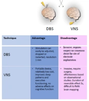
Deep brain stimulation is a surgical treatment in which a device known as brain pacemaker delivers electrical impulses to specific areas in the brain. DBS is an adjustable and reversible approach, which modulates brain function, without causing injury to brain tissues. The DBS system is composed of three components: 1- A battery-operated pulse generator that delivers electrical stimulation to target areas in the brain, 2- An electrode (a thin, insulated wire) which is implanted in a certain brain area, and 3- An insulated wire that connects electrode to pulse generator. After electrode implantation in target site, pulse generator is located under the skin below the clavicle or abdomen. Adverse effects of DBS include depression, apathy, hallucinations, compulsive gambling, cognitive dysfunction and hypersexuality. However, most adverse effects have been reported to be temporary and disappeared by the correction of electrode placement and changing stimulation parameters (3).
2.2. Classification of Seizures and Epilepsies
The term epilepsy includes a number of different syndromes characterized predominantly by recurrent unprovoked seizures and can be symptomatic (with known etiology) or idiopathic (with no clear etiology other than a possible genetic predisposition) (4, 5). Epileptic seizures are characterized as abnormal hyperexcitable, hypersynchronous neuronal activity and their clinical manifestation depends on the place of seizure onset and subsequent spread (6). In general, epilepsy syndromes can be classified into two broad categories: generalized and partial syndromes (7, 8). In generalized epilepsies, seizure activity begins simultaneously over both brain hemispheres and can occur as one of several types; myoclonic, generalized tonic-clonic, absence or atypical absence. Most of these types of epilepsies have a genetic origin and in many cases neurologic function is normal. Absence seizures in which occur brief and sudden loss of consciousness are the most pure type of generalized epilepsy and characterized by bilaterally synchronized spike-and-wave discharges in the electroencephalogram (EEG) over wide cortical areas (9). In partial epilepsies, by contrast, seizures originate only from one brain hemisphere (most commonly from the temporal lobe) and may result from a traumatic brain injury (10) or may have a genetic origin (11). Among partial epilepsy, temporal lobe complex partial epilepsy is the most common type of acquired epilepsy in adult and characterized by recurrent epileptic seizures (12).
2.3. Thalamus and Its Role in Epilepsy
The thalamus is a small structure within the brain situated just above the brain stem between the cerebral cortex and the midbrain. It has reciprocal connections with cerebral cortex and limbic system (13, 14). These connections play a role in relaying sensory and motor information as well as regulation of sleep, alertness and wakefulness in the normal physiological state. In general, the thalamus is divided into two subdivisions: dorsal thalamus and ventral thalamus. The dorsal thalamus contains a number of anatomically and functionally distinct nuclei that are associated with an individual sensory, motor or limbic function. These nuclei receive input from the sensory organs and then transmit it to the functionally corresponding primary sensory cortical areas, from which, in turn, they receive modulatory projections. The thalamus includes also another area that is anatomically and functionally different from the dorsal thalamus (15) and known as the ventral thalamus. Unlike dorsal thalamus, the ventral thalamus does not project to cortex, but it instead targets the dorsal thalamus. The most eminent part of the ventral thalamus is the thalamic reticular nucleus (TRN), which is a relatively thin sheet of thalamus that exclusively contains GABAergic neurons enclosed in the dorsal thalamus (16, 17). The reticular thalamic nucleus is considered to be a generator of various wave rhythms, due to having an intrinsic ability to generate rhythmic oscillatory activity, which can transform into Spike-and-wave discharges. Spike-and-wave discharges are regarded to be a hallmark of idiopathic generalized epilepsy. Moreover, recently an important role of nucleus anterior thalami has been shown in bilateral Spike-and-wave discharges synchrony (18-20). However, the role of thalamus in partial epilepsy has been poorly understood. There are evidences showing the involvement of thalamus in patients with temporal lobe epilepsy (21). For example, Bertram et al. reported that in animal models of temporal Lobe complex partial epilepsy, there is a significant neuronal loss with synaptic changes in the midline thalamic regions, which can enhance the excitability of the thalamolimbic circuits (22). In addition, increased GABA activity in medial dorsal nucleus and medline region of the thalamus reduced seizure duration significantly (22, 23). Furthermore, inactivation of thalamus by tetrodotoxin (TTX) suppressed pilocarpine-induced seizures (24). It is about six decades that the role of thalamic nuclei in the generation of idiopathic generalized seizures has been recognized. Idiopathic generalized epilepsies are characterized by generalized spike and wave discharges in EEG. Involvement of thalamus and its cortical interactions are considered essential in the generation of spike-and-wave discharges. For example, some studies have shown the role of posterolateral and midline thalamic nuclei in the generation of generalized seizures in animal models of absence seizures (25, 26) and in humans with depth electrode recordings (27). In this line, involvement of anterior, centromedian and parafascicular thalamic nuclei during generalized spike-and-wave discharges (28) as well as that of anterior and mediodorsal nuclei of the thalamus in animal models of epilepsy induced by pentylenetetrazol (PTZ) or bicuculline (29) have been demonstrated. Moreover, unilateral thalamic lesions have been revealed in magnetic resonance imaging of two patients with IGE (idiopathic generalized epilepsy) (30). These findings suggest that several nuclei of thalamus have a role in neuronal circuits of seizures.
2.4. Electrical Stimulation of Anterior and Centromedian Thalamic Nuclei in Animal and Human Studies for the Treatment of Epilepsy
In patients with epilepsy, DBS in anterior nucleus of the thalamus (ANT), centromedian nucleus of the thalamus (CMN), caudate nucleus, cerebellum, hippocampus, hippocampal commissure, amygdala, subthalamic nucleus, mammillary bodies, corpus callosum and neocortex has been evaluated for treatment of intractable epilepsy. Here, we reviewed DBS application in ANT and CMN in animal models and patients with epilepsy, since a large body of DBS studies have been conducted on thalamic nuclei, particularly ANT. Thalamic nuclei DBS outcomes in animal epilepsy models and patients with epilepsy have been summarized in Tables 1 and 2.
Table 1.
Deep Brain Stimulation Outcomes in Animal Models of Epilepsy a
| Author | Year | Target | Animal Model of Epilepsy | Stimulation Parameters | Main Findings |
|---|---|---|---|---|---|
| Lado et al. | 2003 | ANT | Electrical kindling | 8 Hz, bilateral stimulation | Increase in seizures frequency |
| Hamani et al. | 2004 | ANT | Pilocarpine model of epilepsy | bilateral high-frequency stimulation | Delay in the onset of SE |
| Hamani et al. | 2004 | ANT | Pilocarpine model of epilepsy | Unilateral stimulation | A nonsignificant reduction in seizure frequency |
| Jou et al. | 2013 | left ANT | Pilocarpine model of epilepsy | Unilateral high frequency stimulation of 200 Hz and low intensity current of 50 µA | A significant reduction in seizure frequency |
a ANT, Anterior nucleus of thalamus.
Table 2.
Deep Brain Stimulation Outcomes in Patients With Epilepsy a
| Author | Year | Target | No. of Patients | Stimulation Parameters | Main Findings |
|---|---|---|---|---|---|
| Upton et al. | 1987 | ANT | 6 patients with intractable partial seizures | NA | A significant reduction in seizure frequency in 4 patients, Lack of seizure in one patient |
| Velasco et al. | 1987 | CMN | 5 patients with refractory tonic–clonic seizures | 60-100 Hz , 100 µs pulses, 200-800 µA, bilateral stimulation | A more than 80% reduction in generalized seizure frequency |
| Fisher et al. | 1992 | CMN | 7 | 5 V, 65 Hz | A mean reduction of 30% in tonic-clonic seizure frequency in 4 patients |
| Hodaie et al. | 2004 | ANT | 5 | 100 Hz, 10 V, and 90-msec, bilateral stimulation | A mean 54% decrease in seizure frequency |
| Kerrigan et al. | 2004 | ANT | 5 | 100 Hz, pulse width of 90 µs, and the intensity of 1-10V, bilateral stimulation | A significantly reduction in seizure frequency in 1 patient, and a less than 50% reduction in frequency of seizures in other 4 patients |
| Lee et al. | 2006 | ANT in 3 patients and STN in 3 patients | 6 | 130 Hz, 3-7 V, 90 µs, bilateral stimulation | 75.4% and 49.1% reduction in seizure frequency in patients with ANT and STN stimulation, respectively |
| Andrade et al. | 2006 | ANT | 6 | NA | A reduction ≥ 50% in five patients |
| Velasco et al. | 2006 | CMN | 13 patients with atypical absence seizures | 130 Hz, 400-600 µA, bilateral stimulation | A significant reduction of seizure frequency in 10 patients |
| Osorio et al. | 2007 | ANT | 4 | 175 Hz, 4.1 V, pulse width of 90 µs, bilateral stimulation | A mean 75.6% reduction in seizure frequency in 4 patients |
| Lim et al. | 2007 | ANT | 4 (one patient with generalized seizures and three patients with partial seizures) | 4 - 5 V, 90-110 Hz, and 60-90 µs, bilateral stimulation | A 49% reduction in seizure in seizure frequency |
| Valentin et al. | 2012 | CMN | 1 patient with myoclonic jerks and generalized tonic-clonic seizures | NA | An immediately disappearance of tonic-clonic seizures |
| Valentin et al. | 2013 | CMN | 11 patients with refractory generalized or frontal lobe epilepsy | bilateral stimulation | A reduction in seizure frequency, particularly in patients with refractory generalized epilepsy |
a Abbreviations: ANT, Anterior nucleus of thalamus; CMN, Centromedian nucleus; NA, Not available.
2.4.1. Animal Studies
Potential usefulness of DBS application for the treatment of epilepsy has been tested in animal models of epilepsy induced by pentylenetetrazol, kainic acid, bicuculline, picrotoxin or using electrical kindling (31-37). Lado FA discovered that bilateral stimulation of the ANT with low frequency (8 Hz) increased seizures frequency (38). Hamani and coworkers reported that bilateral high-frequency stimulation of ANT delayed the onset of pilocarpine-induced status epilepticus (SE), but it failed to prevent SE (in 56% of the animals), while bilateral lesions of ANT prevent SE development in all animals after pilocarpine injection. In this study, animals experienced unilateral or bilateral electrical stimulation of ANT or unilateral or bilateral ANT lesioning. The control group received bilateral placement of electrodes in ANT, but no stimulation (39). Unilateral stimulation or lesioning in these animals did not reduce the occurrence of seizure significantly. They concluded that bilateral stimulation of anterior thalamic nucleus and thalamotomies are protective against pilocarpine-induced SE. In a study, left ANT stimulation with high frequency of 200 Hz and low intensity current of 50 µA, beginning from 1 h before the pilocarpine administration for seizure induction significantly reduced the occurrence rate of seizure activity in rats, while right ANT stimulation had little effect on the occurrence rate of seizures (40).
2.4.2. Human Studies
Osorio and colleagues conducted bilateral DBS of ANT (175 Hz, 4.1 V, pulse width of 90 µs) for four patients with mesial temporal lobe complex partial epilepsy. The stimulation was conducted as intermittent with 1 minute on and 5 minutes off. Results indicated a mean 75.6% reduction in seizure frequency in all patients (41). In a study, bilateral DBS (130 Hz, 3-7 V, 90 µs) was performed for six patients with intractable epilepsy. Electrodes were implanted for three patients in ANT and for three patients in subthalamic nucleus. The patients with DBS of ANT showed a 75.4% reduction in seizure frequency, while in those with DBS of subthalamic nucleus seizure frequency reduced by 49.1% (42). Lim et al. conducted bilateral stimulation of ANT in four patients (one patient with generalized seizures and three patients with partial seizures). Stimulus parameters were 4-5 V, 90 - 110 Hz and 60 - 90 µs. Results showed a 49% reduction in seizure to ANT stimulation. In addition, a 67% reduction in seizure frequency was observed only with bilateral inserting electrodes in ANT (43). In another study, bilateral neurostimulation of ANT (100 Hz, 10 V, and 90-msec) in five patients with medically refractory epilepsy resulted in a mean 54% decrease in seizure frequency. Two patients showed a seizure reduction of ≥ 75%. In this study stimulation was in an intermittent pattern with 1 minute on and 5 minutes off (44). Kerrigan et al. conducted bilateral DBS of ANT (with the frequency of 100 Hz, pulse width of 90 µs and the intensity of 1 - 10 V) for five patients with intractable partial seizures. The voltage was incrementally increased during 12 - 30 weeks, depending on the clinical response of patients. Results indicated that DBS significantly reduced seizure frequency in one patient and reduction in frequency of seizures in four patients was less than 50%, although it was not significant (45). In a study, long-term follow-up of six patients with epilepsy with ANT DBS showed a reduction ≥ 50% in five patients. In these patients, electrode implantation in ANT was followed by seizure reduction 1 to 3 months before stimulation and authors suggested the possibility of a beneficial microthalamotomy effect (46). In another study, Upton et al. evaluated the effect of electrical stimulation of ANT in six patients with medically intractable partial seizures. Results indicated a statistically significant reduction in seizure frequency in four patients. One patient was shown to be seizure-free for the last two years. Measurement of regional cerebral glucose metabolism, blood cortisol levels and blood levels of valproic acid, diphenylhydantoin and carbamazepine in two patients showed a marked change in these factors during periods of stimulation compared to periods without stimulation (47).
Neurostimulation of centromedian-thalamic nucleus for treatment of refractory epilepsy has been performed in several studies. Centromedian thalamic nucleus is the part of the reticulothalamocortical system involved in cerebral cortex excitability (48). In 1987, bilateral DBS CMN was conducted in five patients with refractory tonic–clonic seizures. Stimulation (60 - 100 Hz, 100 µs pulses, 200 - 800 µA) was delivered for 2 hours daily, in a pattern of 1 minute on and 4 minutes off. Results showed more than 80% reduction in generalized seizure frequency (49). In a study, bilateral centromedian thalamic nucleus DBS in 11 patients with refractory generalized or frontal lobe epilepsy improved seizure frequency, particularly in patients with refractory generalized epilepsy (50). In another study, electrical stimulation of CMN was effective in patients with generalized tonic-clonic seizures and atypical absences (51). Furthermore, electrical stimulation of CMN in 49 patients with generalized, frontal and temporal lobe epileptic seizures was highly efficient to control patients with generalized tonic-clonic seizures, atypical absences and tonic seizures, but not in those with partial seizures. Stimulation protocol was 1 minute on and 4 minutes off for 24 hours, at 60-130 Hz, 2.5-5.0 V, 210-450 µs (51). Bilateral stimulation of the parvocellular division of the CMN (130 Hz, 400-600 µA) in 13 patients with atypical absence seizures of the Lennox-Gastaut syndrome, which is characterized by frequent medically intractable seizures, reduced seizure frequency significantly in 10 patients (52). A placebo-controlled study of centromedian thalamic stimulation in seven patients with intractable epilepsy with an intermittent protocol of 1 minute on (5 V, 65 Hz) and 4 minutes off stimulation caused a mean reduction of 30% in tonic-clonic seizure frequency in four patients compared to baseline when stimulator was on and a decrease of 8% when the stimulator was off, but these differences were not statistically significant (53). Moreover, bilateral DBS of the CMN in a patient with myoclonic jerks and generalized tonic-clonic seizures immediately was associated with disappearance of tonic-clonic seizures and generalized periodic epileptiform discharges (54).
3. Results
Neurostimulation of ANT and CMN particularly is effective in improvement of partial and generalized tonic-clonic seizures, respectively. However, the efficiency of thalamic stimulation in treating refractory epilepsy depends on the stimulation parameters, particularly frequency. For example, low frequency stimulation of ANT was epileptogenic, while high frequency stimulation reduced seizure frequency both in experimental and clinical studies. Moreover, most studies of thalamic nuclei DBS demonstrated antiepileptic feature by application of a high-intensity current (320 - 800 µA) or a high-voltage (4 - 10 V). However, electrical stimulation of ANT with high-intensity or high voltage currents might affect the memory, since anterior nucleus of thalamus is a significant nucleus in the Papez circuit involved in memory. This hypothesis is supported by a study in which Hamani et al. demonstrated that ANT stimulation at relatively high current of 500 µA disrupted the acquisition of contextual fear conditioning and impaired performance on a spatial alternating task in rats (55). There is an animal study indicating the efficacy of unilateral stimulation of ANT with low intensity current to control the occurrence of seizure in pilocarpine-induced seizure. In this study, left ANT DBS was applied 1 hour or 30 minutes before seizure induction (40). Difference in timing ANT DBS within the same anatomic target makes it difficult to compare the results of this study to other studies, suggesting the application of ANT DBS after induction of seizure. One the other hand, in contrast to DBS human studies performed in patients with chronic epilepsy, most DBS experimental studies are conducted before starting spontaneous recurrent seizures that might be due to differences resulted from animal and human studies, suggesting DBS application in experimental studies after the animals acquired epilepsy. Results showed that stimulation paradigm of the unilateral or bilateral thalamic nuclei DBS has an impact on the effects of stimulation to prevent seizures. Stimulation protocol in most thalamic nuclei DBS was intermittent and there are reports suggesting that an intermittent stimulation paradigm might be more effective than a continuous paradigm (56). DBS program in CMN in most studies was in an intermittent pattern and associated with a reduction of generalized seizure. DBS in ANT, although, in several studies was in an intermittent model, implantation of DBS electrodes without stimulation in ANT and lesioning of ANT in a few reports appeared to be effective in reduction of seizure frequency and hence, stimulation pattern in ANT DBS for seizure treatment might have no significant effect on the effectiveness of procedure.
3.1. Possible Mechanisms of Thalamic Nuclei DBS in Treatment of Epilepsy
It is well known that certain subtypes of epileptic seizures are associated with oscillatory activity in the thalamus (57) and deep brain stimulation of specific thalamic nuclei has been indicated able to abolish or reduce spindles oscillations in thalamus. Although, the precise mechanism by which DBS is able to eliminate seizures associated with epilepsy remains largely unknown, there are evidences showing that high frequency stimulation of the thalamus leads to neurotransmitter release (58, 59). Indeed, it has been demonstrated that high-frequency stimulation of thalamus increased extracellular adenosine significantly (60). In addition, in a study, Lee and coworkers reported that high-frequency stimulation generates excitatory postsynaptic potentials and the antagonists of glutamate receptor can eliminate the excitatory postsynaptic potentials caused by high frequency stimulation in vitro (61), supporting the hypothesis that glutamate release is involved in high frequency stimulation function. A few years later, Tawfik et al. showed that high-frequency stimulation increased extracellular glutamate and adenosine levels in the thalamic slices and demonstrated that glutamate and adenosine release in slices was able to abolish spontaneous spindle oscillations. In this study, glutamate release was not inhibited by blocking the release of neurotransmitter from neurons. However, authors showed that high frequency stimulation of isolated astrocytes resulted in glutamate release, suggesting the role of astrocytic release in elevating extracellular glutamate levels (62). These findings present evidence consistent with a role of glutamate and adenosine in the modulation of neuronal responses that are essential for the cessation of abnormal spindles oscillations in thalamus. Further studies aimed deep brain stimulation of astrocytes in addition to thalamic neurons might be useful in the treatment of certain subtypes of epileptic seizures.
4. Conclusions
Encouraging results of neurostimulation of ANT in the treatment of refractory epilepsy, particularly in patients with partial epilepsy, make it the most well-established target for the treatment of this type of epilepsy. Although electrical stimulation of CMN is associated with an improvement in generalized tonic clonic seizures, further studies are needed to determine the effectiveness of CMN DBS in this type of seizure. Although lesions or resection of ANT in several studies has been shown to be effective in reducing epileptic seizures, ANT DBS in contrast to surgical resection has the advantages of flexibility and reversibility able to protect against epileptic seizures. Further studies are needed to determine the risk/benefit ratio of thalamic nuclei stimulation in the treatment of limbic and generalized seizures and to recognize the long-term effects of neurostimulation on epileptic brain network and memory, since anterior nucleus of thalamus has a significant role in memory and cognitive function.