Cover image of the article: Primary Immune Deficiency or Infantile Hyaline Fibromatosis? A Case Report

Image Credit:Arch Pediatr Infect Dis

Primary Immune Deficiency or Infantile Hyaline Fibromatosis? A Case Report

Authors

Delara BabaieDelara Babaie ORCID1, Mohammad Qaisari2, Hanieh Sadat Mirzade1, Fatemeh Abdolinejad1, Farahnaz Bidari ZerehpoushFarahnaz Bidari Zerehpoush ORCID3,*
1Mofid Children’s Hospital, Shahid Beheshti University of Medical Sciences, Tehran, Iran
2Skin Research Center, Shahid Beheshti University of Medical Sciences, Tehran, Iran
3Loghman Hakim Hospital, Shahid Beheshti University of Medical Sciences, Tehran, Iran
*Corresponding Author: Loghman Hakim Hospital, Shahid Beheshti University of Medical Sciences, Tehran, Iran. Email: farahnaz_bidari@sbmu.ac.ir

Archives of Pediatric Infectious Diseases:Vol. 6, issue 4; e62182
Published online:Aug 07, 2018
Article type:Case Report
Received:Sep 25, 2017
Accepted:Dec 13, 2017
How to Cite:Babaie D, Qaisari M, Mirzade HS, Abdolinejad F, Bidari Zerehpoush F. Primary Immune Deficiency or Infantile Hyaline Fibromatosis? A Case Report. Arch Pediatr Infect Dis. 2018;6(4):e62182. doi: https://doi.org/10.5812/pedinfect.62182

Abstract

Hyaline fibromatosis syndrome (HFS) is an autosomal recessive (AR) condition featured by abnormal hyaline deposition in the body tissues, primarily skin and mucous membranes. It presents with pearly papules and fleshy nodules on the skin. Additional features include progressive joint contractures, gingival hypertrophy, osteopenia, and osteoporosis. Failure to thrive (FTT) and protein-losing enteropathy (PLE) have also been observed. PLE might lead to some immunodeficiency and infection susceptibility. Herein, we report a 2-year-old girl from consanguineous parents who was first diagnosed as a common variable immunodeficiency (CVID) based on recurrent upper respiratory infections, severe diarrhea, and the decreased level of IgG. Her infections were controlled after monthly IVIG infusions. She developed contracture of shoulders, knees, and hips. She also suffered from gingival hypertrophy, skin nodules, and pearly papules on her face and neck. The histopathological examination of the lesions confirmed the diagnosis of HFS. PLE induced a secondary immunodeficiency due to the low level of IgG, which led to recurrent infections.

Highlights

1. Introduction

Hyaline fibromatosis syndrome is a rare autosomal recessive disease (1). It is characterized by abnormal deposition of amorphous hyaline material into the dermis and other tissues (1). It presents with pearly papules or fleshy nodules on the face, neck, ears, scalp, hands, feet, and perianal regions (1, 2). Additional features may include progressive joint contracture resulting in mobility limitation, severe pain with movement, skeletal muscle atrophy, gingival hypertrophy, thickened skin, coarse facial features, osteopenia, and osteoporosis (3, 4). FTT and protein-losing enteropathy are important complications resulting in decreased levels of immunoglobulins and some cellular and humoral deficiency, which may lead to infection susceptibility and affected survival (5). The severity is variable; individuals with the severe form present in infancy and die in early childhood (previously called infantile systemic hyalinosis); other patients that have later onset of the milder form survive into adulthood (previously called juvenile hyaline fibromatosis) (6, 7). Histopathology of skin lesions demonstrates hyaline material as an amorphous eosinophilic substance in the dermis and spindle-shaped fibroblasts spread in it (8). Molecular genetic testing revealed that mutations in the CMG2 gene (capillary morphogenesis gene 2) are responsible for this autosomal recessive condition (9). CMG2 as a transmembrane protein is expressed during capillary formation and binds to collagen IV through a Von Willebrand factor type A (VWA) domain (10).

2. Case Presentation

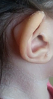
A 2-year-old girl was admitted to Mofid children’s hospital with a history of PID, recently developed whitish skin papules, and subcutaneous nodules around her mouth (Figures 1 and 2). She had been admitted because of upper respiratory infection at the ages of 4, 6, and 9 months and due to severe diarrhea at the ages of 8 and 10 months. Finally, immunologic workup was performed showing the IgG level of 215 mg/dL. The serum level of IgA was 32 mg/dL. The serum levels of IgM and IgE were within the normal ranges. NBT was normal and CD flow cytometry was unremarkable. Then, IVIG was started to control recurrent respiratory infections and poor response to antibiotics. When she was receiving monthly IVIG, her symptoms such as respiratory infections and diarrhea were controlled.
Whitish papules on her neck, face, and ears
Figure 1.
Whitish papules on her neck, face, and ears
Subcutaneous nodules and gingival hypertrophy
Figure 2.
Subcutaneous nodules and gingival hypertrophy
She was the only child of healthy consanguineous parents. Her umbilical cord was detached at 25th of life because of omphalitis. At the age of eight months, her parents complained of excessive crying while they were changing her clothes. The shoulder X-ray was unremarkable. Her mother detected multiple slow growing whitish papules appeared on her neck, face, and gum thickening at the age of 12 months. She was receiving regular IVIG since she was 12 months of age. She referred to our department for further investigation to confirm the diagnosis.
We discontinued IVIG and performed immunologic evaluations. The level of serum immunoglobulins was normal (IgM = 35 g/dL, IgA = 58 g/dL, IgG = 680 g/dL). Antibody titers to tetanus toxin and diphtheria were in the normal range. There was no abnormality in peripheral blood flow cytometry. The NBT test was 100% and LTT was normal. Then, CVID diagnosed was ruled out and excisional biopsy of subcutaneous nodule around her mouth was performed. Fibrosis and hyalinization, cells with abundant granular cytoplasm, and spindle-shaped cells spread in the hyaline material suggesting hyaline fibromatosis syndrome were demonstrated. Immunohistochemical (IHC) panel was compatible with fibromatosis (Table 1).
Table 1.
The IHC Results of the Subcutaneous Nodule
Results
SMAWeakly positive in few dispersed cells
DesminNegative
CD34Negative
CD117Negative (cytoplasmic staining in few dispersed cells)
Ki-67Positive in rare dispersed cell
S-100Positive
She underwent four times of surgical excision of hypertrophied gingiva and rectum prolapses. Imatinib was started and could decrease the growth rate of gum and reduce joints contractures.

3. Discussion

CVID is a heterogeneous group of primary immunodeficiency syndromes described as the low level of immunoglobulins and poor or absent specific antibody responses to vaccination (11), in spite of good antibody response in our patient. The symptoms may present at any age but more frequent between 15 and 40 years of age. The low level of IgG (≤ 2 S.D of the mean of the age-adjusted normal range) along with at least low-level IgA or IgM is found in CVID patients (12, 13). Over 90% of adult patients with CVID have IgA levels under the normal range. The level of serum IgM is more variable but it decreases in more than 50% of CVID patients (11). In this patient, a low level of IgG and IgA, recurrent infections, and optimal response to IVIG lead to the diagnosis of CVID. Although antibody response is not reliable at the age of 11 months, it could distinguish THI (transient hypogammaglobinemia of infancy) from CVID. On the other hand, there was no new infection while some new findings like the reduced range of motions and rectal prolapse were revealed in the course of the disease, which is not common in CVID or THI that needs more investigation.
After biopsy and diagnosis of IHF, the decreased level of IgG could be justified by PLE. Of course, it could predispose the patient to recurrent infection. PLE is a more common cause of profound hypogammaglobulinemia (14). The low level of IgA was justified by incomplete synthesis due to the age of the patient. Indeed, the levels of specific antibody responses and isohemagglutinin titers are helpful in the discrimination of THI and PLE from CVID.
The Hyaline fibromatosis syndrome is an AR disease in which the abnormal amorphous hyaline material is deposited in the dermis and other tissues (1). FTT and protein-losing enteropathy are important complications resulting in the decreased levels of immunoglobulins and some cellular and humoral deficiency, which lead to infection susceptibility (5). Other features of IHF including progressive joint contracture resulting in mobility limitation, severe pain with movement, gingival hypertrophy, thickened skin, coarse facial features, whitish skin papules, and subcutaneous nodules were found in this patient.
Indeed, molecular genetic testing to reveal genetic mutations will be confirmatory. When a patient is suspected to PID, the secondary immune deficiency should be ruled out. Moreover, patient follow-up and revaluation until definite diagnosis should be considered.


References

  • 1.
    Shieh JTC, Hoyme HE, Arbour LT. Hyalinosis, inherited systemic. GeneReviews. 2013.
  • 2.
    Momin YA, Bharambe BM, D'Costa G. Juvenile hyaline fibromatosis: a rare lesion. Indian J Pathol Microbiol. 2011;54(4):838-9. [PubMed ID: 22234127]. https://doi.org/10.4103/0377-4929.91546.
  • 3.
    Aghighi Y, Bahremand S, Nematollahi LR. Infantile systemic hyalinosis: report of three Iranian children and review of the literature. Clin Rheumatol. 2007;26(1):128-30. [PubMed ID: 16328093]. https://doi.org/10.1007/s10067-005-0124-y.
  • 4.
    Lindvall LE, Kormeili T, Chen E, Ramirez MC, Grum-Tokars V, Glucksman MJ, et al. Infantile systemic hyalinosis: Case report and review of the literature. J Am Acad Dermatol. 2008;58(2):303-7. [PubMed ID: 18222328]. https://doi.org/10.1016/j.jaad.2007.06.008.
  • 5.
    Klebanova Y, Schwindt C. Infantile Systemic Hyalinosis: A Case Report of Compromised Cellular and Humoral Branches of the Immune System Leading to Infections. Pediatr Asthma Allergy Immunol. 2009;22(3):127-30. [PubMed ID: 20563226]. [PubMed Central ID: PMC2885719]. https://doi.org/10.1089/pai.2009.0011.
  • 6.
    Nofal A, Sanad M, Assaf M, Nofal E, Nassar A, Almokadem S, et al. Juvenile hyaline fibromatosis and infantile systemic hyalinosis: a unifying term and a proposed grading system. J Am Acad Dermatol. 2009;61(4):695-700. [PubMed ID: 19344977]. https://doi.org/10.1016/j.jaad.2009.01.039.
  • 7.
    Glover MT, Lake BD, Atherton DJ. Infantile systemic hyalinosis: newly recognized disorder of collagen? Pediatrics. 1991;87(2):228-34. [PubMed ID: 1702887].
  • 8.
    Urbina F, Sazunic I, Murray G. Infantile systemic hyalinosis or juvenile hyaline fibromatosis? Pediatr Dermatol. 2004;21(2):154-9. [PubMed ID: 15078358]. https://doi.org/10.1111/j.0736-8046.2004.21214.x.
  • 9.
    Dowling O, Difeo A, Ramirez MC, Tukel T, Narla G, Bonafe L, et al. Mutations in capillary morphogenesis gene-2 result in the allelic disorders juvenile hyaline fibromatosis and infantile systemic hyalinosis. Am J Hum Genet. 2003;73(4):957-66. [PubMed ID: 12973667]. [PubMed Central ID: PMC1180616]. https://doi.org/10.1086/378781.
  • 10.
    Deuquet J, Lausch E, Superti-Furga A, van der Goot FG. The dark sides of capillary morphogenesis gene 2. EMBO J. 2012;31(1):3-13. [PubMed ID: 22215446]. [PubMed Central ID: PMC3252584]. https://doi.org/10.1038/emboj.2011.442.
  • 11.
    Warnatz C C R. Hypogammaglobulinemia and Common Variable Immunodeficiency. In: Kathleen E, Sullivan ERS, editors. Stiehm's Immune Deficiencies. UK: Elsevier; 2014. p. 347-61.
  • 12.
    Conley ME, Notarangelo LD, Etzioni A. Diagnostic criteria for primary immunodeficiencies. Representing PAGID (Pan-American Group for Immunodeficiency) and ESID (European Society for Immunodeficiencies). Clin Immunol. 1999;93(3):190-7. [PubMed ID: 10600329]. https://doi.org/10.1006/clim.1999.4799.
  • 13.
    Al-Herz W, Bousfiha A, Casanova JL, Chapel H, Conley ME, Cunningham-Rundles C, et al. Primary immunodeficiency diseases: an update on the classification from the international union of immunological societies expert committee for primary immunodeficiency. Front Immunol. 2011;2:54. [PubMed ID: 22566844]. [PubMed Central ID: PMC3342372]. https://doi.org/10.3389/fimmu.2011.00054.
  • 14.
    Onigbanjo MT, Orange JS, Perez EE, Sullivan KE. Hypogammaglobulinemia in a pediatric tertiary care setting. Clin Immunol. 2007;125(1):52-9. [PubMed ID: 17631052]. https://doi.org/10.1016/j.clim.2007.05.017.

Copyright

Copyright © 2018, Author(s). This is an open-access article distributed under the terms of the Creative Commons Attribution-NonCommercial 4.0 International License (http://creativecommons.org/licenses/by-nc/4.0/) which permits copy and redistribute the material just in noncommercial usages, provided the original work is properly cited.

Similar Articles

9
Dec
2014
Infantile Systemic Hyalinosis: Report of 17-year Experience

Infantile Systemic Hyalinosis: Report of 17-year Experience

Azadeh Afshin,
Seyed Reza Raeeskarami,
Yahya Aghighi,
Abdolreza Malek,
Ali Zamani,
Vahid Ziaee

Afshin A, Raeeskarami SR, Aghighi Y, Malek A, Zamani A, et al. Infantile Systemic Hyalinosis: Report of 17-year Experience. Inn J Pediatr. 2014;24(6):. doi:

6
Oct
2015
Association of Celiac Disease With Idiopathic Pulmonary Hemosiderosis; Lane Hamilton Syndrome

Association of Celiac Disease With Idiopathic Pulmonary Hemosiderosis; Lane Hamilton Syndrome

Hikmet Tekin Nacaroglu,
Ozlem Sarac Sandal,
Ozlem Bag,
Semiha Bahceci Erdem,
Ozlem Bekem Soylu,
Gulden Diniz
,et al.

Nacaroglu HT, Sandal OS, Bag O, Erdem SB, Bekem Soylu O, et al. Association of Celiac Disease With Idiopathic Pulmonary Hemosiderosis; Lane Hamilton Syndrome. Inn J Pediatr. 2015;25(5):e3312. doi: https://doi.org/10.5812/ijp.3312

31
Jan
2011
Localized Jaw Enlargement due to Fibrous Dysplasia in a Hemodialysis Patient

Localized Jaw Enlargement due to Fibrous Dysplasia in a Hemodialysis Patient

Gargah Tahar,
Ounissi Mondher,
Lakhoua Mohamed Rachid

Tahar G, Mondher O, Rachid L. Localized Jaw Enlargement due to Fibrous Dysplasia in a Hemodialysis Patient. Nephro-Urol Mon. 2011;3(1):. doi:

7
Jan
2014
Chronic Diarrhea as a Presenting Manifestation of Common Variable Immunodeficiency

Chronic Diarrhea as a Presenting Manifestation of Common Variable Immunodeficiency

Amir Hossein Hosseini,
Mehrnaz Mesdaghi,
Ali Akbar Sayyari,
Zahra Chavoshzadeh

Hosseini AH, Mesdaghi M, Sayyari AA, Chavoshzadeh Z. Chronic Diarrhea as a Presenting Manifestation of Common Variable Immunodeficiency. Arch Pediatr Infect Dis. 2014;2(1):180-182. doi: https://doi.org/10.5812/pedinfect.12295

18
Jul
2013
Alexander Disease: Report of Two Unrelated Infantile Form Cases, Identified by GFAP Mutation Analysis and Review of Literature; The First Report from Iran

Alexander Disease: Report of Two Unrelated Infantile Form Cases, Identified by GFAP Mutation Analysis and Review of Literature; The First Report from Iran

Alireza Tavasoli,
Mahmoud-Reza Ashrafi,
Omid Aryani,
Hooman Alizadeh,
Massoud Houshmand

Tavasoli A, Ashrafi M, Aryani O, Alizadeh H, Houshmand M. Alexander Disease: Report of Two Unrelated Infantile Form Cases, Identified by GFAP Mutation Analysis and Review of Literature; The First Report from Iran. Inn J Pediatr. 2013;23(4):. doi:

More by these authors

Delara BabaiePubMedScholar
Mohammad QaisariPubMedScholar
Hanieh Sadat MirzadePubMedScholar
Fatemeh AbdolinejadPubMedScholar
Farahnaz Bidari ZerehpoushPubMedScholar
Share
Cited by
Metrics