Bone Marrow Aplasia: a Rare Complication of Disseminated Tuberculosis

Authors

Ravi Ambey1,*, Veerbhadra CD1
1Department of Pediatrics, G.R. Medical College, Gwalior, India
*Corresponding Author: Corresponding author: Ravi Ambey, Department of Pediatrics, G.R. Medical College, Gwalior, India. Tel.: +91-7512403268, E-mail: Email: [email protected]

Archives of Clinical Infectious Diseases:Vol. 7, issue 2; e13949
Published online:Apr 13, 2012
Article type:Brief Report
Received:Feb 10, 2012
Accepted:Mar 01, 2012
How to Cite:Ambey R, CD V. Bone Marrow Aplasia: a Rare Complication of Disseminated Tuberculosis. Arch Clin Infect Dis. 2012;7(2):e13949. doi: https://doi.org/10.5812/archcid.13949

Abstract

We report 11 year old male child, a case of acquired aplastic anemia as a complication of disseminated tuberculosis with acute onset, rapid downhill course and refractory to both antitubercular drugs as well as immunosuppressive drugs.

1. Introduction

We would like to share our experience with an eleven-year-old male, a case of acquired aplastic anemia as a complication of disseminated tuberculosis with acute onset, rapid downhill course, and refractory to both anti-tubercular as well as immunosuppressive drugs.

2. Short Communication

Tuberculosis is known to cause wide variety of systemic complications when it disseminates in the body. Tuberculosis, suppressing all three cell lines which causes pancytopenia, leading to aplastic anemia has been reported by very few case reports, either as the result of disseminated tuberculosis or as an adverse effect of anti-tubercular medicines (1-3). An eleven-year-old male, weighing 27 kg with poor nutritional status presented to our pediatric intensive care unit with history of fever for one month, headache and vomiting for 15 days and weakness of right half of his body with right-sided deviation of the angle of his mouth for 3 days. He also noticed bluish spots on the chest and trunk for one day. He lived in an overcrowded house with illiterate parents and belonged to a family with low socioeconomic status, but was immunized and had BCG scar with a history of contact with an active tuberculosis patient in his family. He did not have similar complaints in the past or any hospitalization or medication. On examination, the child was afebrile and normotensive, his heart rate was 110 beats per minute, and respiratory rate was 27/min. He was severely pale, and petechiae were observed over the trunk accompanied by subconjunctival hemorrhage. He was drowsy with altered sensorium. Neurological examination revealed meningoencephelitis with right sided hemiparesis with left sided facial palsy. The examination of other systems was within normal limits. Cerebrospinal fluid analysis revealed 432 mg/dL protein, 43 mg/dL glucose, 110 meq/L chloride with 160 cells out of which 75% was lymphocytes and 25% was polymorphs. Gram staining of the CSF was unremarkable and culture was negative. However, CSF PCR was positive for mycobacteria tuberculosis. There were few signs of raised intracranial pressure without active seizure. He was given supportive treatment with intravenous fluids, mannitol, and dexamethasone. When he was stabilized on the second day, antitubercular therapy and nasogastric tube feeding were initiated. Brain CT showed gyral swelling with patchy hemorrhage involving fronto-parieto-temporo-occipital lobe (meningo-encephelitis) with grade 3 perifocal edema leading to mild leftward midline shift (Figure. 1).
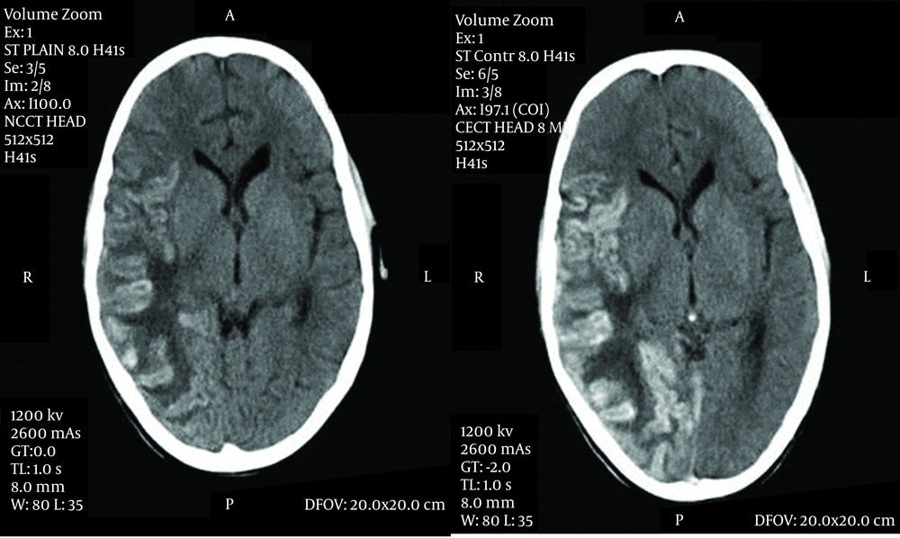
Axial Contrast-Enhanced CT Scan Images of The Brain Demonstrating Patchy Gyral Hemorrhage With Leptomeningeal Enhancement Involving the Right Temporo-Parieto-Occipital Region With Extensive Edema and Midline Shift
Figure 1.
Axial Contrast-Enhanced CT Scan Images of The Brain Demonstrating Patchy Gyral Hemorrhage With Leptomeningeal Enhancement Involving the Right Temporo-Parieto-Occipital Region With Extensive Edema and Midline Shift
Hematological deterioration continued as pancytopenia was persistent on peripheral blood smear examination. Peripheral smear did not show any abnormal cells, hemoparasites, or evidence of hemolysis. Bone marrow aspiration confirmed hypoplastic marrow with epitheloid cell granuloma and focal necrosis consistent with tuberculosis. There were no features suggestive of hemophagocytic syndrome in bone marrow smear examination. The bleeding time was 14 minutes. The clotting, prothrornbin and partial thromboplastin time were normal. The urine and stool examination did not show any abnormality. Urine and blood cultures did not grow any organisms. The direct and indirect Coomb's tests were negative. Blood Widal, HIV ELISA and VDRL were non-reactive. We added anabolic steroids and later cyclosporine for which there was no response. There was no possibility to transfer the patient to another center with facilities for bone marrow transplantation. In spite of two platelet-rich plasma transfusions, the child started bleeding due to severe thrombocytopenia, developed hypovolumic shock and died as the result of massive uncontrollable upper gastrointestinal and pulmonary hemorrhages after 19 days of hospital stay. Autopsy could not be performed.
Literature shows the occurrence of pancytopenia in disseminated tuberculosis is attributed to hypersplenism, histiocytic hyperplasia and indiscriminate phagocytosis of blood cells by histiocytes in the bone marrow, maturational arrest, or infiltration of the bone marrow by caseating or noncaseating tubercular granulomas. Tubercular granulomas may cause pancytopenia by replacement of marrow cells or suppression through release of interferon and lymphotoxin. However, despite presence of tuberculous granulomas in a high proportion of patients with disseminated tuberculosis, pancytopenia is uncommon (1). Pathophysiology of acquired aplastic anemia in tuberculosis is found to be immune-mediated based on observation of recovery after immunosuppressant, but the present case was resistant to this treatment (4).

3. Conclusions

There are several unusual features of interest in this case. Amidst many reports which have not shown association between tuberculosis and aplastic anemia, our case brings back the old view to support the causation of aplastic anemia in tuberculosis (5). Aplasia of the bone marrow was caused by an overwhelming toxemia. Surprisingly no system was able to analyze the effect of the condition on bone marrow, disregarding the neurological aspects. In spite of limiting to two foci, severity of the bone marrow disease was rapidly progressive and did not respond to supportive treatment.


Acknowledgments

Footnotes

  • Implication for health policy/practice/research/medical education:Tuberculosis is known to cause wide variety of systemic manifestations when it disseminate to the body. We are reporting a case of bone marrow aplasia in a child due to disseminated tuberculosis. The case emphasizes that Tuberculosis continues to be a Pandora’s Box with many new surprises for clinicians.

  • Authors’ Contribution:Veerbhadra CD collected the clinical data and prepared the manuscript. Ravi Ambey reviewed the literature, revised the manuscript and is guarantor.

  • Financial Disclosure:We declare that we have no financial interest related to material in manuscript.

  • Funding/Support:The study did not receive any fund.

References

  • 1.
    Yadav TP, Mishra S, Sachdeva KJ, Gupta VK, Siddhu KK, Bakshi G, et al. Pancytopenia in disseminated tuberculosis. Indian Pediatr. 1996;33(7):597-9. [PubMed ID: 8979574].
  • 2.
    Avasthi R, Mohanty D, Chaudhary SC, Mishra K. Disseminated tuberculosis: interesting hematological observations. J Assoc Physicians India. 2010;58:243-4. [PubMed ID: 21046880].
  • 3.
    Kernohan EJ. Aplastic anaemia complicating miliary tuberculosis. Br Med J. 1950;2(4675):399-400. [PubMed ID: 15434400].
  • 4.
    Young NS. Pathophysiologyic mechanism in acquired aplastic anemia. Heamatol. 2006:72-7.
  • 5.
    Hsu HC, Lee YM, Su WJ, Huang CY, Yang CF, Ho CK, et al. Bone marrow samples from patients with aplastic anemia are not infected with parvovirus B19 and Mycobacterium tuberculosis. Am J Clin Pathol. 2002;117(1):36-40. [PubMed ID: 11789728]. https://doi.org/10.1309/B361-LA0V-UX0X-GJPQ.

Copyright

Copyright © 2012, Infectious Diseases and Tropical Medicine Research Center. This is an open-access article distributed under the terms of the Creative Commons Attribution-NonCommercial 4.0 International License (http://creativecommons.org/licenses/by-nc/4.0/) which permits copy and redistribute the material just in noncommercial usages, provided the original work is properly cited.

Similar Articles

19
May
2023

An Unusual Presentation of Pulmonary Mucormycosis as a “Mass-like” Consolidation in an Infant with Chronic Granulomatous: A Case Report Study

Narges Lashkarbolouk,
Mahdi Mazandarani,
Ali Ahani Azari,
Lobat Shahkar

Lashkarbolouk N, Mazandarani M, Ahani Azari A, Shahkar L. An Unusual Presentation of Pulmonary Mucormycosis as a “Mass-like” Consolidation in an Infant with Chronic Granulomatous: A Case Report Study. J Compr Ped. 2023;14(3):e136054. doi: https://doi.org/10.5812/compreped-136054

14
Oct
2018
Visceral Leishmaniosis in Bone Marrow Aspiration

Visceral Leishmaniosis in Bone Marrow Aspiration

Mohammad Naderisorki,
Hossein Karami

Naderisorki M, Karami H. Visceral Leishmaniosis in Bone Marrow Aspiration. Int J Infect. 2018;5(4):e83304. doi: https://doi.org/10.5812/iji.83304

21
Aug
2024
Treatment Challenges of Methimazole-Induced Agranulocytosis in Younger Patients

Treatment Challenges of Methimazole-Induced Agranulocytosis in Younger Patients

Azadeh Kiumarsi,
Reza Mohamadhosseinzadeh Davatgari,
Maryam Habibi,
Mahsa Soti Khiabani

Kiumarsi A, Mohamadhosseinzadeh Davatgari R, Habibi M, Soti Khiabani M. Treatment Challenges of Methimazole-Induced Agranulocytosis in Younger Patients. Inn J Pediatr. 2024;34(5):e140262. doi: https://doi.org/10.5812/ijp-140262

30
Apr
2016

Linezolid Induced Twice Pure Red Cell Aplasia in a Patient with Central Nervous System Infection after Allogeneic Stem Cell Transplantation

Wenqing Hu,
Bing Shi,
Lihui Liu,
Shengke He,
Liping Ye,
DengMei Tian
,et al.

Hu W, Shi B, Liu L, He S, Ye L, et al. Linezolid Induced Twice Pure Red Cell Aplasia in a Patient with Central Nervous System Infection after Allogeneic Stem Cell Transplantation. Iran J Pharm Res. 2016;15(2):e125190. doi: https://doi.org/10.22037/ijpr.2016.1794

10
Feb
2019
Visceral Leishmaniasis with Massive Pulmonary Involvement: A Case Report

Visceral Leishmaniasis with Massive Pulmonary Involvement: A Case Report

Mehdi Dehghani,
Ahmad Monabati,
Mozhdeh Sanei,
Mohammad Ali Davarpanah

Dehghani M, Monabati A, Sanei M, Davarpanah MA. Visceral Leishmaniasis with Massive Pulmonary Involvement: A Case Report. Int J Infect. 2019;6(1):e87993. doi: https://doi.org/10.5812/iji.87993

More by these authors

Ravi AmbeyPubMedScholar
Veerbhadra CDPubMedScholar
Share
Cited by
Metrics