Cover image of the article: Refractory Ulcerative Lesions on Lips and Nose of a Patient With Systemic Lupus Erythematosus

Image Credit:Arch Clin Infect Dis

Refractory Ulcerative Lesions on Lips and Nose of a Patient With Systemic Lupus Erythematosus

Authors

Atyeh Ebadi1, Fahimeh Abdollahimajd1, Sajjad Nazari1, Soheila Nasiri1,*
1Skin Research Center, Shohada-e-Tajrish Hospital, Shahid Beheshti University of Medical Sciences, Tehran, IR Iran
*Corresponding Author: Corresponding author: Soheila Nasiri, Skin Research Center, Shohada-e-Tajrish Hospital, Shahid Beheshti University of medical Sciences, Tehran, IR Iran. Tel.: +98-2122741507, Fax: +98-2122744392, E-mail: Email: nasiri_s@hotmail.com

Archives of Clinical Infectious Diseases:Vol. 8, issue 1; e16033
Published online:Dec 24, 2012
Article type:Discussion
Received:Aug 29, 2012
Accepted:Nov 01, 2012
How to Cite:Ebadi A, Abdollahimajd F, Nazari S, Nasiri S. Refractory Ulcerative Lesions on Lips and Nose of a Patient With Systemic Lupus Erythematosus. Arch Clin Infect Dis. 2012;8(1):e16033. doi: https://doi.org/10.5812/archcid.16033

1. Case Summary

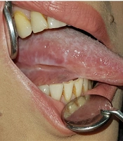
A 37-year-old female was seen as an in-patient on the rheumatology ward of Loghman-e-Hakim Hospital, Shahid Beheshti University of Medical Sciences, Tehran, Iran, for the evaluation of her refractory ulcerative lesions. She had a three year history of systemic lupus erythematosus (SLE) with an insidiously deteriorating nature, which waxed and waned through time. She was hospitalized in order to get thorough treatment though with some potential side effects for the disease flare up using high doses of Cychlophosphamide and Prednisolone. Two days after initiating the treatment, ulcerative lesions developed on the nose and lip. The lesions were painful and appeared to be lupus oral ulcers, but in spite of the difficult process of the therapy regimen, the lesions was insidiously progressing over weeks without any sign of improvement and eventually dermatology consultation was obtained after one month delay. When we visited her, she was free of any systemic symptoms. Physical examination revealed ulcerative lesions on the upper and lower lips as well as on the nose. Ulcers were surrounded by elevated borders admixed with central crusts and necrosis (Figure 1).
Ulcerative Lesions on Upper and Lower Lip and on Nose Admixed With Central Crusts and Necrosis
Figure 1.
Ulcerative Lesions on Upper and Lower Lip and on Nose Admixed With Central Crusts and Necrosis
In addition, the patient had mild, non-tender and right submandibular lymphadenopathy. Other examinations were unremarkable. Laboratory tests showed a leukopenia (WBC: 1300) with 80% neutrophils and 20% lymphocytes.
- To confirm the diagnosis, a skin biopsy was achieved (Figure 2).
- Now, what is your diagnosis?
Histopathology of Lesions
Figure 2.
Histopathology of Lesions

2. Diagnosis: Herpes Simplex Virus Infection

Tzank smear and biopsy specimen were obtained (Figure 2). Microscopic examination revealed a reticular degeneration and spongiosis of the epidermis, ballooning of the cytoplasm of keratinocytes and intranuclear inclusion bodies, multinucleated giant cells (are formed by fusion of the infected keratinocytes) and inflammatory response of lymphocytes, neutrophils and eosinophils infiltrate in the dermis. The results were consistent with the diagnosis of HSV infection. Therapy with oral acyclovir was then initiated (400 mg po × 5/d/14 days). After one week, the lesions were markedly improved (Figure 3) and they were completely healed after two weeks.
One week after Treatment With Acyclovir Therapy, the Lesions Markedly Improved
Figure 3.
One week after Treatment With Acyclovir Therapy, the Lesions Markedly Improved
HSV-1 and HSV-2 produce primarily orolabial and genital herpes infections. Their clinical presentations are influenced by several factors including viral entry root, degree of immunocompetency and nature of the virus. They have a wide range of clinical presentations, ranging from limited mucocutaneous disease to fatal dissemination particularly in immunocompromised conditions. Initial lesions appear as painful vesicles on an erythematous base followed by pustules and/or ulcerations. Resolution of symptoms occurs within 2 - 6 weeks. In general, the most common sites of involvement are mouth and lips. Less common sites are perioral skin, nasal mucosa, hard palates, and cheeks. In immunocompromised patients, recurrent herpes infections can involve intraoral 'soft' mucosa (1-4). Oral herpes simplex virus (HSV) infections may be severe particularly in immunocompromised patients and may be associated with serious morbidity, potential secondary infections (such as bacterial and fungal infections) and systemic dissemination. They may frequently be misdiagnosed due to their atypical presentations (5). Immunocompromising conditions include congenital immunodeficiencies, AIDS, hematological malignancies such as Leukaemia, autoimmune diseases such as SLE and iatrogenically induced immunosuppression as seen in organ transplantation. In hospitalized and/or immunocompromised patients, prompt and accurate diagnosis of infectious diseases such as HSV infection is critical and urgent dermatologic consultation for atypical mucocutaneous presentations may prevent common delay in diagnosis. Hence, it helps physicions to settle appropriate treatments, prohibiting progression to potentially serious and fatal outcomes (6, 7). Characteristics of atypical infections in immunocompromised hosts include deeper and larger size of lesions, more profound ulceration, satellite lesions, long lasting lesions with longer healing time and atypical locations (3). Verrucous, exophytic, pustular or ulcerative lesions can also be seen. Respiratory and gastrointestinal tract can be involved as well. Differential diagnosis of oral lesions in these patients includes candidiasis, other oral infections (such as deep fungal), herpes infection, mucositis, and malignant infiltration. Diagnosis can often be achieved by history, physical examination and a Tzanck smear. In immunocompromised patients, a direct fluorescent antibody test, viral culture or skin biopsy with immunohistochemistry may be required for definitive diagnosis. In these patients, acyclovir therapy should be initiated (8-10). When a clinician evaluates immunocompromised patients with chronic ulcerations and necrosis, it is important to keep herpetic infections high on the differential diagnosis in order to avoid serious and possibly fatal outcomes. Given the patient under study, she also presented with chronic ulceration along with necrotic area within them and she responded well to acyclovir therapy.
Acknowledgments

Footnotes

  • Implication for health policy/practice/research/medical education:When a clinician examines immunocompromised patients with chronic ulcerations and necrosis, it is important to take herpetic infections into consideration in order to avoid serious and possibly fatal consequences.

  • Authors’ Contribution:Data collection and drafting of the manuscript were carried out by Dr Abdollahimajd and Dr Ebadi. Dr Nazari cooperated in writing the manuscript. Critical revision of the manuscript was made by Dr Nasiri.

  • Financial Disclosure:There are no conflicts of interest.

  • Funding/Support:The study did not receive any fund.

References

  • 1.
    Brady RC, Bernstein DI. Treatment of herpes simplex virus infections. Antiviral Res. 2004;61(2):73-81. [PubMed ID: 14670580].
  • 2.
    Leflore S, Anderson PL, Fletcher CV. A risk-benefit evaluation of aciclovir for the treatment and prophylaxis of herpes simplex virus infections. Drug Saf. 2000;23(2):131-42. [PubMed ID: 10945375].
  • 3.
    Meyers JD, Wade JC, Mitchell CD, Saral R, Lietman PS, Durack DT, et al. Multicenter collaborative trial of intravenous acyclovir for treatment of mucocutaneous herpes simplex virus infection in the immunocompromised host. Am J Med. 1982;73(1):229-235.
  • 4.
    Nadelman CM, Newcomer VD. Herpes simplex virus infections. New treatment approaches make early diagnosis even more important. Postgrad Med. 2000;107(3):189-200. [PubMed ID: 10728143].
  • 5.
    Epstein JB, Scully C. Herpes simplex virus in immunocompromised patients: Growing evidence of drug resistance. Or Surg, Or Med, Or Pathol. 1991;72(1):47-50. https://doi.org/10.1016/0030-4220(91)90188-I.
  • 6.
    Lieb JA, Brisman S, Herman S, Macgregor J, Grossman ME. Linear erosive herpes simplex virus infection in immunocompromised patients: the "knife-cut sign". Clin Infect Dis. 2008;47(11):1440-1. [PubMed ID: 18937574]. https://doi.org/10.1086/592976.
  • 7.
    Burgoyne M, Burke W. Atypical herpes simplex infection in patients with acute myelogenous leukemia recovering from chemotherapy. J Am Acad Dermatol. 1989;20(6):1125-27.
  • 8.
    Barrett AP. Chronic indolent orofacial herpes simplex virus infection in chronic leukemia: A report of three cases. Or Surg, Or Med, Or Pathol. 1988;66(3):387-390. https://doi.org/10.1016/0030-4220(88)90251-4.
  • 9.
    Smith E, Hallman JR, Pardasani A, McMichael A. Multiple herpetic whitlow lesions in a patient with chronic lymphocytic leukemia. Am J Hematol. 2002;69(4):285-8. [PubMed ID: 11921024].
  • 10.
    Wakui H, Togashi M, Omokawa A, Okuyama S, Masai R, Komatsuda A, et al. Simultaneous herpes simplex virus esophagitis and lupus enteritis in a patient with systemic lupus erythematosus. Mod Rheumatol. 2010;20(1):98-101. [PubMed ID: 19802650]. https://doi.org/10.1007/s10165-009-0237-2.

Copyright

Copyright © 2012, Infectious Diseases and Tropical Medicine Research Center. This is an open-access article distributed under the terms of the Creative Commons Attribution-NonCommercial 4.0 International License (http://creativecommons.org/licenses/by-nc/4.0/) which permits copy and redistribute the material just in noncommercial usages, provided the original work is properly cited.

Similar Articles

15
Mar
2023
Management of Lichen Planus in a Post Liver Transplant Patient with Lupus Erythematosus, and Ulcerative Colitis; A Case Report

Management of Lichen Planus in a Post Liver Transplant Patient with Lupus Erythematosus, and Ulcerative Colitis; A Case Report

Mohaddeseh Davari,
Sara Pourshahidi,
Mohammad Javad Shokrian

Davari M, Pourshahidi S, Shokrian MJ. Management of Lichen Planus in a Post Liver Transplant Patient with Lupus Erythematosus, and Ulcerative Colitis; A Case Report. Middle East J Rehabil Health Stud. 2023;10(4):e131509. doi: https://doi.org/10.5812/mejrh-131509

27
Feb
2017
Methylprednisolone Pulse Therapy Can Be Life-Saving in Neonatal Lupus Erythematous: A Case Report

Methylprednisolone Pulse Therapy Can Be Life-Saving in Neonatal Lupus Erythematous: A Case Report

Maliheh Kadivar,
Razieh Sangsari,
Leila Shahbaznejad,
Vahid Ziaee

Kadivar M, Sangsari R, Shahbaznejad L, Ziaee V. Methylprednisolone Pulse Therapy Can Be Life-Saving in Neonatal Lupus Erythematous: A Case Report. Inn J Pediatr. 2017;27(3):e9004. doi: https://doi.org/10.5812/ijp.9004

16
May
2014
Neonatal Lupus erythematosus Following Rheumatoid Arthritis: Case Report and Literature Review

Neonatal Lupus erythematosus Following Rheumatoid Arthritis: Case Report and Literature Review

Raheleh Assari,
Vahid Ziaee,
Mohammad-Hassan Moradinejad,
Arash Mirmohammadsadeghi

Assari R, Ziaee V, Moradinejad M, Mirmohammadsadeghi A. Neonatal Lupus erythematosus Following Rheumatoid Arthritis: Case Report and Literature Review. Inn J Pediatr. 2014;24(4):. doi:

2
Dec
2013
Severe, Recurrent Mesenteric Vasculitis as the Initial Presentation of Systemic Lupus Erythematosus

Severe, Recurrent Mesenteric Vasculitis as the Initial Presentation of Systemic Lupus Erythematosus

Mohammadreza Seyyedmajidi,
Jamshid Vafaeimanesh

Seyyedmajidi M, Vafaeimanesh J. Severe, Recurrent Mesenteric Vasculitis as the Initial Presentation of Systemic Lupus Erythematosus. Zahedan J Res Med Sci. 2014;16(11):55-56. doi:

20
Jul
2013
Disseminated Tuberculosis Imitating Systemic Lupus Erythematosus

Disseminated Tuberculosis Imitating Systemic Lupus Erythematosus

Mahboubeh Hajiabdolbaghi,
Negin Esmailpour,
Mehrnaz Rasoolinejad,
Mojtaba Hedayat Yaghoobi,
Seyed Ali Dehghan Manshadi

Hajiabdolbaghi M, Esmailpour N, Rasoolinejad M, Hedayat Yaghoobi M, Dehghan Manshadi SA. Disseminated Tuberculosis Imitating Systemic Lupus Erythematosus. Arch Clin Infect Dis. 2013;8(3):e17288. doi: https://doi.org/10.5812/archcid.17288

More by these authors

Atyeh EbadiPubMedScholar
Fahimeh AbdollahimajdPubMedScholar
Sajjad NazariPubMedScholar
Soheila NasiriPubMedScholar
Share
Cited by
Metrics