Full Text
Full text is available in PDF
Official Journal of Research Center for Gastroenterology and Liver Diseases
Authors
A solitary necrotic nodule (SNN) of the liver is an uncommon lesion, which is different from primary and metastatic liver cancers.
To analyze the classification, CT and MR manifestation, and the pathological basis of solitary necrotic nodule of the liver (SNN) in order to evaluate CT and MRI as a diagnosing tool.
This study included 29 patients with liver SNNs, out of which 14 had no clinical symptoms and were discovered by routine ultrasound examinations, six were found by computed tomography (CT) due to abdominal illness, four had ovarian tumors, and five had gastrointestinal cancer surgeries, previously. Histologically, these SNNs can be divided into three subtypes, i.e., type I, pure coagulation necrosis (14 cases); type II, coagulation necrosis mixed with liquefaction necrosis (five cases); and type III, multi-nodular fusion (10 cases). CT and magnetic resonance imaging (MRI) patterns were shown to be associated with SNN histology. All patients were treated surgically with good prognosis.
CT and MRI appearance and correlation with pathology types: three subtypes of lesions were hypo-density on both pre contrast and post contrast CT, 12 lesions were found the enhanced capsule and 1 lesion of multi- nodular fusion type showed septa enhancement. The lesions were hypo-intensity on T2WI and the lesions of type II showed as mixed hyperintensity on T2WI. The capsule showed delayed enhancement in all cases, and all lesions of multi- nodular fusion type showed delayed septa enhancement on MR images. 15 cases on CT were misdiagnosed and Four cases on MRI were misdiagnosed and the accuracy of CT and MRI were 48.3% and 86.2% respectively.
In conclusion, CT and MRI are useful tools for SNN diagnosis.
© 2012, Author(s). This open-access article is available under the Creative Commons Attribution 4.0 (CC BY 4.0) International License (https://creativecommons.org/licenses/by/4.0/), which allows for unrestricted use, distribution, and reproduction in any medium, provided that the original work is properly cited.
Zeinali-Rafsanjani B. Diagnosis of Solitary Pulmonary Nodules Using MRI: A Feasibility Study. I J Radiol. 2017;14(Special Issue):e48010. doi: https://doi.org/10.5812/iranjradiol.48010
Jalli R, Jafari SH, Sefidbakht S, Kazemi K. Comparison of the Accuracy of DWI and Ultrasonography in Screening Hepatocellular Carcinoma in Patients With Chronic Liver Disease. I J Radiol. 2015;12(1):e37958. doi: https://doi.org/10.5812/iranjradiol.12708
Radmard AR. Imaging of Hepatic Metastasis. I J Radiol. 2017;14(Special Issue):e48396. doi: https://doi.org/10.5812/iranjradiol.48396
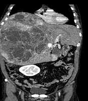
Ayoobi Yazdi N, Dashti H, Safaei M, Salahshour F, Fotouhi M, et al. Radiologic and Pathologic Findings of a Huge Solitary Fibrous Tumor of the Liver with Malignant Transformation: A Case Report. I J Radiol. 2020;17(1):e68449. doi: https://doi.org/10.5812/iranjradiol.68449
Yoon J, Kim J. Atypical Findings of Focal Nodular Hyperplasia with Gadoxetic Acid (Gd-EOB-DTPA)-Enhanced Magnetic Resonance Imaging. I J Radiol. 2014;11(1):e9269. doi: https://doi.org/10.5812/iranjradiol.9269
Last Update: 2 weeks ago
Last Update: 2 weeks ago
Last Update: 2 weeks ago
Last Update: 2 hours ago
Last Update: 8 hours ago
Last Update: 5 days ago