Incidentally Detected Persistent Left Superior Vena Cava With an Absent Right Superior Vena Cava in a Neonate

Authors

Young Earl Choi1, Hwa Jin Cho1,*, Jae Sook Ma1
1Department of Pediatrics, Chonnam National University Hospital, Gwangju, South Korea
*Corresponding Author: Corresponding author: Hwa Jin Cho, Department of Pediatrics, Chonnam National University Hospital, Gwangju, South Korea. Tel: +82-622206646, Fax: +82-622236103, E-mail: Email: [email protected]

Innovative Journal of Pediatrics:Vol. 26, issue 3; e4692
Published online:May 15, 2016
Article type:Letter
Received:Nov 24, 2015
Accepted:Dec 08, 2015
How to Cite:Earl Choi Y, Jin Cho H, Sook Ma J. Incidentally Detected Persistent Left Superior Vena Cava With an Absent Right Superior Vena Cava in a Neonate. Inn J Pediatr. 2016;26(3):e4692. doi: https://doi.org/10.5812/ijp.4692

Dear Editor,
A persistent left superior vena cava with an absent right superior vena cava is extremely rare and only a few cases have been reported in adults (1-3), while no cases have been reported in neonates or infants. Here, we report a 3-week-old female who was diagnosed with this condition using transthoracic echocardiography and confirmed with thoracic computed tomography (CT) angiogram.
A 3-week-old female underwent transthoracic echocardiography (iE33, Philips medical system, Andover, MA, USA) to evaluate a grade 1 systolic murmur. In the subcostal view, blood flow in the inferior vena cava drained to the right atrium, while no blood flow from the superior vena cava to the right atrium was noted (Figure 1A). In the four-chamber view, no superior vena caval blood flow was seen, but an enlarged coronary sinus drained to the right atrium. Except for a patent foramen ovale, there were no congenital heart defects. In the parasternal long-axis view, a dilated coronary sinus was noted posterior to the left atrium (Figure 1B) and the suprasternal view showed a persistent left superior vena cava with a right innominate vein draining toward the left superior vena cava and an absent right superior vena cava (Figure 1C). The baby girl was diagnosed with a persistent left superior vena cava with an absent right superior vena cava. Since there was no obvious problem with venous blood draining to the right atrium, her parents were reassured. Six months later, thoracic CT (somatome definition flash, Siemens healthcare, Forchheim, Germany) angiogram was performed to confirm the diagnosis and find any unrevealed anomalies. CT confirmed absence of the right superior vena cava and the right jugular and subclavian veins drained to the left superior vena cava through the innominate vein (Figure 2). The left superior vena cava drained to the right atrium through the dilated coronary sinus with no stenosis. The baby girl did not undergo any medical or surgical procedure to correct this anomaly and she is healthy during 2 years of follow up period.
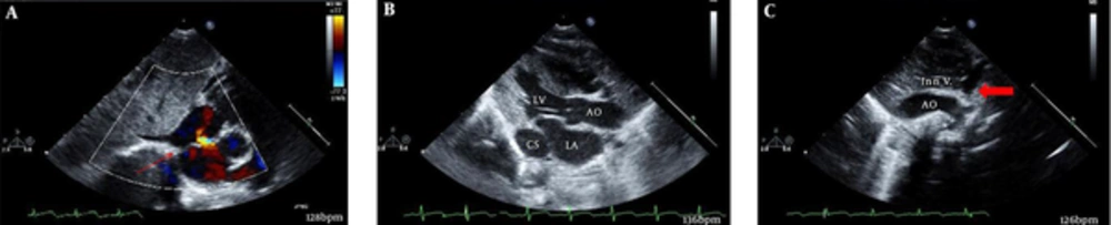
A, The subcostal view shows no right superior vena cava blood flow draining to the right atrium (arrow); B, In the parasternal long axis view, an enlarged coronary sinus is seen; C, In the suprasternal view, the left superior vena cava and innominate vein are seen. AO, aorta; CS, coronary sinus; Inn V, innominate vein; LA, left atrium; LV, left ventricle.
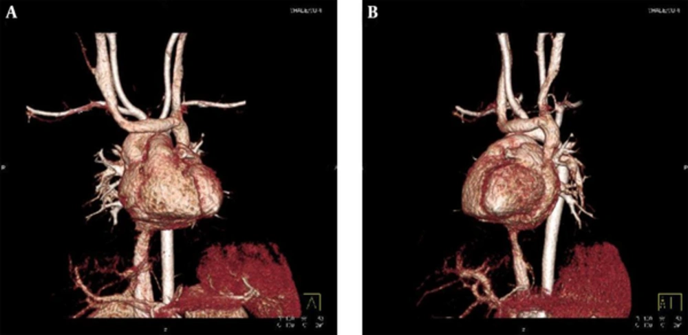
Figure 1.
A, The subcostal view shows no right superior vena cava blood flow draining to the right atrium (arrow); B, In the parasternal long axis view, an enlarged coronary sinus is seen; C, In the suprasternal view, the left superior vena cava and innominate vein are seen. AO, aorta; CS, coronary sinus; Inn V, innominate vein; LA, left atrium; LV, left ventricle.
A, Left superior vena cava draining to; B, The coronary sinus, with an absent right superior vena cava.
Figure 2.
A, Left superior vena cava draining to; B, The coronary sinus, with an absent right superior vena cava.
Although a persistent left superior vena cava with a right superior vena cava is relatively common, a persistent left superior vena cava without a right superior vena cava is extremely rare. A persistent left superior vena cava with an absent right superior vena cava is generally asymptomatic, but has clinical implications. It is associated with an increased incidence of arrhythmias and conduction disturbance (4), and also has high incidence of accompanying congenital heart disease (5). Careful management is needed while inserting central venous catheter (2), cannulating in open-heart surgery (6), and also while performing electrophysiological studies and pacemaker implantation at any age (3).
In neonates and infants, the subcostal and suprasternal views of echocardiography with CT images give much information on the venous anatomy which should not miss the important diagnosis. By knowing important clinical implication, future risks for patients can be expected.
Footnotes

  • Authors’ Contribution:Hwa Jin Cho and Jae Sook Ma were responsible for reported research and all authors have participated in the concept and design, Young Earl Choi was responsible in drafting manuscript and all authors revised the manuscript, and we all have approved the manuscript as submitted. Each author listed on the manuscript has seen and approved the submission of this version of the manuscript and takes full responsibility for the manuscript.

References

  • 1.
    Ucar O, Pasaoglu L, Cicekcioglu H, Vural M, Kocaoglu I, Aydogdu S. Persistent left superior vena cava with absent right superior vena cava: a case report and review of the literature. Cardiovasc J Afr. 2010;21(3):164-6. [PubMed ID: 20532458].
  • 2.
    Azocar RJ, Narang P, Talmor D, Lisbon A, Kaynar AM. Persistent left superior vena cava identified after cannulation of the right subclavian vein. Anesth Analg. 2002;95(2):305-7. table of contents. [PubMed ID: 12145039].
  • 3.
    Rizkallah J, Burgess J, Kuriachan V. Absent right and persistent left superior vena cava: troubleshooting during a challenging pacemaker implant: a case report. BMC Res Notes. 2014;7:462. [PubMed ID: 25047923]. https://doi.org/10.1186/1756-0500-7-462.
  • 4.
    Morgan DR, Hanratty CG, Dixon LJ, Trimble M, O'Keeffe DB. Anomalies of cardiac venous drainage associated with abnormalities of cardiac conduction system. Europace. 2002;4(3):281-7. [PubMed ID: 12134974].
  • 5.
    Postema PG, Rammeloo LA, van Litsenburg R, Rothuis EG, Hruda J. Left superior vena cava in pediatric cardiology associated with extra-cardiac anomalies. Int J Cardiol. 2008;123(3):302-6. [PubMed ID: 17391785]. https://doi.org/10.1016/j.ijcard.2006.12.020.
  • 6.
    Luo ZQ, Liu KY, Han Z, Zhou C, Liu FL, Zhou XD. Surgical management of persistent left superior vena cava associated with an absent right superior vena cava. J Card Surg. 2012;27(1):117-8. [PubMed ID: 22321116]. https://doi.org/10.1111/j.1540-8191.2011.01376.x.

Copyright

Copyright © 2016, Growth & Development Research Center. This is an open-access article distributed under the terms of the Creative Commons Attribution-NonCommercial 4.0 International License (http://creativecommons.org/licenses/by-nc/4.0/) which permits copy and redistribute the material just in noncommercial usages, provided the original work is properly cited.

Similar Articles

18
Aug
2011

Absent Right Superior Vena Cava with Atrial Septal Defect and Unroofed coronary sinus

Mahmood Hosseinzadeh maleki,
Molavipoor Alireza

Hosseinzadeh maleki M, Alireza M. Absent Right Superior Vena Cava with Atrial Septal Defect and Unroofed coronary sinus. Multidiscip Cardio Annal. 2011;3(3):e8754. doi:

31
Dec
2013

Implantation of Transvenous Permanent Pacemaker in a Patient with Persistent Left Superior Vena Cava and Absent Right Superior Vena Cava

Mohammad Javad Alemzadeh-Ansari,
Akbar Shafiee,
Ahmad Yamini Sharif

Alemzadeh-Ansari MJ, Shafiee A, Yamini Sharif A. Implantation of Transvenous Permanent Pacemaker in a Patient with Persistent Left Superior Vena Cava and Absent Right Superior Vena Cava. Int Cardiovasc Res J. 2017;7(4):e12380. doi:

30
Sep
2009

Superior Vena Cava Syndrome Following Central Venous Cannulation

M Abbasi,
G Soltani,
A Karamroudi,
H Javan

Abbasi M, Soltani G, Karamroudi A, Javan H. Superior Vena Cava Syndrome Following Central Venous Cannulation. Int Cardiovasc Res J. 2009;3(3):e68045. doi:

30
Sep
2015

Single Chamber Pacemaker Implantation in a Patient with Persistent Left Superior Vena Cava and Right Superior Vena Cava Occlusion: A Technical Challenge Solved with a Particular Right Ventricular Lead

Patrizio Mazzone,
Fabrizio Guarracini,
Andrea Radinovic,
Silvio Romano,
Federico Migliore,
Maria Penco
,et al.

Mazzone P, Guarracini F, Radinovic A, Romano S, Migliore F, et al. Single Chamber Pacemaker Implantation in a Patient with Persistent Left Superior Vena Cava and Right Superior Vena Cava Occlusion: A Technical Challenge Solved with a Particular Right Ventricular Lead. Int Cardiovasc Res J. 2017;9(3):e11242. doi:

22
May
2018
Unusual Pattern of Left Inferior Vena Cava: A Case Report

Unusual Pattern of Left Inferior Vena Cava: A Case Report

Ah Reum Park,
Kyung Eun Bae

Park AR, Eun Bae K. Unusual Pattern of Left Inferior Vena Cava: A Case Report. I J Radiol. 2018;15(3):e60669. doi: https://doi.org/10.5812/iranjradiol.60669

More by these authors

Young Earl ChoiPubMedScholar
Hwa Jin ChoPubMedScholar
Jae Sook MaPubMedScholar
Share
Cited by
Metrics