
Dear Editor,
Papillon-Lefevre syndrome (PLS) is a rare genetic disease inherited in an autosomal recessive manner. This syndrome is characterized by hyperkeratosis of the palms and soles, early loss of teeth due to infection and inflammation, and susceptibility to infections, especially liver abscesses and soft tissue infections. In this syndrome, the production of cathepsin C, a lysosomal protease, is disturbed (1). The infections are caused by Citrobacter, which is an enteric gram-negative organism that causes septicemia as well as infections of the urinary tract, CNS, soft tissues, bones and joints, pneumonia and abscess formation in the lungs (2).
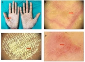
We provided a report on a 7.5-year-old boy, who was admitted to the pediatric ward of Mousavi hospital, Zanjan, Iran, with history of fever, malaise, and an exudative plaque-like lesion on his hand (Figure 1). The disease started few days prior to admission. He had good growth and development, but had a history of frequent hospitalizations due to severe infections, including liver abscess, preseptal cellulitis and severe septicemia at the ages of 4, 5 and 7 years, respectively. Furthermore, the patient had a recurrent infection with giardiasis and received treatment. There was a history of premature loss of many primary teeth at 4 years of age. There was also a severe palmoplantar hyperkeratosis. Consultations with dermatologists and pediatric infections specialists led to Papillon-Lefevre syndrome diagnosis, based on history and physical examination. Cultures of the wound aspiration and blood were obtained, and initial antibiotic therapy was carried out on the cutaneous lesions and underlying disease with ceftazidime (50 mg/kg/6h), Vancomycin (15 mg/kg/8h) and amphotericin B. The blood culture was negative, but the culture of the wound aspiration was positive for Citrobacter. Based on the antibiogram results, the antibiotic treatment regime was changed to meropenem (20 mg/kg/8h) and amikacin (5 mg/kg/8h). Two days after the onset of the new antibiotic therapy, fever subsided and the size of cutaneous lesions gradually decreased. One week after the treatment, new culture of the lesions was negative for Citrobacter. Finally, after receiving treatment for two weeks, he was discharged in good general condition and improved cutaneous lesions. At the initial evaluation of the immune system, the levels of serum immunoglobulins (IgG, IgM, IgA, and IgE), flow cytometry and NBT test were normal.
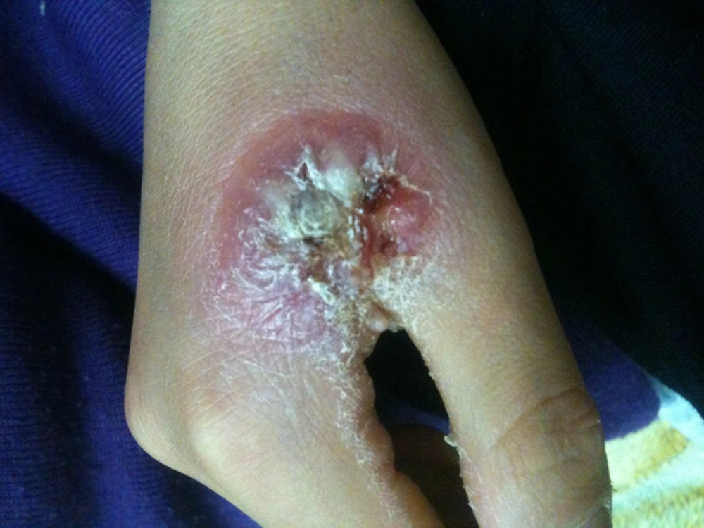
Figure 1.
A 7.5-Year-Old Boy, Who Was Admitted to the Pediatric Ward of Mousavi Hospital, Zanjan, Iran, With History of Fever, Malaise, and an Exudative Plaque-Like Lesion on His Hand
This was the first case report on dangerous cutaneous infection caused by Citrobacter in a child with PLS, based on our search of the Internet and PubMed. This syndrome is often characterized by palmoplantar hyperkeratosis, dental and liver infections, but serious cutaneous infection with an unusual organism is completely unexpected. Zandieh et al. (3) reported a 15-year-old boy with PLS, with history of nocturnal fever, chills, sweating, and pain sensation of the right upper abdomen and right flank. The child had a history of liver abscess about two years earlier. Liver biopsy showed a granulomatous lesion caused by Mycobacterium tuberculosis that was confirmed by PCR. Contrary to this study, no serious cutaneous infection was observed. However, in both studies, immunological studies of the patients showed that the phagocytic system (NBT) and flow cytometry for T-cells and B-Cells were normal. Infection caused by Citrobacter can be seen in many different forms, and is occasionally associated with abscess formation. Patients with PLS are prone to dental and liver abscess (4, 5). The most common organisms associated with dental and liver infections are anaerobic bacteria (such as Actinobacillus actinomycetem comitans) and Staphylococcus aureus (5, 6). In this study, the patient had a history of pyogenic liver abscess, preseptal cellulitis and loss of many teeth secondary to dental infections, and hyperkeratosis of the palms and soles. Diagnosis of PLS in this patient was confirmed clinically, in which soft tissue infection and hand cellulitis were observed. Cellulitis caused by Citrobacter is very rare in healthy individuals, but occur in patients with this syndrome due to the existence of a degree of disorders in the cytotoxicity of natural killer cells and neutrophil chemotaxis. Therefore, it is recommended that unusual infectious organisms, including Citrobacter should be suspected in patients with PLS and serious infections (local or systemic). Also, Citrobacter organism and its antibiotic sensitivity pattern should be considered when starting an empirical antibiotic treatment.