
Image credit: pxhere

Image Credit:Image credit: pxhere

Variations of Cesarean Scar Pregnancy in Practice

Author(s):
Mojgan BaratiMojgan BaratiMojgan Barati ORCID1, Farideh MorameziFarideh Moramezi2,*, Sara MasihiSara MasihiSara Masihi ORCID1, Razieh Mohamad JafariRazieh Mohamad JafariRazieh Mohamad Jafari ORCID1
1Associate Professor of Obstetrics and Gynecology, Imam Khomeini Hospital, Ahvaz Jundishapur University of Medical Sciences, Ahvaz, Iran
2Associate Professor of Obstetrics and Gynecology, Ahvaz Jundishapur University of Medical Sciences, Ahvaz, Iran
*Corresponding Author: Associate Professor of Obstetrics and Gynecology, Ahvaz Jundishapur University of Medical Sciences, Ahvaz, Iran. Email: [email protected]

Jentashapir Journal of Cellular and Molecular Biology:Vol. 9, issue 6; e86355
Published online:Jan 02, 2019
Article type:Letter
Received:Nov 13, 2018
Accepted:Nov 17, 2018
How to Cite:Barati M, Moramezi F, Masihi S, Mohamad Jafari R. Variations of Cesarean Scar Pregnancy in Practice. Jentashapir J Cell Mol Biol. 2018;9(6):e86355. doi: https://doi.org/10.5812/jjhr.86355

Dear Editor,
Due to the increasing number of cesareans, cesarean scar pregnancies are also increasing.
It is not intended to reemphasize the importance of diagnosis of this life threatening event since it is well known. In the literature, practical diagnosis of cesarean scar pregnancy is based on the history of cesarean delivery, a positive pregnancy test result, and the following sonographic criteria: (1) Empty uterine cavity, and closed and empty endocervical canal, (2) chorionic/gestational sac and/or placenta located in low and anterior walls of the uterus, below the bladder, in close proximity, and at the level of the internal os of the site of a previous hysterotomy scar (niche), with or without fetal or embryonic pole and/or yolk sac, with or without heart beats (depending on gestational age), (3) absence or thin myometrial layer between the chorionic/gestational sac and unusually close proximity to the bladder wall, and (4) abundant blood flow around the chorionic/gestational sac concentrated on the anterior side of the chorionic sac. The typical sonographic appearance of an arteriovenous malformation (AVM) is rarely observed (1). There were some cases of cesarean scar pregnancy in which placenta was low, but not necessarily located in the anterior wall of uterus. Placenta in some cesarean scar pregnancies is dominantly located in lateral wall or even posterior wall of uterus. But lateral parts of placenta extended to lateral sides of previous scar and invaded from lateral side of scar to outside of uterus. Therefore, if low-lying placenta/pregnancy is considered fewer cases of cesarean scar pregnancy are missed.
Based on gestational age, and size of fetus and placenta, there are different types of scar pregnancy: Site of placenta, site of the whole pregnancy (sac and fetus), and appearance of endometrium in upper part of uterus in early weeks of pregnancy.
In some cases it seems that with growing placenta and fetus, intrauterine pregnancy and later morbidly adherent placenta occur.
Different types of scar pregnancy
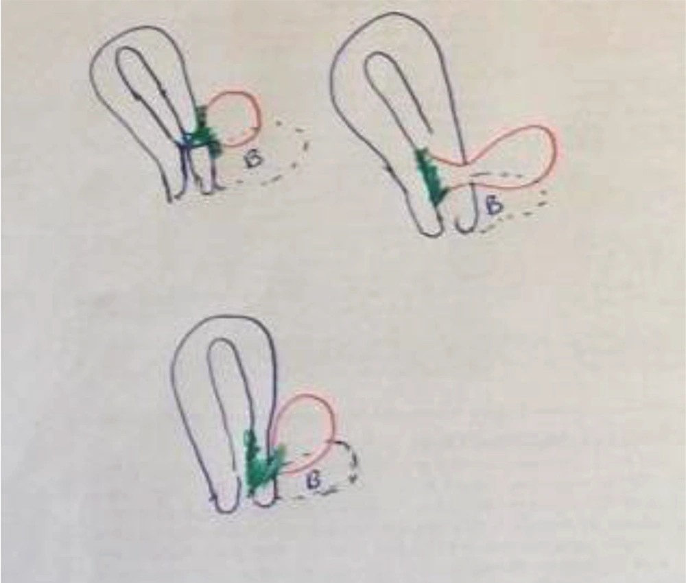
Extrauterine scar pregnancy; placenta (green), pregnancy (red).

Figure 1.

Extrauterine scar pregnancy; placenta (green), pregnancy (red).

Scar line pregnancy. (A) lateral view, small sac and HCG level↑; (B) anterior view.
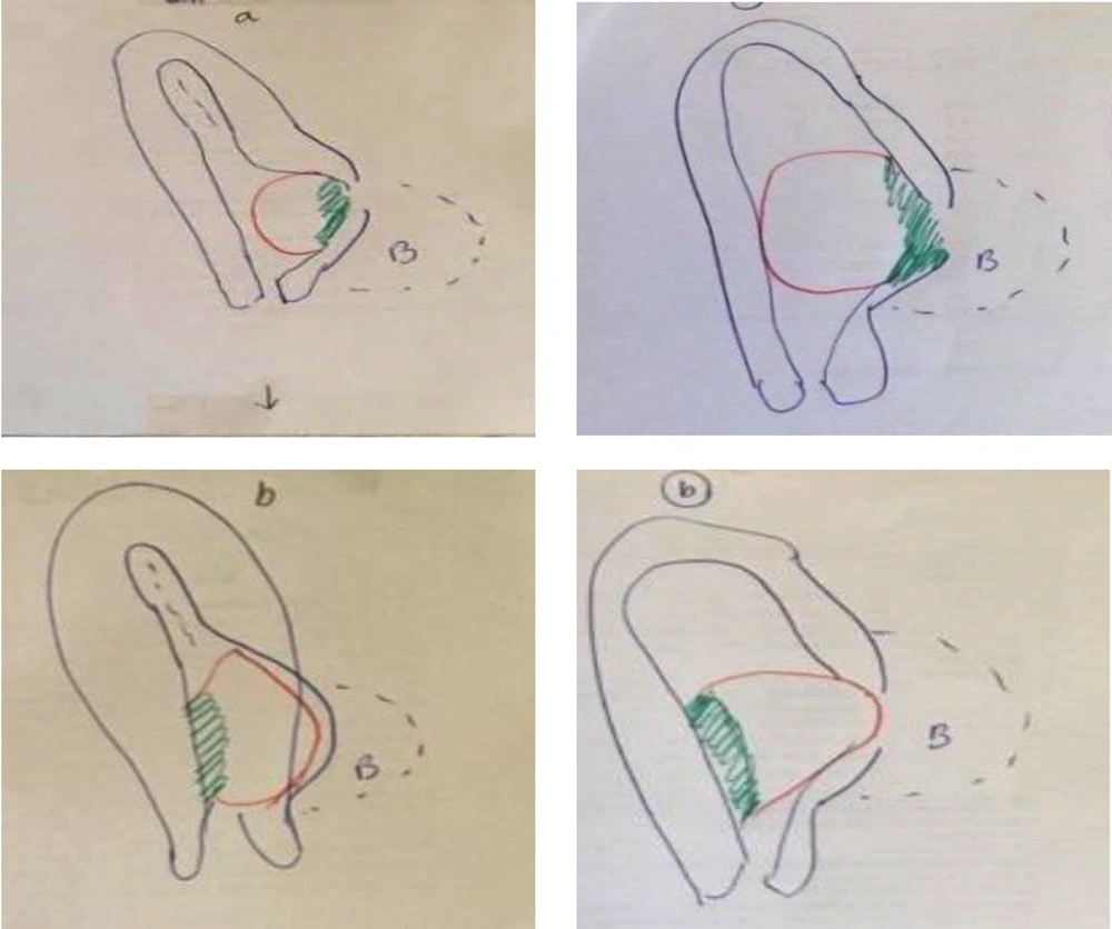
Figure 2.

Scar line pregnancy. (A) lateral view, small sac and HCG level↑; (B) anterior view.

Intrauterine scar pregnancy
Figure 3.

Intrauterine scar pregnancy

For a practical approach, scar pregnancy is divided into two groups:
A: Death scar pregnancy
B: Live scar pregnancy
Based upon authors' opinion, in group A, nature by itself passed more than half of the way of treatment. Decision between medical or surgical management depends on the site of scar pregnancy. In case of an extrauterine placenta and extrauterine pregnancy, curettage and intrauterine procedures are not enough and maybe dangerous.
In group B, decision about the kind of management is a per-case decision. In live extrauterine and line embedded scar pregnancy, both nonsurgical and/or surgical approaches are suggested. Management and decision in live intrauterine scar pregnancy is very obscure. It is not predictable which pregnancy reaches more than 24 weeks of gestation.

Footnotes

  • Conflict of Interests: The authors declared no conflict of interest.

  • Funding/Support: The study was supported by Ahvaz Jundishapur University of Medical Sciences.

References

  • 1.
    Timor-Tritsch IE, Monteagudo A, Agten AK. Cesarean scar pregnancy diagnosis and management.Obstetrics-Gynecology. 2015 Nov 24.

Copyright

Copyright © 2019, Jentashapir Journal of Health Research. This is an open-access article distributed under the terms of the Creative Commons Attribution-NonCommercial 4.0 International License (http://creativecommons.org/licenses/by-nc/4.0/) which permits copy and redistribute the material just in noncommercial usages, provided the original work is properly cited.

Similar Articles

27
Jun
2015

Non-Surgical Treatment of Cesarean Scar Pregnancy: Two Case Reports

Mojgan Barati,
Kobra Shojayi,
Farideh Moramezi,
Nafiseh Hoseiny Moghadam

Barati M, Shojayi K, Moramezi F, Hoseiny Moghadam N. Non-Surgical Treatment of Cesarean Scar Pregnancy: Two Case Reports. Jentashapir J Cell Mol Biol. 2015;6(3):e28703. doi: https://doi.org/10.5812/jjhr.28703v2

28
Feb
2014

Various Types of Niche Imaging by Sonohysterography

Firoozeh Ahmadi,
Farnaz Akhbari,
Fatemeh Niknejad

Ahmadi F, Akhbari F, Niknejad F. Various Types of Niche Imaging by Sonohysterography. I J Radiol. 2014;11(30th Iranian Congress of Radiology):e21411. doi: https://doi.org/10.5812/iranjradiol.21411

20
May
2013

Appearance of Uterine Scar Due to Previous Cesarean Section on Hysterosalpingography: Various Shapes, Locations and Sizes

firoozeh ahmadi,
Farnaz Akhbari,
Leila Torbati,
Gholam Shahrzad

ahmadi F, Akhbari F, Torbati L, Shahrzad G. Appearance of Uterine Scar Due to Previous Cesarean Section on Hysterosalpingography: Various Shapes, Locations and Sizes. I J Radiol. 2013;10(2):e93635. doi: https://doi.org/10.5812/iranjradiol.5143

31
Dec
1998

Studying the rate of cesarean section in Qazvin city in the first six months of 2015

MA Joafshani

Joafshani M. Studying the rate of cesarean section in Qazvin city in the first six months of 2015. J Inflamm Dis. 1998;2(2):e154626. doi:

31
Dec
1997

Prevalence of cesarean section in Kowsar Hospital

F Almasi,
M Pishghahi

Almasi F, Pishghahi M. Prevalence of cesarean section in Kowsar Hospital. J Inflamm Dis. 1997;1(3):e154525. doi:

Download PDF3.02 MB

Crossmark

Crossmark

Checking

Share on
Cited by
Metrics

Ordering Reprints

Articles are published under the Creative Commons license stated on each article. No permission or royalty fee is required for uses permitted by that license. CCC handles optional bulk and customized reprint orders. Any quotation covers production and delivery services only, not copyright permission. > Request Reprints from CCC 

Search Relations

Author(s):

Related Articles