
1. Background
Phototherapy is commonly used to manage neonatal jaundice (1-4). Hypocalcemia is a serious complication of phototherapy in newborns (5-7). Premature infants are at higher risk of phototherapy-induced hypocalcemia (8). Hypocalcemia is defined as either a total serum calcium level of ≤ 8 mg/dL and an ionized calcium level of less than 4.4 mg/dL in a newborn over 37 weeks of gestation, or a total calcium level of ≤ 7 mg/dL or an ionized calcium level of less than 4 mg/dL in a newborn under 37 weeks (2). Infants with hypocalcemia may be asymptomatic or present with irritability, high-pitched crying, jitteriness, seizures, apnea, cyanosis, hypotonia, tetany, laryngeal spasm, bronchospasm, lethargy, poor feeding, or vomiting; the presentation may even mimic sepsis (2). Hypocalcemia leads to prolongation of the QT interval on the electrocardiogram (ECG). Measurement of the corrected QT (QTc) interval is also a noninvasive indicator of ionized calcium levels (9).
The mechanism of hypocalcemia during phototherapy is not well understood. Proposed mechanisms include:
1) Increased urinary calcium excretion.
2) An immature parathyroid hormone response.
3) Light transmission through the skull, disruption of the circadian rhythm, inhibition of the pineal gland, and a subsequent reduction in melatonin secretion. Reduced melatonin secretion increases the effect of cortisol on bone and calcium uptake, leading to hypocalcemia (2, 10).
Monitoring serum calcium is recommended during phototherapy (11). Some researchers recommend covering the infant's head with a hat to prevent light penetration (1, 10, 12). Because preterm infants have thinner skin and greater light penetration, this effect may be more prominent in this group; however, studies in preterm infants are very limited (12).
2. Objectives
This study aimed to investigate the prevalence of phototherapy-induced hypocalcemia in term and late-preterm icteric infants and to evaluate the prophylactic effect of head covering on serum calcium levels and QTc intervals.
3. Methods
This prospective, parallel-group randomized clinical trial included infants with a gestational age of ≥ 35 weeks and evaluated the effect of head covering during phototherapy on serum calcium levels compared with routine phototherapy without a hat.
The study included 89 term and late preterm infants with a gestational age of ≥ 35 weeks and serum bilirubin levels of ≥ 14 mg/dL who were admitted for phototherapy in Rasht, Iran, between July 2024 and July 2025. The infants were otherwise healthy.
The sample size was calculated based on the results of the study by Asghar et al. (1), with a confidence level of α = 95%, a power of β = 90%, and a dropout rate of 10%. The initial sample size was determined to be 39 individuals in each group. Therefore, the final sample size was determined to be at least 43 individuals in each group (for a total of 86 infants).
1-∝=mg/dl, expected clinical difference.
Consecutive sampling was performed during daily visits to the neonatal ward. Eligible infants were included after written consent was obtained from their parents (Figure 1).
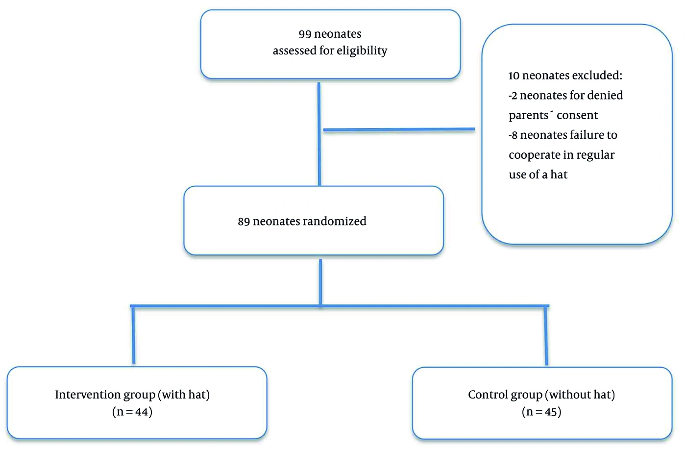
Figure 1.
Flow diagram of patients
Randomization was performed using permuted blocks of four participants. A total of 22 blocks of four were generated, resulting in 88 allocation slots. Subsequently, one additional eligible neonate was enrolled after completion of the planned allocation sequence, resulting in a total sample of 89 neonates. The additional participant was allocated according to the same predefined randomization procedure. The random sequence list was generated using software. Allocation was concealed using sequentially numbered, opaque, sealed envelopes. The sequence list was kept at the Children's Research Center. After ethics committee approval and clinical trial registration (IRCT 20180228038895N3), the envelopes were opened daily to allocate infants to the study groups.
3.1. Inclusion Criteria
- Gestational age of ≥ 35 weeks.
- Indirect hyperbilirubinemia (bilirubin levels ≥ 14 mg/dL).
- Normal clinical examination findings except for jaundice.
3.2. Exclusion Criteria
- Bilirubin levels near the exchange transfusion threshold.
- Sepsis.
- Respiratory distress.
- Facial dysmorphism.
- Congenital heart problems.
- Maternal diabetes.
- A maternal history of parathyroid problems.
- Maternal use of anticonvulsant medication.
- Birth asphyxia or a low Apgar score.
- Intrauterine growth retardation.
- Parenteral nutrition.
- Hypocalcemia on initial testing before phototherapy.
All neonates underwent conventional phototherapy according to the standard clinical protocol (AAP 2022) (13). Phototherapy was administered using a Ningbo David Medical device (China) with blue fluorescent lamps emitting light at wavelengths of 410 - 470 nm. The number of lamps (4 or 8) was adjusted by the pediatrician based on the infant's age, jaundice risk factors, and bilirubin levels. Neonates were placed supine under the phototherapy unit, with the light source positioned 25 cm away. The maximum possible body surface area was exposed, while the eyes and genital area were protected. Phototherapy was administered continuously, except for brief interruptions for feeding, routine nursing care, and clinical assessment.
Intervention group (44 infants): Phototherapy with a blindfold, a small diaper, and a dark hat.
Control group (45 infants): Phototherapy with a blindfold and a small diaper, without a hat.
The hat was made of dark cotton fabric and covered the occiput, ears, and back of the neck. The same shape, material, and color were used for all infants. The hat was available in three sizes to accommodate the infants' head circumferences (Figure 2).
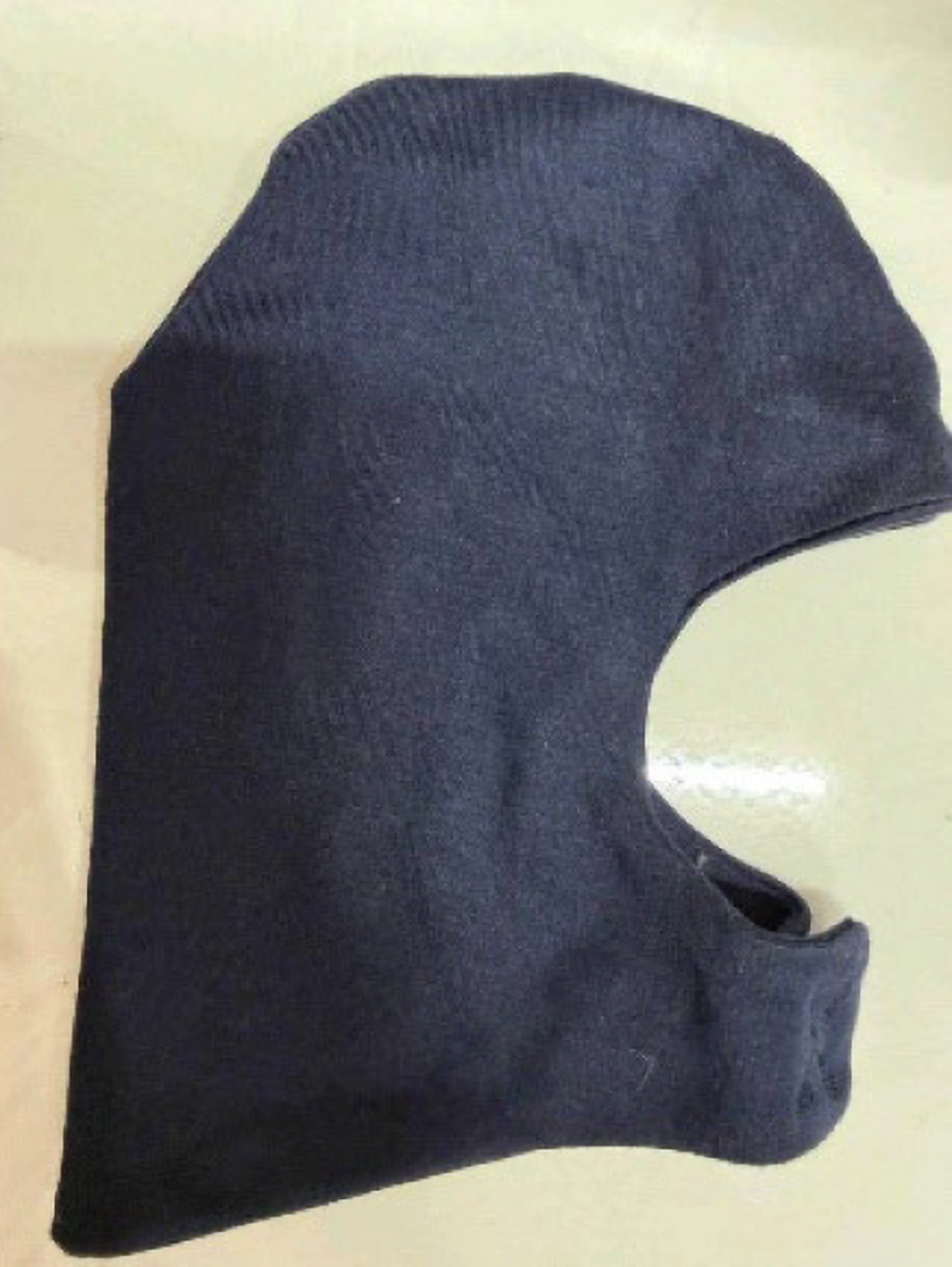
Figure 2.
The hat used was dark and made of cotton fabric that covered the occiput, ears, and back of the neck.
Blood samples (2 mL) were collected from a peripheral vein to measure bilirubin and total serum calcium at baseline and 48 hours after the start of phototherapy. A Padco kit was used to measure total serum calcium. Electrocardiograms were obtained at admission and after 48 hours. The pediatrician calculated the QTc interval using (QTc = QT / √RR). Lead 2 was used to calculate QTc. Total calcium ≤ 8 mg/dL in infants older than 37 weeks and ≤ 7 mg/dL in infants 35 to 37 weeks was considered hypocalcemia. QTc > 450 ms was considered prolonged. Bilirubin measurement intervals were determined at the physician's discretion. Symptoms of hypocalcemia were closely monitored.
Because of the nature of the intervention, participants and clinical staff could not be blinded. However, statistical analysis was performed by an analyst blinded to group allocation using a single-blind approach.
Variables included infant age (days), gestational age (weeks), weight, sex, type of milk consumed, serum calcium levels at admission and 48 hours later, QTc intervals at admission and 48 hours later, serum bilirubin levels at baseline and 48 hours later, and symptoms of hypocalcemia.
Primary outcome: Comparison of changes in serum calcium levels and QTc intervals between the two groups during 48 hours of phototherapy.
Secondary outcome: Comparison of the frequency of laboratory-confirmed hypocalcemia between the two groups.
3.3. Statistical Analysis
Statistical analysis was performed using SPSS version 27 (IBM Corp., Armonk, NY, USA). Continuous variables were assessed for normality using the Kolmogorov–Smirnov test and visual inspection of histograms. Data were expressed as mean ± standard deviation (SD), median, interquartile range (IQR; 25th - 75th percentiles), minimum, and maximum, as appropriate.
Continuous variables were compared between the two independent groups using the independent-samples t-test for normally distributed data and the Mann–Whitney U test when normality assumptions were not met. Categorical variables were compared using the chi-square test or Fisher's exact test, as appropriate.
Within-group comparisons between baseline and 48 hours after phototherapy were performed using the paired-samples t-test for normally distributed variables or the Wilcoxon signed-rank test for nonnormally distributed variables. To account for potential baseline differences between groups, analysis of covariance was performed for serum calcium levels, QTc intervals, and bilirubin levels at 48 hours. The corresponding baseline value for each outcome was included as a covariate. Gestational age, body weight, and preterm/term status were also included as covariates. Adjusted between-group mean differences with 95% confidence intervals (CIs) were reported. P < 0.05 was considered statistically significant.
4. Results
Table 1 compares the demographic and general characteristics of the two groups. Infants were 3 - 18 days old. Age and sex distributions were similar between the groups with and without hats. Late preterm infants accounted for approximately 20.2% of the sample. Gestational age (P = 0.046), weight (P = 0.029), and the percentage of preterm infants (P = 0.01) differed between groups. The control group included more late preterm neonates and had a lower mean weight.
Table 1.
Demographic Variables in the Two Study Groups a
| Variables | Without hat | with hat | Total | P value * |
|---|---|---|---|---|
| Sex | 0.245 | |||
| Female | 17 (37.8) | 22 (50.0) | 39 (43.8) | |
| Male | 28 (62.2) | 22 (50.0) | 50 (56.2) | |
| Total | 45 (100.0) | 44 (100) | 89 (100.0) | |
| Age (d) | 0.090 | |||
| Mean | 4.64 | 5.89 | 5.26 | |
| SD | 3.08 | 3.71 | 3.45 | |
| Minimum | 3.00 | 3.00 | 3.00 | |
| Maximum | 12.00 | 18.00 | 18.00 | |
| Gestational age (wk) | 0.046 | |||
| Mean | 37.51 | 38.00 | 37.75 | |
| SD | 1.32 | 0.91 | 1.16 | |
| Minimum | 35.00 | 36.00 | 35.00 | |
| Maximum | 40.00 | 40.00 | 40.00 | |
| Weight (g) | 0.029 | |||
| Mean | 2947.47 | 3221.10 | 3082.76 | |
| SD | 704.87 | 415.77 | 593.15 | |
| Minimum | 2050.00 | 2440.00 | 2050.00 | |
| Maximum | 4075.00 | 4120.00 | 4120.00 | |
| Term/preterm status | 0.010 | |||
| Term | 31 (68.9) | 40 (90.9) | 71 (79.8) | |
| Preterm | 14 (31.1) | 4 (9.1) | 18 (20.2) | |
| Feeding | 0.412 | |||
| Breast milk | 37 (82.2) | 37 (84.1) | 74 (83.1) | |
| Formula | 5 (11.1) | 2 (4.5) | 7 (7.9) | |
| Both | 3 (6.7) | 5 (11.4) | 8 (9.0) |
a Values are expressed as No. (%) unless otherwise indicated. Continuous variables were compared using the independent-samples t-test or Mann–Whitney U test, as appropriate. Categorical variables were compared using the chi-square test or Fisher's exact test. Abbreviations: SD, standard deviation; IQR, interquartile range.
Table 2 compares total serum calcium levels between the two groups. Total calcium levels at baseline (9.78 ± 1.84 vs. 9.92 ± 1.79 mg/dL, P = 0.366), after 48 hours (9.61 ± 0.63 vs. 9.50 ± 0.54 mg/dL, P = 0.255), and changes over 48 hours (0.18 ± 1.83 vs. 0.42 ± 1.79 mg/dL, P = 0.103) did not differ significantly between the group without hats and the group with hats, respectively. Changes in total calcium from baseline to 48 hours were also not significant within the group without hats (P = 0.233), the group with hats (P = 0.275), or the overall sample (P = 0.919). Although calcium levels decreased slightly during 48 hours of phototherapy in both groups, the decreases from baseline were not statistically significant.
Table 2.
Comparison of Total Serum Calcium Levels in the Two Groups a
| Variables | Without Hat | with Hat | Total | P Value b |
|---|---|---|---|---|
| Calcium (baseline) (mg/dL) | 0.366 | |||
| Mean ± SD | 9.78 ± 1.84 | 9.92 ± 1.79 | 9.85 ± 1.80 | |
| Median | 9.40 | 9.65 | 9.50 | |
| 25th percentile | 9.00 | 9.20 | 9.00 | |
| 75th percentile | 10.10 | 10.05 | 10.10 | |
| Calcium (after 48 hours) (mg/dL) | 0.255 | |||
| Mean ± SD | 9.61 ± 0.63 | 9.50 ± 0.54 | 9.56 ± 0.58 | |
| Median | 9.60 | 9.45 | 9.60 | |
| 25th percentile | 9.30 | 9.10 | 9.20 | |
| 75th percentile | 10.00 | 9.90 | 9.90 | |
| Changes in calcium levels for 48 hours (mg/dL) | 0.103 | |||
| Mean ± SD | 0.18 ± 1.83 | 0.42 ± 1.79 | 0.30 ± 1.80 | |
| Median | -0.10 | 0.10 | 0.00 | |
| 25th percentile | -0.50 | -0.20 | -0.30 | |
| 75th percentile | 0.20 | 0.35 | 0.30 | |
| P value c | 0.233 | 0.275 | 0.919 |
a Abbreviation: SD, standard deviation.
b Independent-samples t-test/Mann–Whitney U test.
c Paired-samples t-test/Wilcoxon signed-rank test.
Table 3 compares QTc intervals between the two groups. QTc intervals at baseline (P = 0.447) and after 48 hours (P = 0.158), as well as changes in QTc intervals (P = 0.774), did not differ significantly between groups. Changes from baseline to 48 hours were also not significant within the group without hats (P = 0.918), the group with hats (P = 0.933), or the overall sample (P = 0.940).
Table 3.
Comparison of QTc Intervals in the Two Groups a
| Variables | Without hat | with hat | Total | P Value b |
|---|---|---|---|---|
| QTc (baseline) (ms) | 0.447 | |||
| Mean ± SD | 423.78 ± 21.52 | 427.77 ± 17.70 | 425.75 ± 19.71 | |
| Median | 422.00 | 429.50 | 427.00 | |
| 25th percentile | 404.00 | 416.00 | 404.00 | |
| 75th percentile | 443.00 | 443.00 | 443.00 | |
| QTc (after 48 hours) (ms) | 0.158 | |||
| Mean ± SD | 423.36 ± 20.13 | 428.77 ± 18.31 | 426.03 ± 19.33 | |
| Median | 422.00 | 432.50 | 424.00 | |
| 25th percentile | 404.00 | 421.00 | 404.00 | |
| 75th percentile | 443.00 | 443.00 | 443.00 | |
| Changes in QTc interval for 48 hours (ms) | 0.774 | |||
| Mean ± SD | 0.42 ± 21.12 | -1.00 ± 16.97 | -0.28 ± 19.09 | |
| Median | 0.00 | 0.00 | 0.00 | |
| 25th percentile | -16.00 | -1.50 | -2.00 | |
| 75th percentile | 13.00 | 4.00 | 6.00 | |
| P value c | 0.918 | 0.933 | 0.940 |
a Abbreviations: QTc, corrected QT; SD, standard deviation.
b Independent-samples t-test/Mann–Whitney U test.
c Paired-samples t-test/Wilcoxon signed-rank test.
As shown in Table 4, bilirubin levels at baseline (P = 0.332), after 48 hours (P = 0.375), and changes over 48 hours (P = 0.828) did not differ significantly between groups. However, bilirubin levels decreased significantly from admission to 48 hours in the group without hats (P < 0.001), the group with hats (P < 0.001), and the overall sample (P < 0.001).
Table 4.
Comparison of Bilirubin Levels in the Two Groups a
| Variables | Without hat | with hat | Total | P Value b |
|---|---|---|---|---|
| Bilirubin (baseline) (mg/dL) | 0.332 | |||
| Mean ± SD | 16.49 ± 2.09 | 17.15 ± 2.75 | 16.82 ± 2.45 | |
| Median | 16.00 | 16.40 | 16.20 | |
| 25th percentile | 14.70 | 15.25 | 15.20 | |
| 75th percentile | 17.70 | 18.70 | 18.50 | |
| Bilirubin (after 48 hours) (mg/dL) | 0.375 | |||
| Mean ± SD | 9.69 ± 2.51 | 9.83 ± 1.66 | 9.76 ± 2.12 | |
| Median | 9.30 | 9.50 | 9.40 | |
| 25th percentile | 8.60 | 8.70 | 8.60 | |
| 75th percentile | 10.50 | 10.30 | 10.30 | |
| Changes in Bilirubin (mg/dL) | 0.828 | |||
| Mean ± SD | 6.80 ± 3.88 | 7.32 ± 3.32 | 7.06 ± 3.60 | |
| Median | 7.30 | 7.15 | 7.30 | |
| 25th percentile | 4.50 | 5.50 | 5.20 | |
| 75th percentile | 9.40 | 9.80 | 9.70 | |
| P value c | < 0.001 | < 0.001 | < 0.001 |
a Abbreviation: SD, standard deviation.
b Independent-samples t-test/Mann–Whitney U test.
c Paired-samples t-test/Wilcoxon signed-rank test.
No cases of laboratory-confirmed hypocalcemia were observed during the 48-hour study period in either group. No convulsions, seizures, apnea, hypotonia, or other symptoms of hypocalcemia were reported.
As shown in Table 5, calcium levels, changes in calcium levels over 48 hours, and QTc intervals did not differ significantly between the two groups within gestational-age subgroups (P > 0.05).
Table 5.
Comparison of Calcium Levels in the Two Groups a
| Variables | Term: Without hat | Term: with hat | Term: Total | Term: P | Preterm: Without hat | Preterm: with hat | Preterm: Total | Preterm: P b |
|---|---|---|---|---|---|---|---|---|
| Calcium (baseline) (mg/dL) | 0.609 | 0.957 | ||||||
| Number | 31 | 40 | 71 | 14 | 4 | 18 | ||
| Median | 9.50 | 9.75 | 9.60 | 9.30 | 9.10 | 9.30 | ||
| 25th percentile | 9.00 | 9.25 | 9.20 | 8.50 | 8.80 | 8.80 | ||
| 75th percentile | 10.10 | 10.10 | 10.10 | 9.70 | 9.60 | 9.70 | ||
| Calcium (after 48 hours) (mg/dL) | 0.293 | 0.831 | ||||||
| Median | 9.60 | 9.55 | 9.60 | 9.70 | 9.30 | 9.60 | ||
| 25th percentile | 9.30 | 9.10 | 9.20 | 9.10 | 9.20 | 9.20 | ||
| 75th percentile | 10.00 | 9.90 | 9.90 | 10.20 | 10.10 | 10.20 | ||
| Changes in calcium levels for 48 hours (mg/dL) | 0.231 | 0.740 | ||||||
| Median | -0.10 | 0.10 | 0.10 | -0.20 | -0.50 | -0.20 | ||
| 25th percentile | -0.40 | -0.20 | -0.20 | -1.00 | -0.80 | -1.00 | ||
| 75th percentile | 0.30 | 0.40 | 0.40 | 0.10 | -0.10 | 0.10 | ||
| QTc (baseline) (ms) | 0.355 | 0.306 | ||||||
| Median | 422.00 | 426.50 | 423.00 | 435.00 | 443.00 | 443.00 | ||
| 25th percentile | 404.00 | 407.00 | 404.00 | 422.00 | 439.00 | 422.00 | ||
| 75th percentile | 443.00 | 443.00 | 443.00 | 443.00 | 443.00 | 443.00 | ||
| QTc (after 48 hours) (ms) | 0.665 | 0.104 | ||||||
| Median | 422.00 | 432.00 | 428.00 | 404.00 | 438.00 | 421.00 | ||
| 25th percentile | 422.00 | 417.50 | 422.00 | 400.00 | 426.50 | 400.00 | ||
| 75th percentile | 443.00 | 443.00 | 443.00 | 437.00 | 443.00 | 443.00 | ||
| Changes in QTc interval for 48 hours (ms) | 0.265 | 0.325 | ||||||
| Median | 0.00 | 0.00 | 0.00 | 12.00 | 5.00 | 8.00 | ||
| 25th percentile | -18.00 | -2.00 | -18.00 | 0.00 | 0.00 | 0.00 | ||
| 75th percentile | 0.00 | 3.00 | 1.00 | 22.00 | 12.50 | 21.00 |
a Abbreviation: QTc, corrected QT.
b Mann–Whitney U test.
As shown in Table 6, data are presented as mean differences with 95% CIs. The between-group effect represents the difference in each variable between the two groups. Adjusted estimates were obtained after controlling for relevant baseline characteristics and baseline outcome values.
Table 6.
Adjusted Between Group Differences in Changes in Serum Calcium, QTc Interval, and Bilirubin Levels with 95% CI Based on Gestational Age, Weight and Term/Preterm Status a
| Outcome (at 48 h) | Adjusted Mean Difference (Hat-No Hat) | 95% Confidence Interval | P Value |
|---|---|---|---|
| Serum total calcium(mg/dl) | -0.066 | (-0.324 to 0.192) | 0.611 |
| QTc interval(ms) | +1.28 | (-5.80 to 8.37) | 0.720 |
| Total bilirubin(mg/dL) | +0.03 | (-0.81 to 0.87) | 0.947 |
a The adjusted mean difference represents the estimated effect of the hat group. A negative difference indicates a lower value in the hat group relative to the non-hat group. Adjusted for gestational age, weight and term/preterm status. Abbreviations: ANCOVA, analysis of covariance; CI, confidence interval; QTc, corrected QT.
5. Discussion
Phototherapy-induced hypocalcemia is hypothesized to result from blue light penetrating the infant’s skull, affecting the pineal gland, and reducing melatonin secretion (14). Some studies have confirmed that head covering during phototherapy prevents changes in serum calcium levels (1, 10, 12, 15, 16), whereas others have not (3). Evidence from studies of preterm infants is very limited (12, 17).
The absence of laboratory-confirmed hypocalcemia contrasts with previous findings (1, 7, 12). In a study by Barekatain et al. that included preterm infants with a gestational age < 35 weeks, the prevalence of hypocalcemia among infants without head covering was approximately 30%, and head covering significantly reduced this prevalence (12). Similarly, Ezzeldin reported prevalences of 24% among infants without head covering and 10% among infants with head covering (10). Muhssin et al. found hypocalcemia in 6% of neonates undergoing phototherapy (18). Our findings were consistent with those of Jain Puja, who reported a 0% incidence of hypocalcemia in term infants (19).
Several physiological factors may explain this finding.
First, the present study included infants with a gestational age ≥ 35 weeks, who are less susceptible to hypocalcemia than populations in studies focused on very preterm infants, such as those born at < 32 weeks (12, 17). Premature infants are at higher risk of hypocalcemia because of reduced renal tubular calcium reabsorption, inadequate absorption of calcium from milk, lower calcium stores, and thinner skin that allows greater light transmission (19). Late preterm infants are also more susceptible to hypocalcemia, although their risk is lower than that of infants born at < 35 weeks (3). The greatest transfer of calcium to the fetus occurs in the last trimester. The hypocalcemia threshold is also lower in preterm and late preterm infants. In this study, only 20.2% of infants were preterm, and their distribution was unbalanced: 31.1% in the group without hats versus 9.1% in the group with hats (P = 0.01). The difference in gestational age (37 weeks without hats vs. 38 weeks with hats; P = 0.046) and the difference in the number of preterm infants could introduce bias. However, subgroup analyses showed no significant differences (P > 0.05).
Second, phototherapy intensity and duration were standardized in this study: 48 hours with four to a maximum of eight fluorescent lamps. However, treatment may have been less intense and shorter than in other studies, such as that by Tawfik (4), in which phototherapy continued for up to 72 hours, or studies in which calcium was rechecked on the day of discharge (2, 21). Hypocalcemia may therefore appear after 72 hours of continuous phototherapy. With advances in phototherapy devices and light-emitting diode (LED) lamps, recent studies have reported numerically lower rates of hypocalcemia than older studies, including a rate of 2.5% in the 2023 study by Pereira et al. (21). The present study used fluorescent lamps, whereas some studies used LED lamps (2, 21). However, LED lamps may cause hypocalcemia to the same extent as fluorescent lamps, and monitoring infants receiving LED phototherapy is also reasonable (2).
Third, hypocalcemia in this study was defined as total calcium levels ≤ 8 mg/dL for term infants and ≤ 7 mg/dL for preterm infants (22), consistent with most previous studies and recent references. However, Barekatain et al. used a hypocalcemia threshold of 8 mg/dL in preterm infants (12).
Total calcium measurements can be misleading when albumin concentrations are abnormal (14, 23). Ionized calcium is the active fraction of circulating calcium (24). Ionized calcium, a more accurate indicator, was not measured because of its limited availability at this center and its high cost. This may have led to an underestimation of hypocalcemia. However, the absence of clinical signs of hypocalcemia or QTc prolongation may partially, but not definitively, support the laboratory findings. Pereira et al. found no difference in total calcium levels between the first day and the day of discharge in the two groups, but ionized calcium levels differed significantly before and after phototherapy (21). Tawfik et al. also found no difference between infants with and without hats despite measuring both total and ionized calcium (4).
Race, climate, and maternal or infant nutrition may have had protective effects in this study. A total of 83.1% of infants were breastfed (P = 0.412), which may have been protective. Breast milk contains calcium-regulating factors, such as PTH-related peptide. Although breast milk and formula do not differ in calcium content, breast milk provides greater calcium availability to infants. Formula may increase the risk of hypocalcemia because of its higher phosphorus content (8, 25).
The present findings suggest that head covering had no effect on serum calcium levels. This is consistent with the findings of Bibi and Tawfik (4) but differs from those of other studies (1, 10, 15, 16). The hat may not have completely blocked light, as light penetration was not assessed. In addition, light penetration through other areas of the body, such as the back, could circumvent the proposed mechanism, particularly in preterm infants (10).
The QTc interval is an indicator of abnormal ventricular myocardial repolarization (26) and can be used as a noninvasive marker for calcium monitoring. In this study, QTc intervals remained within the normal range, with a mean of approximately 426 ms, and no cases of QTc prolongation were observed. Similar studies did not assess QTc intervals. Studies have demonstrated a stronger association between QTc prolongation and declining ionized calcium levels than between QTc prolongation and total calcium levels, supporting ionized calcium as the preferred biochemical marker (27, 28).
The absence of a between-group difference in bilirubin reduction over 48 hours alleviated concerns that covering the infant's head and neck—which constitute a large proportion of the body surface area, particularly in preterm infants—might reduce phototherapy effectiveness.
Overall, these findings suggest that head covering during phototherapy had no effect on serum calcium levels, QTc intervals, or the prevention of hypocalcemia in term and near-term infants after 48 hours of phototherapy. Thus, under conditions similar to those in this study, including a low-risk population, adequate nutrition, and short-term, controlled phototherapy, routine use of a hat to prevent hypocalcemia is not justified.
5.1. Conclusions
Among icteric infants born at ≥ 35 weeks of gestation, head covering during phototherapy was not associated with a statistically significant difference in total calcium levels or QTc intervals compared with phototherapy without head covering after 48 hours of phototherapy. No clinical or biochemical hypocalcemia was observed in either group. No benefit of routine head covering during phototherapy was detected in similar low-risk populations. Calcium monitoring may nevertheless be considered in high-risk neonates based on their clinical condition.
5.2. Limitations
Limitations included the small sample size, the limited number and unbalanced distribution of preterm infants, the lack of ionized calcium measurements, low-intensity phototherapy, the lack of measurement of light penetration through the hat, and the limited monitoring period.
5.3. Recommendations
Multicenter studies with larger samples; simultaneous measurement of ionized calcium, melatonin, and PTH; longer monitoring periods; standardized evaluation of hats in terms of type, material, and light-blocking capacity; and studies specifically involving preterm infants born at < 35 weeks of gestation may help clarify the role of head covering in preventing hypocalcemia and provide more precise clinical guidance. Meta-analyses and systematic reviews are also recommended to obtain more precise results.