Journal of Semnan University of Medical Sciences
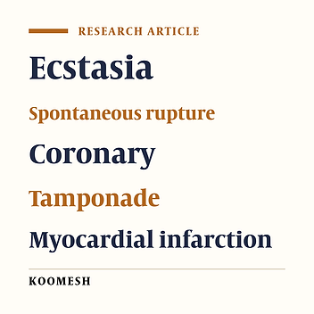
Image Credit:Koomesh
Outline
A patient with myocardial infarction and tamponade as a result of spontaneous rupture of an ectatic coronary artery
Authors
 ,*
,*Abstract
Introduction: Spontaneous rupture of ectatic vessels is possible in different vascular beds and it can be life-threatening. Rapid diagnosis and initiation of appropriate treatment are essential. Case report: A 63-year-old man presented with chest pain in a state where he was sleepy. Vital signs at the time of admission included a systemic blood pressure of 90/70 mm Hg, heart rate of 100 beats per minute, tachypnea, and arterial blood oxygen saturation of 92%. Muffled heart sounds, normal lung and abdominal examination, coldness of all four end organs, and weak and filiform pulses were recorded. Electrocardiogram showed ST segment elevation in leads I and avL. The patient underwent echocardiography, which reported moderate pericardial effusion and possible hematoma inside the pericardial fluid. Emergency CT angiography showed only pericardial effusion and intrapericardial hematoma. Pericardectomy was performed and the patient's systemic pressure increased to 150/ 90 mm Hg. The patient was transferred to the angiography unit due to the persistence of chest pain, where severe ectasia of the coronary arteries and 100% occlusion of the first diagonal were suggested. Importantly, the general condition was good and full consciousness. After eight hours, the patient suddenly suffered from severe melena, and after half an hour, a cardiorespiratory arrest occurs. Conclusion: Rapid treatment is necessary in possible cases of rupture of ectatic vessels and may prevent death and irreversible complications. Ecstatic vessels may be present in different vascular beds and this question is raised whether it is necessary to check other systems after determining the presence of ectasia in one system.
Highlights
References
- 1.Luo Y, Tang J, Liu X, Qiu J, Ye Z, Lai Y, et al. Coronary artery aneurysm differs from coronary artery ectasia: Angiographic characteristics and cardiovascular risk factor analysis in patients referred for angiography. Angiology 2017; 68: 823-8230.
- 2.Sultana R, Sultana N, Ishaq M, Samad A. The prevalence and clinical profile of angiographic coronary ectasia. J Pak Med Assoc 2011; 61: 372-375.
- 3.Bahremand M, Zereshki E, Karami Matin B, Rezaei M, Omrani H. Hypertension and coronary artery ectasia: a systematic review and meta-analysis study. Clin Hypertens 2021; 27: 14.
- 4.Qin Y, Tang C, Ma C, Yan G. Risk factors for coronary artery ectasia and the relationship between hyperlipidemia and coronary artery ectasia. Coron Artery Dis 2019; 30: 211-215.
- 5.Ipek G, Gungor B, Karatas MB, Onuk T, Keskin M, Tanik O, Hayiroglu MI, et al. Risk factors and outcomes in patients with ectatic infarct-related artery who underwent primary percutaneous coronary intervention after ST elevated myocardial infarction. Catheter Cardiovasc Interv 2016; 88: 748-753.
- 6.Doi T, Kataoka Y, Noguchi T, Shibata T, Nakashima T, Kawakami S, et al. Coronary artery ectasia predicts future cardiac events in patients with acute myocardial infarction. Arterioscler Thromb Vasc Biol 2017; 37: 2350-2355.
- 7.Kaljusto ML, Koldsland S, Vengen A, Woldbk PR, Tnnessen T. Cardiac tamponade caused by acute spontaneous coronary artery rupture. J Card Surg 2006; 21: 301-303.
- 8.Longobardi A, Iesu S, Baldi C, Di Maio M, Panza A, Mastrogiovanni G, et al. Spontaneous coronary artery rupture presenting as an acute coronary syndrome evolved in pseudoaneurysm and cardiac tamponade: Case report and literature review. Eur Heart J Acute Cardiovasc Care 2017; 6: 666-669.
- 9.Spontaneous coronary artery rupture without a pericardial effusion: a diagnostic challenge. BMJ Case Rep 2016; 2016: bcr2016214424.
- 10.Jiang Li, Cao J, Chen M. Coronary artery aneurysm combined with other multiple aneurysms at multiple locations. A case report and systematic review. Medicine (Baltimore). 2017; 96: e9230.
Copyright
© 2023, Author(s). This open-access article is available under the Creative Commons Attribution 4.0 (CC BY 4.0) International License (https://creativecommons.org/licenses/by/4.0/), which allows for unrestricted use, distribution, and reproduction in any medium, provided that the original work is properly cited.
Similar Articles
Mitral Stenosis, LV Aneurysm, Myocardial Bridge, and Myocardial Infarction: The Mystery Demystified
Chaurasia AK, Harikrishnan S, Kumar Ajith V, Tharakan JM. Mitral Stenosis, LV Aneurysm, Myocardial Bridge, and Myocardial Infarction: The Mystery Demystified. Int Cardiovasc Res J. 2017;7(3):e12428. doi:
Sudden Cardiac Arrest in A Young Adult Caused by Anomalous Origin of the Left Anterior Descending Artery from the Right Sinus of Valsalva: A Case Report
Alhouri A, Al Houri H, Obeidat O, Baniissa B, Juweid M. Sudden Cardiac Arrest in A Young Adult Caused by Anomalous Origin of the Left Anterior Descending Artery from the Right Sinus of Valsalva: A Case Report. Int Cardiovasc Res J. 2020;14(2):e102070. doi:
Strenuous Exercise Induced Syncope Due to Coronary Artery Anomaly
Yavuz V, Cetin N, Tuncer E, Dalgic O, Taskin U, et al. Strenuous Exercise Induced Syncope Due to Coronary Artery Anomaly. Int Cardiovasc Res J. 2017;8(3):e11105. doi:
Acute Myocardial Infarction in a Patient With Elevated ST-Segment in aVR Lead and Diffuse ST Segment Depression in Other Leads: A Case Report
Safi M, Khaheshi I, Memaryan M. Acute Myocardial Infarction in a Patient With Elevated ST-Segment in aVR Lead and Diffuse ST Segment Depression in Other Leads: A Case Report. Int J Cardiovasc Pract. 2016;1(1):e130021. doi: https://doi.org/10.20286/ijcp-010102
Transcatheter Coil Embolization of Coronary Artery Fistula
Zamani J, Tavassoli M, Mahmoody Y. Transcatheter Coil Embolization of Coronary Artery Fistula. Int Cardiovasc Res J. 2010;4(1):e67673. doi:
- Scopus by DOI: 0
Last Update: 3 weeks ago
- Scopus by Title: 0
Last Update: 3 weeks ago
- Scopus by Title (Ref): 0
Last Update: 3 weeks ago
- CrossRef: 0
Last Update: 8 hours ago
- OpenAlex: 0
Last Update: 10 hours ago
- OpenCitations: 0
Last Update: 4 days ago

