Full Text
Full text is available in PDF
Authors
Background and Aims: To discuss sources of error and pitfalls of color duplex sonography (CDUS) in screening for renovascular hypertension (RVH).
Methods: We retrospectively reviewed 47 patients with positive CDUS in patients with suspected RVH from May 1, 2007 to February 28, 2009. Manifestations of RVH on CDUS were analyzed. The results of main renal artery stenosis on CDUS were confirmed with that of magnetic resonance angiography (MRA), computed tomographic angiography (CTA), or digital subtraction angiography (DSA). Pitfalls and sources of error of CDUS in screening for RVH were identified. RVH was classified into three groups: 1) main renal artery stenosis (RAS); 2) intrarenal artery stenosis; and 3) intrarenal arteriovenous fistula (AVF).
Results: Two cases of false positive and two cases of false negative main RAS on CDUS were corrected by MRA or DSA. Three cases with intrarenal artery stenosis and four cases with intrarenal AVF were not visualized at the initial CDUS but detected on repeat studies. Peak systolic velocity > 2 m/s at the stenotic artery was the most sensitive parameter for detecting RAS in either main renal artery or intrarenal artery. Acceleration time > 0.07 m/s in the intrarenal artery was seen in only 10 cases (10 /20, 50%) with hemodynamically significant main RAS (>60% arterial lumen reduction).
Conclusions: Some pitfalls and sources of error of CDUS can be corrected and minimized with proper scanning and interpretation. Intrarenal RAS and AVF should be investigated with optimized Doppler settings. CDUS, with its advantages outweighing its limitations, should be considered a first line study in screening for RVH by experienced ultrasound professionals.
© 2010, Author(s). This open-access article is available under the Creative Commons Attribution 4.0 (CC BY 4.0) International License (https://creativecommons.org/licenses/by/4.0/), which allows for unrestricted use, distribution, and reproduction in any medium, provided that the original work is properly cited.
Yilmaz C, Nart A, Uslu A, Aykas A. Splenic Arteriovenous Fistula: An Unusual Cause of Portal Hypertension Primarily Diagnosed by Color Duplex Sonography. I J Radiol. 2010;7(1):e78849. doi:
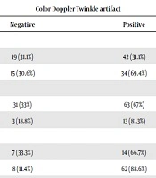
Letafati M, Tarzamni MK, Hajalioghli P, Taheri SM, Vaseghi H, et al. Diagnostic Accuracy of Twinkling Artifact Sign Seen in Color Doppler Ultrasonography in Detecting Microlithiasis of Kidney. Nephro-Urol Mon. 2020;12(2):e102860. doi: https://doi.org/10.5812/numonthly.102860
Firouzi A, Dohaei A, Heidarali M, Mortazavian H. Renal Arteriovenous Fistula; Possibility of Misdiagnosis and Successful Management: A Case Report. Multidiscip Cardio Annal. 2016;7(1):e9126. doi: https://doi.org/10.17795/mcardia.9126

Asadi P, Sabouri S. Comparison of Color Doppler Ultrasound and CT Angiography in Carotid Artery Stenosis: A Four Years’ Experience from a Single Medical Center. I J Radiol. 2014;11(30th Iranian Congress of Radiology):e93648. doi: https://doi.org/10.5812/iranjradiol.21439
Banaei A. The Comparison between Digital Subtraction Angiography, CT Angiography, and Doppler Ultrasonography in Evaluation and Assessment of Carotid Artery Stenosis. Ann Mil Health Sci Res. 2017;15(1):e61661. doi: https://doi.org/10.5812/amh.61661
Last Update: 4 days ago
Last Update: 4 days ago
Last Update: 4 days ago
Last Update: 4 days ago
Last Update: 2 days ago
Last Update: 1 day ago
