


Dear editor,
The severe acute respiratory syndrome coronavirus 2 (SARS-CoV-2), previously called nCoV-2019, has become a public health emergency. Multiple cases of primary infection have been reported from different countries, but there are few cases of recurrent infection. (1, 2) Here, two COVID-19 cases are reported who developed COVID-19 recurrent infection.
Case one: A 43-year-old man presented with a sore throat on June 7, 2020, with no fever, headache, or cough. He had no positive past medical history. The PCR test for COVID-19 was done, which was positive. Preliminary tests were normal, and no pulmonary lesion was observed in the initial CT scan. The patient was treated for one week with hydroxychloroquine as an outpatient, and the PCR test conducted after two weeks was negative. The patient returned to his daily activities with no problems. A week later, he became lethargic with a cough and headache. The PCR test for COVID-19 was conducted again on July 6, 2020, which was positive. The patient was hospitalized with the exacerbation of lethargy and continued fever. A CT scan showed bilateral lung involvement (Figure 1). He was treated with favipiravir and discharged after seven days in good health. The PCR test was not negative one week after treatment with favipiravir but became negative after one month on August 6, 2020. The laboratory tests during the first and second infections are shown in Table 1.
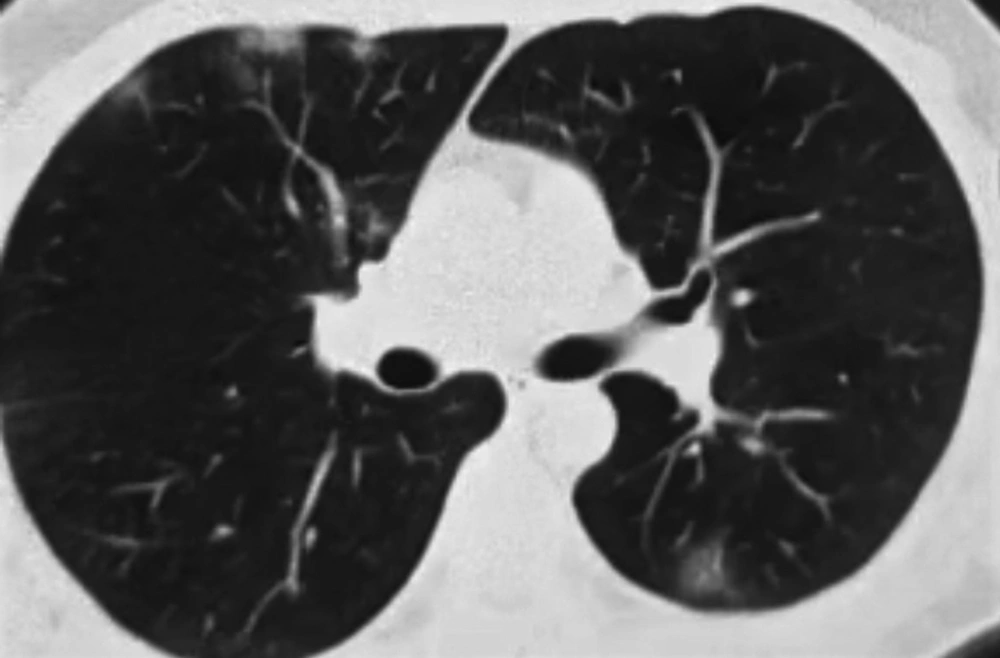
Figure 1.
Chest CT scan showing bilateral lung involvement in favor of COVID-19
Table 1.The Laboratory Tests During the First and Second Infections in Both Patients
| First Time | Second Time | |
|---|---|---|
| Case 1 | ||
| WBC lymphocyte (%) | 4.9×10⁹/L Lymph (21%) | 8.2×10⁹/L Lymph (25%) |
| Troponin | Negative | Negative |
| LDH | 196 IU/L | 896 IU/L |
| D-dimer | 165 ng/mL | 368 ng/mL |
| Case 2 | ||
| WBC lymphocyte (%) | 2000 ×109/L Lymph (5%) | 6.2×10⁹/L Lymph (27%) |
| Troponin | Negative | Negative |
| LDH | 154 IU/L | 276 IU/L |
| D-dimer | 210 ng/mL | 155 ng/mL |
Case two: A 41-year-old woman with no previous history of disease referred with the onset of symptoms from June 7, 2020, as mild, dry coughs and low-grade fever. The chest CT scan was conducted on June 9, 2020, which showed patchy involvement in the right pulmonary base (Figure 2). The initial PCR test was reported positive. The patient was hospitalized on June 12, 2020, for severe episodes of weakness and received corticosteroids, Kaletra, and interferon. She was discharged in good health. She had no problems until July 8, 2020, when she suffered dry coughs and mild lethargy. The PCR test was positive. Favipiravir was initiated, and lethargy episodes were fewer than before. She had no fever or headache and the chest CT scan was normal. A week after treatment with favipiravir, her PCR test became negative, and 24 hours later, the PCR test was negative again.
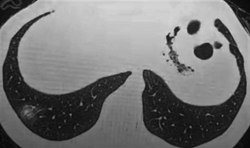
Figure 2.
Patchy involvement in the right pulmonary base
The laboratory tests during the first and second infections are shown in Table 1.
The possibility of COVID-19 reactivation is considered an important global health problem because it can lead to the rapid spread of the virus. Home isolation is required for all patients with COVID-19 for 14 days after discharge from the hospital, but the possible time of infection transmission and the duration of virus shedding are not yet known (1).
We presented two cases of the recurrence of COVID-19 infection, but the differentiation of reinfection from reactivation was not possible since we had no genetic study on SARS-CoV-2 isolates from these two patients. Some factors can influence the likelihood of false-negative results in molecular tests. The presence of the virus in an infected patient can provide different results depending on the viral load, the operator’s experience in collecting samples, and the sampling site (2). The host condition, virological characteristics, and steroid-induced suppression of the immune system have also been proposed as reactivation risk factors (3). Ye et al. reported that the reactivation of the disease in patients with COVID-19 after discharge from the hospital was 9.19% (3). Carriers may be infectious without symptoms and before becoming symptomatic (4). However, it should be noted that the transmission of the virus is possible during recovery (2). In the serum of a patient in this study, the SARS-CoV-2 IgG antibody was reported, indicating that the acute phase of the disease was passed. Initial evidence shows that antibody response happens in those who have been infected (5). It is not yet clear whether these antibodies are protective, and if they are, how long this protection lasts.
Hence, immediate and extensive studies are needed to control the COVID-19 pandemic. The current policy must prevent the spread of the virus to populations, as well as further infections and deaths.



