
Cover image of the article: Oncocytic Cystic Lesions of False and True Vocal Cords: An Uncommon Occurrence

Image Credit:Shiraz E-Med J

Oncocytic Cystic Lesions of False and True Vocal Cords: An Uncommon Occurrence

Authors

Masoumeh Gharib1, Mohadeseh ShahrakiMohadeseh Shahraki ORCID1,*
1Department of Pathology, Faculty of Medicine, Mashhad University of Medical, Mashhad, Iran
*Corresponding Author: Department of Pathology, Faculty of Medicine, Mashhad University of Medical, Mashhad, Iran. Email: m.shahraki1991@gmail.com

Shiraz E-Medical Journal:Vol. 24, issue 5; e136171
Published online:Jun 13, 2023
Article type:Case Report
Received:Mar 23, 2023
Accepted:May 28, 2023
How to Cite:Gharib M, Shahraki M. Oncocytic Cystic Lesions of False and True Vocal Cords: An Uncommon Occurrence. Shiraz E-Med J. 2023;24(5):e136171. doi: https://doi.org/10.5812/semj-136171

Abstract

Introduction:

Oncocytic cysts of the larynx are benign, uncommon, and slow-growing lesions that usually occur in elderly people, especially in those with a history of smoking and chronic irritation. These lesions cause various clinical symptoms depending on their location and size, requiring physicians to consider a wide range of symptoms.

Case Presentation:

Here we present two men who were referred to us because of prolonged voice hoarseness.

Conclusions:

Medical assessments led us to the diagnosis of oncocytic cysts in the false and true vocal cords. Both patients were followed up after removing their laryngeal cysts and have had no clinical complaints since then.

Highlights

1. Introduction

Oncocytic cysts of the larynx are benign, uncommon, and slow-growing lesions that mostly or exclusively consist of oncocytes (1-4). The most common locations of these lesions are false vocal cords and laryngeal ventricles (5). These cysts most frequently occur in middle-aged and elderly people (6-8) and cause various clinical symptoms depending on their location and size (9, 10). We present two male patients referring with the chief complaint of prolonged voice hoarseness, who were finally diagnosed with oncocytic laryngeal cysts.

2. Case Presentation

2.1. Case 1

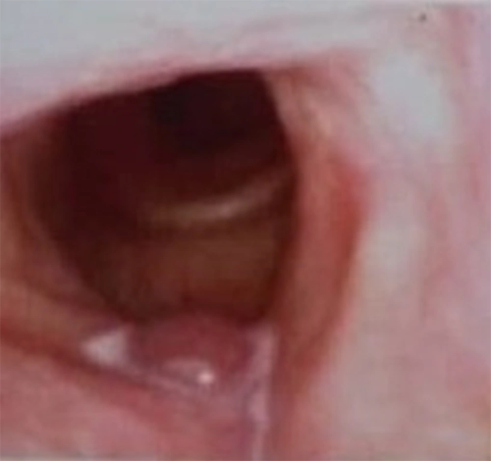
The first patient was a 61-year-old man with the chief complaint of voice hoarseness from six months prior to referral. He was a heavy smoker (150 packs per year) but did not report any history of other respiratory symptoms, medical illnesses, or surgery. Video laryngoscopy revealed a laryngeal mass in the right false vocal cord (Figure 1). Laryngeal Computerized Tomography Scan (CT scan) showed a 14 * 26 * 28 mm mass with a density equivalent to cystic lesions in the right false vocal cord, extending beyond the supraglottis to the level of the hyoid.
Video laryngoscopic view of a cyst in the right false vocal cord of a 61-year-old man
Figure 1.
Video laryngoscopic view of a cyst in the right false vocal cord of a 61-year-old man
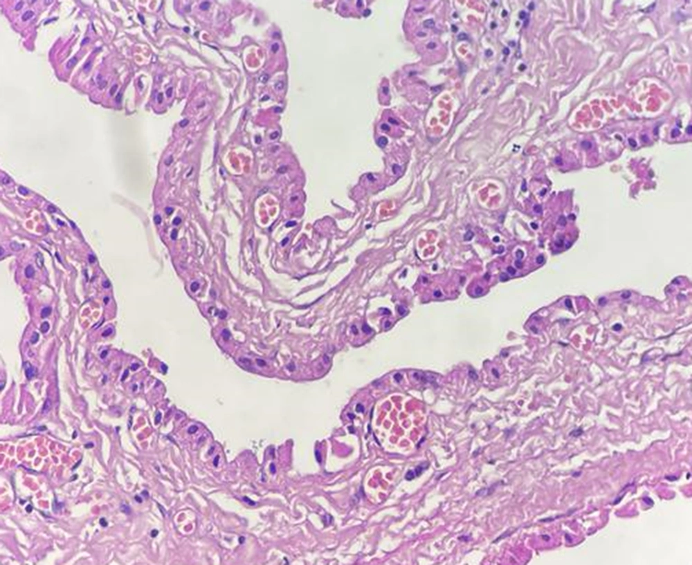
After excision, the mass appeared as a creamy-brown tissue with dimensions of 2.1 * 1.2 * 1.1 cm, a diameter of 3.2 cm, and a thickness of 1.5 cm covered by mucosa. The pathological assessment revealed the false vocal cord epithelium with seromucinous glands and multiple cysts lined by bland oncocytic columnar cells. The final diagnosis was a benign oncocytic cyst. The patient had no hoarseness or other clinical symptoms six months after the procedure (Figure 2).
The cyst lining by bland oncocytic columnar cells (H&E, 400×)
Figure 2.
The cyst lining by bland oncocytic columnar cells (H&E, 400×)

2.2. Case 2

The second patient was a 76-year-old man referred due to voice hoarseness for three months. He did not report any other respiratory symptoms, past medical history, undergoing surgery, or smoking. A neck CT scan without contrast showed hypodense and homogenous foci with a dimension of 37 * 20 * 17 mm and a mass in the lateral side of the left false vocal cord. So, the radiologic diagnosis of a cyst or laryngocele was made (Figure 3). After the surgical removing the mass, the patient had a good condition with no voice hoarseness after three months of follow-up.
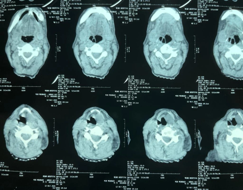
Neck CT scan without contrast
Figure 3.
Neck CT scan without contrast
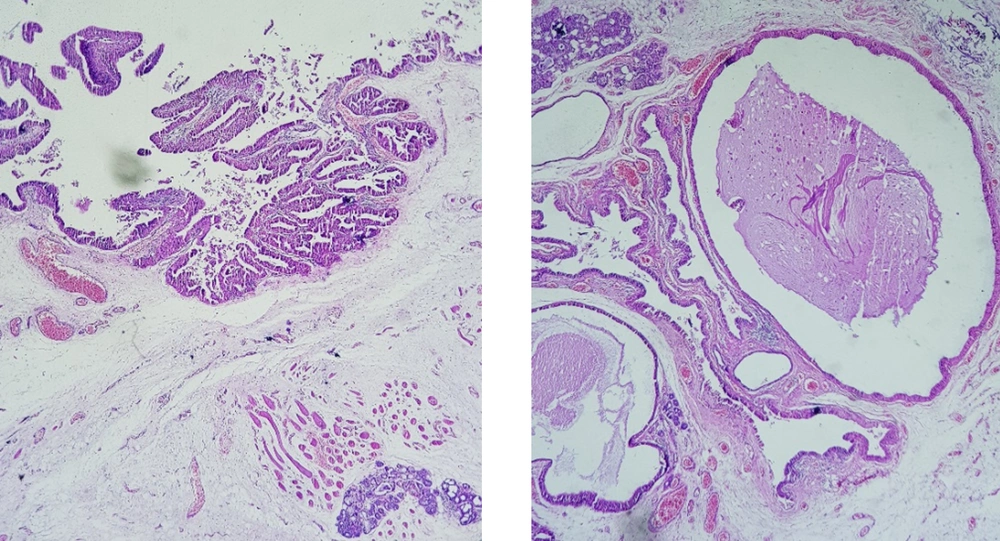
Video laryngoscopy showed three masses on the laryngeal surface of the epiglottis, the right true vocal cord, and the right false vocal cord. A biopsy was performed (Figure 4), and the histopathologic examination showed a cystic structure with papillary growth lined by bland oncocytic cells (Figure 5).
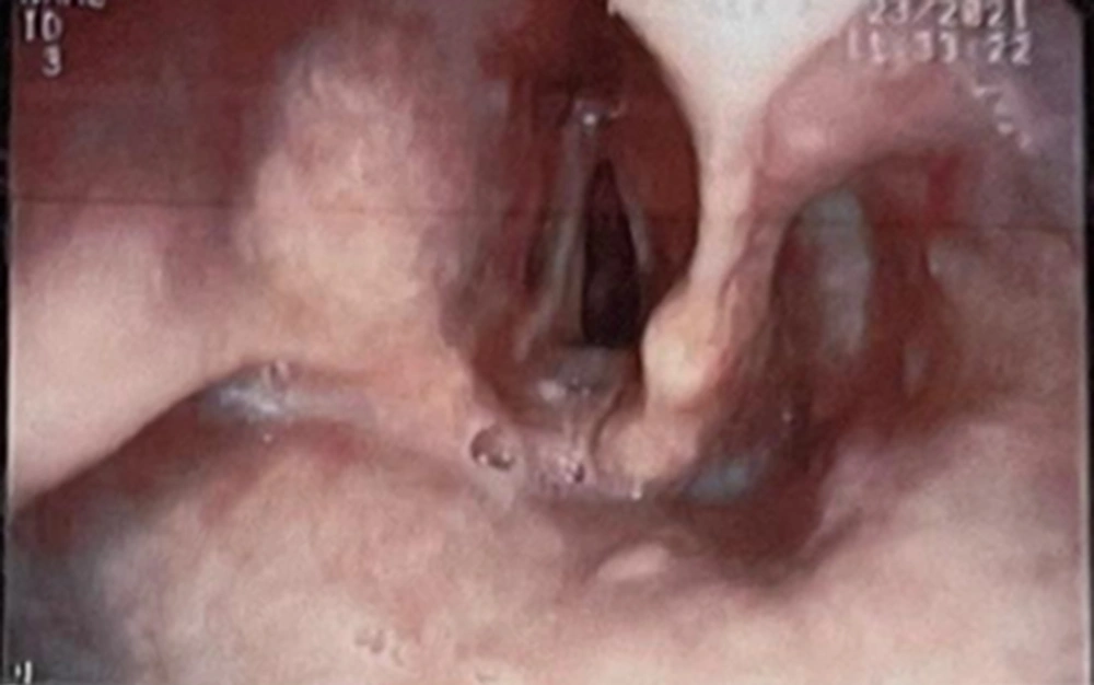
Video laryngoscopic view
Figure 4.
Video laryngoscopic view
The microscopic view shows acystic structure with papillary growth lined by bland oncocytic cells (H&E, 100×).
Figure 5.
The microscopic view shows acystic structure with papillary growth lined by bland oncocytic cells (H&E, 100×).

3. Discussion

Oncocytes consist of polygonal epithelial cells found in salivary glands and the lining epithelium of the respiratory tract (11). Oncocytic metaplasia in the larynx is uncommon, and oncocytic laryngeal cysts are rare, comprising less than 1% of all benign masses of the larynx (12). The oncocytic lesions of the larynx usually present as solitary lesions; however, some multifocal lesions have been described so far (3), including the second case presented in this paper.
Oncocytic metaplasia is a senile process that commonly appears in the epithelial endocrine cells possessing a high metabolic activity (13). Oncocytic cysts usually occur in ages higher than 50 years, unlike other laryngeal cysts that involve younger individuals (3).
Smoking predisposes to oncocytic lesions of the larynx due to increased oxidative stress and laryngeal inflammation. A long history of heavy smoking is reported in most patients with these lesions (14, 15), including the first case presented here. Consistently, Mettias et al. reported an oncocytic cyst in the vocal cord of a 62-year-old man with a history of smoking 20 cigarettes per day for 50 years (16). Nevertheless, the second patient described in the present paper declared no history of smoking.
The common clinical presentation of these lesions is voice hoarseness; however, lesions of different sizes and locations can present with various symptoms (17). Erfanian et al. reported a case of severe respiratory distress due to two large masses on the false vocal cord, which was identified via video laryngoscopy. This patient had aggravating dyspnea for two years prior to diagnosis and reported dysphonia for more than one year. In physical examination, inspiratory stridor was obvious, and the masses were disclosed by fiberoptic nasotracheal intubation (18).
The management of these lesions is conservative and local excision, where endoscopic removal is considered the treatment of choice (1). Oncocytic laryngeal cysts are benign lesions, but follow-up is recommended due to the risk of recurrence, especially in patients with multiple lesions (2).

3.1. Conclusions

In this paper, we reported two men referring with prolonged voice hoarseness. Medical assessments led to the diagnosis of oncocytic cysts in their vocal cords, which were removed by video laryngoscopy. Both patients were followed up after removing their laryngeal cysts and had no clinical symptoms since then.

Footnotes

  • Authors' Contribution: Mohadeseh Shahraki designed the study, participated in patient evaluation, and drafted the manuscript. Masoumeh Gharib participated in study design and patient evaluation, gathering data such as search in medical records and imaging, and helped draft the manuscript. Mohadeseh Shahraki re-evaluated the clinical data, revised the manuscript, performed the statistical analysis, and revised the manuscript. Mohadeseh Shahraki collected the clinical data, interpreted them, and revised the manuscript. Masoumeh Gharib re-analyzed the clinical and statistical data and revised the manuscript. All authors read and approved the final manuscript.

  • Conflict of Interests: The authors declare no conflict of interests.

  • Funding/Support: No Funding was declared.

  • Informed Consent: Written informed consent was obtained from the patients.

References

  • 1.
    Salerno G, Mignogna C, Cavaliere M, D'Angelo L, Galli V. Oncocytic cyst of the larynx: An unusual occurrence. Acta Otorhinolaryngol Ital. 2007;27(4):212-5. [PubMed ID: 17957853]. [PubMed Central ID: PMC2640029].
  • 2.
    Dhingra JK, Aqel NM, McEwen J, Bleach NR. Multiple oncocytic cysts of the larynx. J Laryngol Otol. 1995;109(12):1226-8. [PubMed ID: 8551166]. https://doi.org/10.1017/s0022215100132529.
  • 3.
    Peeters A, Schmelzer B, Beernaert A, Fannes H. Oncocytic laryngeal cysts: A case report and literature review. Acta Otorhinolaryngol Belg. 2001;55(1):71-5. [PubMed ID: 11256195].
  • 4.
    Ugur Kilinc AN, Unlu Y, Yucel A. Laryngeal Oncocytic Cystadenoma with Rare Location and Clinicopathological Aspects. Head Neck Pathol. 2021;15(3):1004-6. [PubMed ID: 33048303]. [PubMed Central ID: PMC8384988]. https://doi.org/10.1007/s12105-020-01232-1.
  • 5.
    Robinson AC, Kaberos A, Cox PM, Stearns MP. Oncocytoma of the larynx. J Laryngol Otol. 1990;104(4):346-9. [PubMed ID: 2196316]. https://doi.org/10.1017/s0022215100112678.
  • 6.
    Holm-Jensen S, Jacobsen M, Thommesen N, Ferreira O. Oncocytic cysts of the larynx. Acta Otolaryngol. 1977;83(3-4):366-71. [PubMed ID: 857607]. https://doi.org/10.3109/00016487709128858.
  • 7.
    Ferlito A, Recher G. Oncocytic lesions of the larynx. Arch Otorhinolaryngol. 1981;232(2):107-15. [PubMed ID: 7271583]. https://doi.org/10.1007/BF00505029.
  • 8.
    Puttasiddaiah PM, Berry S, Whittet HB, Kumar M. Laryngeal oncocytic cyst presenting with an acute onset of stridor. Ear, Nose & Throat Journal. 2009;88(7):1003-4.
  • 9.
    Feinstein AJ, Peng KA, Bhuta SM, Abemayor E, Mendelsohn AH. Laryngeal oncocytic cystadenomas masquerading as laryngoceles. Am J Otolaryngol. 2016;37(1):17-21. [PubMed ID: 26700253]. https://doi.org/10.1016/j.amjoto.2015.09.010.
  • 10.
    Shaik FB, Chowdary KK. A Clinical Study of Laryngeal Cysts. International Journal of Phonosurgery & Laryngology. 2012;6(2):53-6.
  • 11.
    Burgos Sanchez AJ, Lafarga J, Galvan B, Talavera J, Trigueros M. [Presentation of multiple oncocytic type laryngeal cysts. Clinical and histopathological findings]. Acta Otorrinolaringol Esp. 2000;51(4):353-6. [PubMed ID: 10984961].
  • 12.
    Johns ME, Regezi JA, Batsakis JG. Oncocytic neoplasms of salivary glands: An ultrastructural study. Laryngoscope. 1977;87(6):862-71. [PubMed ID: 865204]. https://doi.org/10.1288/00005537-197706000-00002.
  • 13.
    Baris O, Savagner F, Nasser V, Loriod B, Granjeaud S, Guyetant S, et al. Transcriptional profiling reveals coordinated up-regulation of oxidative metabolism genes in thyroid oncocytic tumors. J Clin Endocrinol Metab. 2004;89(2):994-1005. [PubMed ID: 14764826]. https://doi.org/10.1210/jc.2003-031238.
  • 14.
    Newman BH, Taxy JB, Laker HI. Laryngeal cysts in adults: A clinicopathologic study of 20 cases. Am J Clin Pathol. 1984;81(6):715-20. [PubMed ID: 6731351]. https://doi.org/10.1093/ajcp/81.6.715.
  • 15.
    Stenner M, Muller KM, Koopmann M, Rudack C. Squamous cell carcinoma of the larynx arising in multifocal pharyngolaryngeal oncocytic papillary cystadenoma: A case report and review of the literature. Medicine (Baltimore). 2014;93(12). e70. [PubMed ID: 25211046]. [PubMed Central ID: PMC4616269]. https://doi.org/10.1097/MD.0000000000000070.
  • 16.
    Mettias B, Usanov A, Osborne J. Oncocytic cyst of the larynx: Precipitating factors and mode of treatment (case report). Egyptian Journal of Ear, Nose, Throat and Allied Sciences. 2012;13(2):91-3. https://doi.org/10.1016/j.ejenta.2012.03.002.
  • 17.
    Pilch BZ. Larynx and hypopharynx. In: Pilch BZ, editor. Head and neck surgical pathology. Philadelphia: Lippincott Williams & Wilkins; 2001.
  • 18.
    Erfanian R, Shakiba S, Nourbakhsh SMA, Sohrabpour S. Laryngeal oncocytic cyst: An uncommon incident. Case Reports in Clinical Practice. 2020. https://doi.org/10.18502/crcp.v4i4.2384.

Copyright

Copyright © 2023, Author(s). This is an open-access article distributed under the terms of the Creative Commons Attribution-NonCommercial 4.0 International License (http://creativecommons.org/licenses/by-nc/4.0/) which permits copy and redistribute the material just in noncommercial usages, provided the original work is properly cited.

Similar Articles

1
Jun
2013
Symptomatic Cervical Thymic Cyst: A Case Report and Literature Review

Symptomatic Cervical Thymic Cyst: A Case Report and Literature Review

Rahim Mahmodlou,
Shahsanam Gheibi,
Arash Aghajani Nargesi,
Razieh Mahmoodzadeh,
Mirataollah Salabati

Mahmodlou R, Gheibi S, Aghajani Nargesi A, Mahmoodzadeh R, Salabati M. Symptomatic Cervical Thymic Cyst: A Case Report and Literature Review. J Compr Ped. 2013;4(3):147-50. doi: https://doi.org/10.17795/compreped-9379

24
Aug
2019
Primary Laryngeal Neuroendocrine Carcinoma: Two Cases of an Unusual Tumor in a Rare Location

Primary Laryngeal Neuroendocrine Carcinoma: Two Cases of an Unusual Tumor in a Rare Location

Matin Ghazizadeh,
Shervin Ghavami,
Mahboobe Asadi

Ghazizadeh M, Ghavami S, Asadi M. Primary Laryngeal Neuroendocrine Carcinoma: Two Cases of an Unusual Tumor in a Rare Location. Int J Cancer Manag. 2019;12(8):e82626. doi: https://doi.org/10.5812/ijcm.82626

22
Apr
2015
Ultrasound, CT and MRI Appearances of a Rare Symptomatic Laryngeal Chondrometaplasia: A Case Report

Ultrasound, CT and MRI Appearances of a Rare Symptomatic Laryngeal Chondrometaplasia: A Case Report

Ryan Ka Lok Lee,
Edmond Yuen Hok Yuen,
Victor James Abdullah,
Yolanda Yim Ping Lee,
Anil Tejbhan Ahuja

Lok Lee RK, Hok Yuen EY, Abdullah VJ, Ping Lee YY, Ahuja AT. Ultrasound, CT and MRI Appearances of a Rare Symptomatic Laryngeal Chondrometaplasia: A Case Report. I J Radiol. 2015;12(2):e8276. doi: https://doi.org/10.5812/iranjradiol.8276

1
Apr
2015
The Face is the Index of the Mind: Laryngoscopy in a Multiple Endocrine Neoplasia Patient With Mucosal Involvement

The Face is the Index of the Mind: Laryngoscopy in a Multiple Endocrine Neoplasia Patient With Mucosal Involvement

Mojgan Rahimi,
Pooya Kalani

Rahimi M, Kalani P. The Face is the Index of the Mind: Laryngoscopy in a Multiple Endocrine Neoplasia Patient With Mucosal Involvement. Arch Neurosci. 2015;2(2):e16498. doi: https://doi.org/10.5812/archneurosci.16498

31
Mar
2000
A report on ear Teratoma

A report on ear Teratoma

MA Mojabi

Mojabi M. A report on ear Teratoma. J Inflamm Dis. 2000;3(4):e154689. doi:

More by these authors

Masoumeh GharibPubMedScholar
Mohadeseh ShahrakiPubMedScholar
Share
Cited by
Metrics
Indexed in