1. Background
We aimed to determine the association between upright T wave in lead V1 and coronary artery disease (CAD) in patients who presented with chest pain or positive non-invasive tests in emergency department.
Cardiovascular disease is a worldwide great burden on the health care system [1, 2] early diagnosis and treatment of CAD can reduce the mortality and morbidity and thereby increase the life expectancy. Several non-invasive methods have been developed to identify CAD before catastrophic events. Coronary angiography is still the gold standard. Less than 50% of patients with typical chest pain have diagnostic ECG at the first presentation [3].
Electrocardiography is the easy, cost benefit and accessible screening tool for CAD diagnosis [4-6].
T wave changes are associated with myocardial ischemia [7] and upright T wave in right leads is associated with right ventricular (RV) involvement in postero-inferior acute myocardial infarction (AMI) [8]. Uptight T wave in avR was reported to be associated with cardiovascular death [9]. Lone T wave inversion in aVL has been proposed as an important marker of inferior ischemia [10].
In the present study, we aim to identify the association of upright T wave in lead V1 and the presence of CAD in patients with no remarkable ECG pattern in favor of acute myocardial infarction. All patients underwent coronary angiography (CAG) due to typical chest pain or positive non-invasive tests such as myocardial perfusion scan (MPI scan) and/or exercise tolerance test (ETT).
2. Methods
In this cross sectional study, data of 200 patients who underwent coronary angiography at our center between January and September 2013 were evaluated. Inclusion criteria were as follows; age > 18 years, T wave more than 1.5 mm in V1 lead [11] complaint of typical chest pain in favor of acute coronary syndrome and/or positive non-invasive tests. The exclusion criteria were history of cardiac surgery or intervention, history of cardiac arrhythmia or catheter ablation, Pathological Q waves, having pacemaker or other cardiac devices, severe valvular disease, ventricular hypertrophy, inter-ventricular conduction delay, inverted T wave in leads other than V1 or the limb leads and unreadable ECG. A written informed consent was signed by all patients at the time of admission to use clinical data anonymously for research purposes. The local institutional committee of Medical ethics and the research board of Tehran University of Medical Sciences approved the study protocol. This study conforms to the principles outlined in the declaration of Helsinki.
We selected the first 100 patients with normal ECG and upright T wave in lead V1 and another 100 patients with normal ECG without upright T wave in V1 .The ECGs were interpreted by two cardiologists who were blinded to the CAG reports. Demographic and clinical data including age, gender, height, weight, history of previous disease and cardiovascular risk factors as well as the laboratory data were retrieved from the databank of Tehran Heart Center. CAG reports were extracted from Tehran heart center data registry.
Significant atherosclerotic coronary artery lesion was defined according to the guideline of the American college of cardiology/American heart association as ≥ 70% narrowing of the lumen diameter in at least one major coronary artery or stenosis more than 50% on the left main artery [12].
2.1. Statistical Analysis
Continuous variables were described with mean and standard deviation (SD) or with median and 25th and 75th percentiles for skewed data; and were compared between patients with and without upright T wave using student’s t or Mann-Whitney U test were appropriate. Categorical variables were expressed as frequency and percentage and were compared among groups using chi-square test. Variables, which were simultaneously associated with CAD and upright T wave with P values less than 0.2, were considered as potential confounders. P value less than 0.05 were considered significant in this study. The association of upright T wave with CAD was adjusted for detected possible confounders applying logistic regression model. The effect of upright T wave on occurrence of CAD was reported with odds ratio (OR) with 95% confidence interval (CI). All statistical analyses were performed using IBM SPSS statistics for Windows version 22.0 (Armonk, NY: IBM Corp.).
3. Results
In this study, 100 patients with upright T wave in lead V1 and 100 patients without this finding were compared. In the upright T wave group, frequency of males and frequency of patients with CAD was significantly more than the normal ECG group. Other study variables, particularly the frequencies of cardiovascular risk factors were same between the groups. Comparison of the general characteristics of the study groups is shown in Table 1. Univariate binary logistic regression was done to evaluate the association between traditional risk factors of coronary artery disease and T wave deviation in lead V1, Table 1.
Table 1.
Association Between Basic Demographic Feature and Traditional Risk Factors of CAD with T Wave Deviation in V1
| Characteristic | Normal ECG (N = 100) | Upright TV1 ECG (N = 100) | Odds Ratio (95% CI) | P Valuea |
|---|---|---|---|---|
| Age, y | 55.4 ± 9.3 | 57.2 ± 10.8 | 1.04 (0.87 - 1.23) | 0.219 |
| Male gender | 45b | 84 | 1.51 (1.12 - 2.1) | < 0.001 |
| CAD | 26 | 62 | 2.38(1.78 - 3.12) | < 0.001 |
| Diabetes mellitus | 27 | 26 | 0.97 (0.76 - 1.23) | 0.877 |
| Hypertension | 35 | 27 | 0.77 (0.56 - 0.98) | 0.221 |
| Dyslipidemia | 48 | 36 | 0.75 (0.56 - 98) | 0.086 |
| Smoking | 15 | 23 | 1.53 (1.07 - 1.98) | 0.149 |
| Family history of CAD | 14 | 14 | 1.04 (0.87 - 1.16) | 0.999 |
Abbreviations: CAD, Coronary Artery Disease; CI, Confidence Interval.
aP value < 0.05 was considered as statistically significant.
bNumbers represent both the frequency and percentage.
Based on uni-variate binary logistic regression, age and coronary artery disease were significantly related to up right T wave in patients with chest pain, odds ratio 1.51 and 2.38 respectively. Older patients with chest pain and upright T wave in lead V1 probably have significant CAD.
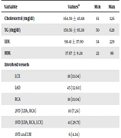
Association between different angiographic findings and upright T wave in V1 has been shown, Table 2. Patients with chest pain and significant coronary artery disease (> 50%) are more likely to have upright T wave in their baseline ECG. However, majority of patients with chest pain and normal serial ECGs in emergency department have normal or minimal CAD. Among 100 patients with upright T wave, 21 patients had two vessels disease; 17 of them had LCx and/or RCA significant obstruction. However, precise data about other culprit lesions are lacking.
Table 2.
Angiographic Findings in Relation to T Wave Polarity in Lead V1
| Characteristic | Normal ECG (N = 100) | Upright TV1 ECG (N = 100) | P Valuea |
|---|---|---|---|
| Results | < 0.001 | ||
| Normal/minimal CAD | 74b | 38 | 0.005 |
| Significant CAD (> 50% stenosis at least in one vessel) | 26 | 62 | |
| Single vessel disease | 14 | 31 | 0.045 |
| 2-vessel disease | 9 | 12 | 0.048 |
| 3-vessel disease | 3 | 19 | 0.003 |
Abbreviation: CAD, Coronary Artery Disease.
aP value < 0.05 was considered as statistically significant.
bNumbers represent both the frequency and percentage.
Base on the uni-variate regression model, presence of upright T wave could predict the presence of CAD (OR = 4.64, 95% CI: 2.54 - 8.48; P < 0.001). To better evaluate the association between upright T wave in V1 on CAD, we performed multivariate logistic regression model, Table 3. All variables with P value less than 0.15 (in uni-variate analysis) were included. In the multivariate binary logistic regression model and after adjustment for confounding variables (including gender, dyslipidemia and smoking) presence of upright T wave in V1 lead could still predict CAD (4.6 CI 95% 3.1 - 6.3); P < 0.001). Male Gender was also statistically related to significant CAD (odds ratio 3.4, CI 95% 2.2 - 4.8).
Table 3.
Determinants of Significant Coronary Artery Disease
| Important Risk Factors | Significant CAD (N=88), No. (%) | Odds Ratio (95% Confidence Interval) |
|---|---|---|
| Upright T wave in lead V1 | 62 (70) | 4.6 (3.1 - 6.3) |
| Dyslipidemia | 45 (51) | 1.9 (0.9 - 3.1) |
| Cigarette smoking | 20 (22) | 1.5 (0.7 - 1.8) |
| Gender (male) | 70 (80) | 3.4 (2.2 - 4.8) |
4. Discussion
In present study, accordant to recent similar studies, upright T wave in V1 may identify significant CAD in patients with suspicious cardiac symptoms.
Pathophysiologic basis is buried in the concept of different ventricular repolarization during ischemic [13, 14]. Myocardial ischemia causes electro physiologic changes in action potential. It causes the difference not only between normal and ischemic area but also between the epicardium and the endocardium [15]. These slight changes can be revealed on superficial ECG [16].
The association between upright T wave in v1 and proximal right coronary artery stenosis was reported [8]. Based on their findings, presence of upright T wave in these patients could represent the ischemia of the right ventricle. Another similar study emphasized on association between upright T wave in V1 and left circumflex artery disease [13]. Another report showed that almost half of the patients with posterior MI-as detected by autopsy or ECG- had upright T wave [17]. It should be noted that the capability of ECG for predicting the presence of CAD in the absence of apparent ischemia or myocardial damage is limited [7, 18, 19]. It has been shown that upright T-wave in ECG will increase the specificity and diagnostic accuracy of CAD detection, despite the reduction in sensitivity [20]. In 2012 Stankovic et al. conducted similar study on smaller sample size, and reported patients with positive T-wave in V1 are 4 times more likely to have significant stenosis in coronary angiogram [11].
Based on our findings and the body of literature, slight changes in the ECG, such as upright T wave in V1 can be used as a good alarming sign for detecting significant CAD earlier in patients who complain from chest pain.
4.1. Conclusion
Overall, this study showed that utilization of ECG as a screening tool by considering the changes of T wave in precordial leads, particularly Upright T wave in V1 lead could predict the presence of CAD in suspicious patients and decrease the threshold for performing coronary angiography.