Fulltext
The full text of this article is available on the PDF file.
Zahedan Journal of Research in Medical Sciences
The Official Journal of Zahedan University of Medical Sciences
Image Credit:Zahedan J Res Med Sci
Authors
Background: Various anomalies, especially Down syndrome impose heavy emotional and financial burden on families and communities by development of mental and behavioral retardation. The purpose of this study is to assess the value of Nuchal Translucency (NT) to diagnose Down syndrome in the first trimester of pregnancy.
Materials and Methods: This prospective study was conducted through evaluating 102 pregnant women of 17-44 years old with gestational age of 11 to 13 weeks and 3 days, in a private radiology clinic in Zahedan during May 2008 to June 2010. All embryos were ultra-sounded by a radiologist, their NT and crown-rump length were measured. After birth, all infants were examined by a pediatrician and cases suspected with Down syndrome were diagnosed through karyotype test.
Results: The mean of NT was 1.62 mm. NT was above 1.7 mm for women older than 35 years and it was 3.1 to 3.8 mm for women older than 40 years. 6 out of 7 cases diagnosed postnatal with Down syndrome, had above 95% normal NT and 4 out of 7 mothers who had baby with Down syndrome, were older than 35 years.
Conclusion: Through measurement of NT during 11th to 13th weeks of pregnancy and consideration of base risk factors, the possibility of Down syndrome can be assessed and necessary diagnostic evaluations can be performed for risky cases.
© 2012, Author(s). This open-access article is available under the Creative Commons Attribution 4.0 (CC BY 4.0) International License (https://creativecommons.org/licenses/by/4.0/), which allows for unrestricted use, distribution, and reproduction in any medium, provided that the original work is properly cited.
Niknejadi M, Haghighi H. Chromosomally and Anatomically Normal Fetuses With Increased First Trimester Nuchal Translucency Conceived by ICSI. I J Radiol. 2015;12(2):e7157. doi: https://doi.org/10.5812/iranjradiol.7157
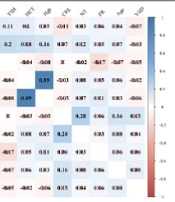
Nassiri S, Hantoushzadeh S, Eshraghi N, Ghaemi M, Panahi Z. Are Fetal Nuchal Translucency Thickness and Crown-rump Length Associated with Maternal Hematological and Biochemical Profile?. Fertil Gynecol Androl. 2023;3(1):e138371. doi: https://doi.org/10.5812/fga-138371
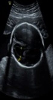
Uzun I, Sayin C, Erzincan S, Inan C, Sutcu H, et al. Analysis of Antenatal Sonographic Features of the Fetuses with Trisomy 21. I J Radiol. 2018;15(2):e62473. doi: https://doi.org/10.5812/iranjradiol.62473
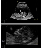
Dorostghoal M, Barati M, Harfsheno M. Combined First-trimester Screening of Trisomy 21 in Women of Different Ages in Southwest Iran. Gene Cell Tissue. 2022;9(3):e113112. doi: https://doi.org/10.5812/gct.113112
Zare Mehrjardi M, Keshavarz E, Sanei Taheri M. Normative Values of Intracranial Translucency (IT) Thickness as an Ultrasonographic Marker for Neural Tube Defect (NTD) Screening in the Iranian Singleton Pregnancies. I J Radiol. 2017;14(Special Issue):e48210. doi: https://doi.org/10.5812/iranjradiol.48210
Last Update: 3 weeks ago
Last Update: 3 weeks ago
Last Update: 3 weeks ago
Last Update: 3 hours ago
Last Update: 6 hours ago
Last Update: 4 days ago