Journal of Kermanshah University of Medical Sciences
Peer-reviewed Medical Quarterly
e-ISSN 2588-2570
p-ISSN 2588-2562
Outline
The comparison of CT-scan and laboratory measures in the assessment of tibial torsion
Authors
1Dept. of Physical Education and Sport Science, School of Literature and Humanities, Bu Ali University, Hamedan, Iran
2Dept. of Orthopaedic, School of Medicine, Kermanshah University of Medical Science, Kermanshah, Iran
3Dept. of Public Health, School of Health, Kermanshah University of Medical Science, Kermanshah, Iran
4Dept. of Sport Physiology, School of Physical Education, Razi University, Kermanshah, Iran
*Corresponding Author: Corresponding author: Farzaneh Gandomi, Dept. of Physical Education and Sport Science, School of Literature and Humanities, Bu Ali University, Hamedan, Iran, Tel: +989185697032 Email: [email protected]
Published online:Jan 29, 2013
Article type:Research Article
Received:Dec 21, 2011
Accepted:Apr 03, 2012
How to Cite:Gandomi F, Saeb M, Karami Matin B, Behpoor N. The comparison of CT-scan and laboratory measures in the assessment of tibial torsion. J Kermanshah Univ Med Sci. 2013;16(6):e77338. doi:
Abstract
Background: Tibial torsion is one of the torsion density which rotating in the longitudes axis of the leg. To measure this deformity, different methods such as radiographic or clinical methods were presented and introduced.
Methods: In this quasi-experimental study, 30 male athletes were selected randomly. Their tibial torsion angle was measured using CT scan, Lavand, Lokomso, Foot print and thigh-maleoli methods. To analyze the data , one way ANOVA and LSD test were used.
Results: The results of this study showed that there was a significant difference between CT scan and Lavand, CT scan and Lokomso, CT scan and Foot print, CT scan and tigh-maleolli angels, while no significant difference was obtained using laboratory methods.
Conclusion: The results of this study showed that though there existed significant differences between laboratory and CT scan methods, laboratory methods did not have reliability and validity for measuring tibial torsion, and it can only be used for the recognition of tibial torsion.
Highlights
References
- 1.The references of this article is available on PDF.
Copyright
© 2013, Journal of Kermanshah University of Medical Sciences. This open-access article is available under the Creative Commons Attribution-NonCommercial 4.0 (CC BY-NC 4.0) International License (https://creativecommons.org/licenses/by-nc/4.0/), which allows for the copying and redistribution of the material only for noncommercial purposes, provided that the original work is properly cited.
Similar Articles
12
Apr
2017
The Normal Value of Tibial Tubercle Trochlear Groove Distance in Patients with Normal Knee Examinations Using MRI
Reza Nafisi Moghadam,
Zoheira Arefmanesh
Nafisi Moghadam R, Arefmanesh Z. The Normal Value of Tibial Tubercle Trochlear Groove Distance in Patients with Normal Knee Examinations Using MRI. I J Radiol. 2017;14(Special Issue):e48000. doi: https://doi.org/10.5812/iranjradiol.48000
31
Dec
2011
Non Invasive Estimation of Left VentrNon Invasive Estimation of Left Ventricular Normalized Torsion Angle in Healthy Persons by Echo Tracking Algorithm: Short Axis Viewicular Normalized Torsion Angle in Healthy Persons by Echo Tracking Algorithm: Short Axis View
Zahra Arab Baferani,
Manijhe Mokhtari-Dizaji,
Farideh Roshanali
Arab Baferani Z, Mokhtari-Dizaji M, Roshanali F. Non Invasive Estimation of Left VentrNon Invasive Estimation of Left Ventricular Normalized Torsion Angle in Healthy Persons by Echo Tracking Algorithm: Short Axis Viewicular Normalized Torsion Angle in Healthy Persons by Echo Tracking Algorithm: Short Axis View. Int Cardiovasc Res J. 2017;5(4):e11214. doi:
30
Dec
2005
CT Patellar Cortex Tilt Angle: A Radiological Method to Measure Patellar Tilt
Fardin Mirza Tolouei
Mirza Tolouei F. CT Patellar Cortex Tilt Angle: A Radiological Method to Measure Patellar Tilt. I J Radiol. 2005;3(1):e79071. doi:
14
Apr
2020
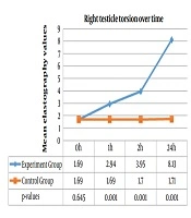
Does Elastography Have a Place in Evaluating Tissue Stiffness in Testicular Torsion?
Kadir Oymen Hancerliogullari
Hancerliogullari KO. Does Elastography Have a Place in Evaluating Tissue Stiffness in Testicular Torsion?. I J Radiol. 2020;17(2):e99758. doi: https://doi.org/10.5812/iranjradiol.99758
2
Feb
2013
Assessment of Left Ventricular Torsion in Short Axis View between Healthy Subjects and Significant Coronary Artery Disease Patients
Zahra Arab,
Manijhe Mokhtari-Dizaji,
Faride Roshanali,
Hassan Moladoust
Arab Z, Mokhtari-Dizaji M, Roshanali F, Moladoust H. Assessment of Left Ventricular Torsion in Short Axis View between Healthy Subjects and Significant Coronary Artery Disease Patients. Zahedan J Res Med Sci. 2013;15(9):e92857. doi:
Cited by
- Scopus by DOI: 0
Last Update: 3 weeks ago
- Scopus by Title: 0
Last Update: 3 weeks ago
- Scopus by Title (Ref): 0
Last Update: 3 weeks ago
- CrossRef: 0
Last Update: 1 day ago
Comments
Number of Comments:0