Cover image of the article: CT Patellar Cortex Tilt Angle: A Radiological Method to Measure Patellar Tilt

Image Credit:I J Radiol

Outline

CT Patellar Cortex Tilt Angle: A Radiological Method to Measure Patellar Tilt

Authors

Fardin Mirza Tolouei1,*
1Assistant professor, Department of Orthopedic Surgery, Motahari Hospital, Urmia University of Medical Sciences, Urmia, Iran
*Corresponding Author: Corresponding author: Fardin Mirza Tolouei, Assistant professor, Department of Orthopedic Surgery, Motahari Hospital, Urmia University of Medical Sciences, Urmia, Iran, Tel: +98-4412237077, Fax: +98-4412234125 Email: fardin_tolouei@yahoo.com

Iranian Journal of Radiology:Vol. 3, issue 1; e79071
Published online:Dec 30, 2005
Article type:Research Article
Received:May 07, 2005
Accepted:Dec 06, 2005
How to Cite:Mirza Tolouei F. CT Patellar Cortex Tilt Angle: A Radiological Method to Measure Patellar Tilt. I J Radiol. 2005;3(1):e79071. doi:

Fulltext

The authors' name, abstract and full text are available in PDF.

Copyright

© 2005, Author(s). This open-access article is available under the Creative Commons Attribution 4.0 (CC BY 4.0) International License (https://creativecommons.org/licenses/by/4.0/), which allows for unrestricted use, distribution, and reproduction in any medium, provided that the original work is properly cited.

Similar Articles

12
Apr
2017
The Normal Value of Tibial Tubercle Trochlear Groove Distance in Patients with Normal Knee Examinations Using MRI

The Normal Value of Tibial Tubercle Trochlear Groove Distance in Patients with Normal Knee Examinations Using MRI

Reza Nafisi Moghadam,
Zoheira Arefmanesh

Nafisi Moghadam R, Arefmanesh Z. The Normal Value of Tibial Tubercle Trochlear Groove Distance in Patients with Normal Knee Examinations Using MRI. I J Radiol. 2017;14(Special Issue):e48000. doi: https://doi.org/10.5812/iranjradiol.48000
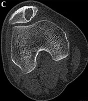
26
Sep
2020
Patellofemoral Morphology and Imaging Features of Patellar Tumors and Tumor-Like Lesions

Patellofemoral Morphology and Imaging Features of Patellar Tumors and Tumor-Like Lesions

Tian-Hao Wu,
Shu-Man Han,
Tao Sun,
Lei Cao,
Yun-Heng Shi,
Bao-Hai Yu
,et al.

Wu T, Han S, Sun T, Cao L, Shi Y, et al. Patellofemoral Morphology and Imaging Features of Patellar Tumors and Tumor-Like Lesions. I J Radiol. 2020;17(4):e94690. doi: https://doi.org/10.5812/iranjradiol.94690

29
Jan
2013
The comparison of CT-scan and laboratory measures in the assessment of tibial torsion

The comparison of CT-scan and laboratory measures in the assessment of tibial torsion

Farzaneh Gandomi,
Morteza Saeb,
Behzad Karami Matin,
Naser Behpoor

Gandomi F, Saeb M, Karami Matin B, Behpoor N. The comparison of CT-scan and laboratory measures in the assessment of tibial torsion. J Kermanshah Univ Med Sci. 2013;16(6):e77338. doi:
15
Aug
2004
A study of results of reconstructing posterior cruciate ligament using two-bundle graft from quadriceps muscle tendon

A study of results of reconstructing posterior cruciate ligament using two-bundle graft from quadriceps muscle tendon

Khalilollah Nazem,
Khashayar JabalAmeli,
AbdolReza Tavakoli

Nazem K, JabalAmeli K, Tavakoli A. A study of results of reconstructing posterior cruciate ligament using two-bundle graft from quadriceps muscle tendon. koomesh. 2004;5(3):e153724. doi:

28
Oct
2015
Effect of Quadriceps Exercise Training on Muscle Fiber Angle in Patients With Patellofemoral Pain Syndrome

Effect of Quadriceps Exercise Training on Muscle Fiber Angle in Patients With Patellofemoral Pain Syndrome

Roshanak Honarpishe,
Amir Hoshang Bakhtiary,
Gholamreza Olyaei

Honarpishe R, Bakhtiary AH, Olyaei G. Effect of Quadriceps Exercise Training on Muscle Fiber Angle in Patients With Patellofemoral Pain Syndrome. Middle East J Rehabil Health Stud. 2015;2(4):e32216. doi: https://doi.org/10.17795/mejrh-32216

More by these authors

Fardin Mirza ToloueiPubMedScholar
Share
Cited by
Metrics