A Forty-Six-Year-Old Man With Tracheomegaly and Bronchiectasis

Author(s):
Abbas FadaiiAbbas Fadaii1,*, Bahador BagheriBahador Bagheri2
1Department of Internal Medicine, Faculty of Medicine, Shahid Beheshti University of Medical Sciences, Tehran, IR Iran
2Department of Pharmacology, Faculty of Medicine, Semnan University of Medical Sciences, Semnan, IR Iran

Archives of Clinical Infectious Diseases:Vol. 7, issue 3; 102-3
Published online:Jul 02, 2012
Article type:Case Report
Received:Apr 06, 2012
Accepted:May 01, 2012
How to Cite:Fadaii A, Bagheri B. A Forty-Six-Year-Old Man With Tracheomegaly and Bronchiectasis. Arch Clin Infect Dis. 2012;7(3):102-3. doi: https://doi.org/10.5812/archcid.14644

Abstract

1. Introduction

Mounier-Kuhn Syndrome is a rare and genetic disorder which is described by dilatation of the trachea and the main bronchus (1, 2). The patients suffering from this disease mainly complain about chronic coughs, purulent sputum and recurrent infection of the lower respiratory tract (3). It was not until 1932 that Mounier-Kuhn Syndrome was first described. It usually initiates primarily with no specific cause hitherto. Autopsy findings suggest a congenital defect or atrophy of elastic tissues or smooth muscles of the trachea and main bronchus (3-5). Moreover, tracheobronchomegaly is also described in association with Ehlers-Danlos syndrome, Marfan syndrome, Kenny-Caffey syndrome, and Brachmann-de Lange syndrome (6, 7). However, the disease typically discloses sporadically. There exists a male predominance , additionally patients are usually adults (1). The symptoms of tracheomegaly are sputum production due to bronchiectasis and infection of the lower respiratory tract. The blockade in mucociliary clearance and inefficient cough mechanism are likely to result in recurrent pneumonia, bronchiectasis, and fibrosis in airways which are markedly dilated. Patients may develop dyspnea and respiratory failure due to extensive damage to the lungs. Furthermore, pneumothorax, hemoptysis and finger clubbing may develop (4, 8). The diagnosis of syndrome is based on chest roentgenogram or CT scan. In females, the transverse diameter of trachea exceeding 21mm and the diameter of the right and left main bronchus exceeding 19.8 mm and 17.4 mm is suggestive for Mounier-Kuhn syndrome. In males, the cut-off is 25 mm for trachea, 21.1 mm and 18.4 mm for right and left main bronchus, respectively (9, 10).

2. Case Presentation

A forty-six-year-old man was referred to Sasan Hospital, Tehran, Iran to evaluate a chronic cough which has been going on for 20 years. He complained about mucoid sputum which was sometimes purulent and bloody. He is a farmer who does not smoke. He denied fever, dyspnea, or weight loss. He had no history of admission, allergy or systemic diseases. There was also no history of respiratory diseases, TB or similar symptoms in his family. Physical examinations proved that the patient was conscious and well-nourished without respiratory distress. The vital signs were normal whereas respiratory rate was 25/min. Respiratory examination revealed crackles at both lungs which was changed by coughing. Finger clubbing was not found. Chest X-ray (Figure 1) and Chest CT scans (Figure 2 and 3) were performed. The results showed tracheomegaly with dramatic increase in the diameters of the trachea and a modest increase in the diameters of the right and left-main bronchi.
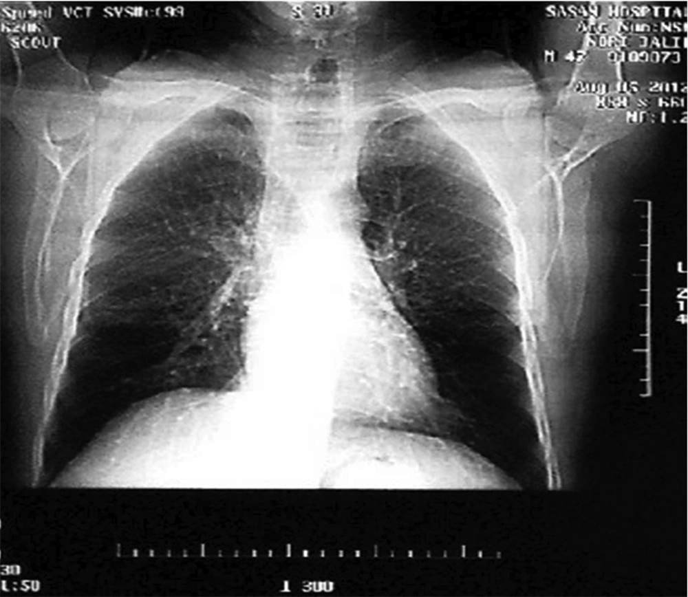
Chest X-Ray Shows Dilatation of the Trachea and Bronchomegaly
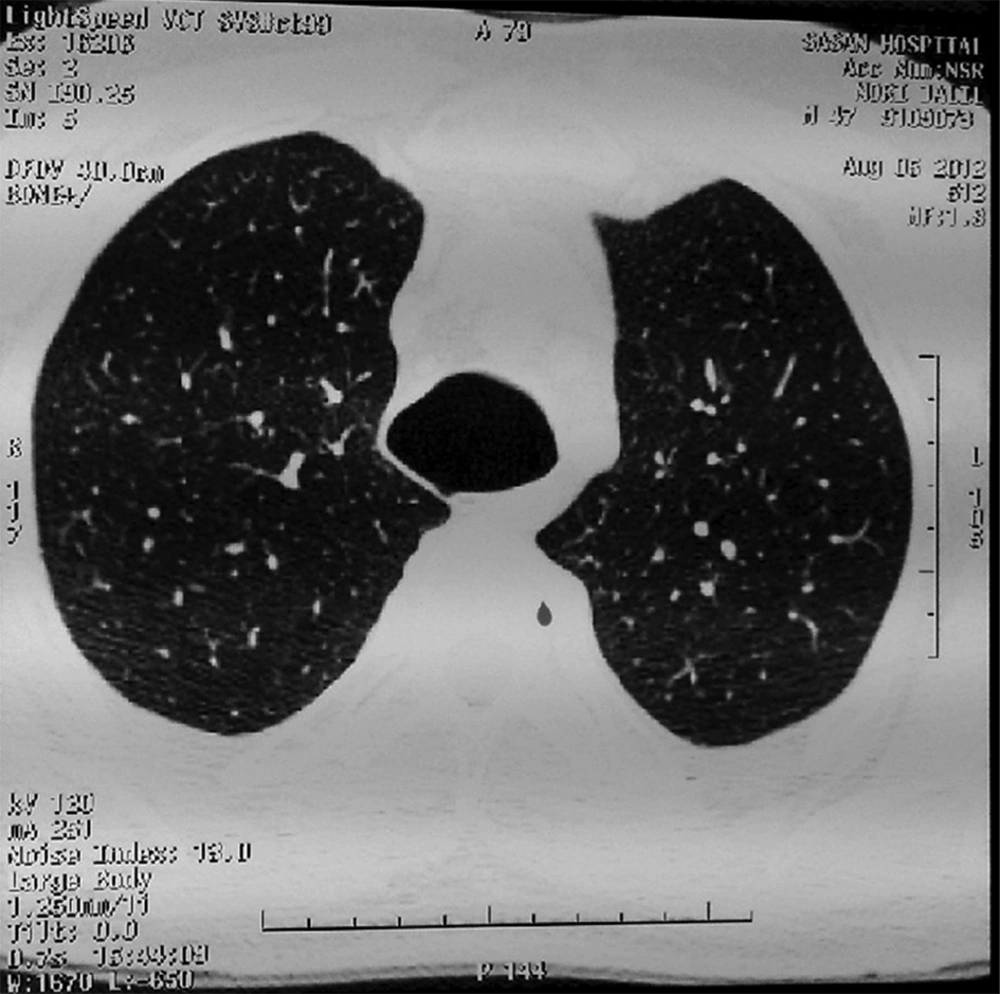
Figure 1.

Chest X-Ray Shows Dilatation of the Trachea and Bronchomegaly

Chest CT Scan in Axial Plane Shows Tracheomegaly
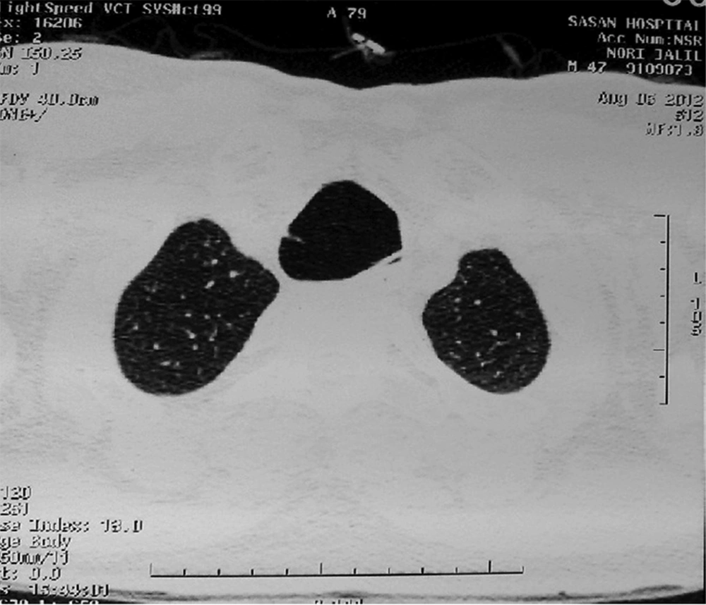
Figure 2.

Chest CT Scan in Axial Plane Shows Tracheomegaly

Chest CT Scan Shows Bronchomegaly
Figure 3.

Chest CT Scan Shows Bronchomegaly

Mild bronchiectasis was also present in right lung. Body plathysmogrophy disclosed a slight decrease in pulmonary volumes with a restrictive pattern. A bronchoscopy was performed and revealed complete atrophy of the trachea membranes. No diverticular outpunching was found. Sputum and tracheal samples were negative for TB.

3. Conclusions

There is currently no effective therapy for this disease. However, treatment is chiefly supportive to relieve dyspnea and other exhausting symptoms. Asymptomatic patients require no specific therapy while symptomatic patients have need of antibiotic therapy and postural drainage. Besides, bronchoscopy and tracheostomy can be used for the management of secretions. Last but not least, it should be pointed out that patients with marked respiratory collapse may undergo endothelial stenting (10, 11).

Acknowledgments

Footnotes

References

  • 1.
    Bateson EM, Woo-Ming M. Tracheobronchomegaly. Clin Radiol. 1973;24(3):354-8.
  • 2.
    Gay S, Dee P. Tracheobronchomegaly--the Mounier-Kuhn syndrome. Br J Radiol. 1984;57(679):640-4. [PubMed ID: 6733410].
  • 3.
    Shin MS, Jackson RM, Ho KJ. Tracheobronchomegaly (Mounier-Kuhn syndrome): CT diagnosis. AJR Am J Roentgenol. 1988;150(4):777-9. [PubMed ID: 3258088]. https://doi.org/10.2214/ajr.150.4.777.
  • 4.
    Bourne TM, Raphael JH, Tordoff SG. Anaesthesia for a patient with tracheobronchomegaly (Mounier-Kuhn syndrome). Anaesthesia. 1995;50(6):545-6. https://doi.org/10.1111/j.1365-2044.1995.tb06049.x.
  • 5.
    Johnston RF, Green RA. Tracheobronchiomegaly. Report of Five Cases and Demonstration of Familial Occurrence. Am Rev Respir Dis. 1965;91:35-50. [PubMed ID: 14259999].
  • 6.
    Blake MA, Clarke PD, Fenlon HM. Thoracic case of the day. Mounier-Kuhn syndrome (tracheobronchomegaly). AJR Am J Roentgenol. 1999;173(3):822-5. [PubMed ID: 10470941]. https://doi.org/10.2214/ajr.173.3.10470941.
  • 7.
    Sane AC, Effmann EL, Brown SD. Tracheobronchiomegaly. The Mounier-Kuhn syndrome in a patient with the Kenny-Caffey syndrome. Chest. 1992;102(2):618-9. [PubMed ID: 1643956].
  • 8.
    Parris WC, Johnson AC. Tracheomegaly. Anesthesiology. 1982;56(2):141-3. [PubMed ID: 7055325].
  • 9.
    Messahel FM. Tracheal dilatation followed by stenosis in Mounier–Kuhn syndrome. Anaesthesia. 1989;44(3):227-9. https://doi.org/10.1111/j.1365-2044.1989.tb11229.x.
  • 10.
    Woodring JH, Barrett PA, Rehm SR, Nurenberg P. Acquired tracheomegaly in adults as a complication of diffuse pulmonary fibrosis. AJR Am J Roentgenol. 1989;152(4):743-7. [PubMed ID: 2784256]. https://doi.org/10.2214/ajr.152.4.743.
  • 11.
    Collard P, Freitag L, Reynaert MS, Rodenstein DO, Francis C. Respiratory failure due to tracheobronchomalacia. Thorax. 1996;51(2):224-6. [PubMed ID: 8711665].

Similar Articles

16
May
2016

Tracheal Bronchus Associated With Unilateral Absence of Pulmonary Artery and Recurrent Pneumonia

Iraj Sedighi,
Mehrnaz Olfat,
Neda Pak,
Maryam Nasr Esfahany

Sedighi I, Olfat M, Pak N, Nasr Esfahany M. Tracheal Bronchus Associated With Unilateral Absence of Pulmonary Artery and Recurrent Pneumonia. J Compr Ped. 2016;7(2):e37092. doi: https://doi.org/10.17795/compreped-37092

14
Dec
2013

Case of Tracheal Diverticulum and Bronchiectasis

Payam Mehrian,
Ali Cheraghvandi,
Firouzeh Talischi,
Saeid Fallah-Tafti

Mehrian P, Cheraghvandi A, Talischi F, Fallah-Tafti S. Case of Tracheal Diverticulum and Bronchiectasis. Zahedan J Res Med Sci. 2015;17(1):e1945. doi:

1
Jun
2013

Mucoepidermoid Carcinoma (MEC): A Rare Cause of Chronic Cough in Children

Soheila Khalilzadeh,
Mohammad Reza Boloursaz,
Shirin Karimi,
Maryam Hassanzad,
Yahya Jand,
Seyed Reza Saghebi
,et al.

Khalilzadeh S, Boloursaz MR, Karimi S, Hassanzad M, Jand Y, et al. Mucoepidermoid Carcinoma (MEC): A Rare Cause of Chronic Cough in Children. J Compr Ped. 2013;4(3):151-4. doi: https://doi.org/10.17795/compreped-10202

9
Mar
2023
Iran J Pediatr

An 11-Year-Old Boy with Dry Cough and Respiratory Distress (from Children’s Medical Center Grand Rand)

Narges Khazaei,
Mansoureh Shariat,
Mohammad Effatpanah,
Mohammad Ali Pourmirzaiee,
Mehrnaz Olfat,
Seyed Hossein Mirlohi

Khazaei N, Shariat M, Effatpanah M, Pourmirzaiee MA, Olfat M, et al. An 11-Year-Old Boy with Dry Cough and Respiratory Distress (from Children’s Medical Center Grand Rand). Inn J Pediatr. 2023;33(1):e127928. doi: https://doi.org/10.5812/ijp-127928

13
Mar
2024
J Compr Ped

A Twelve-Year-Old-Girl with Intra-Tracheal Mucous Gland Adenoma: A Case Report

Ghamartaj Khanbabaee,
Ahmad Safapour,
Zahra Taghizadeh Herat,
Mohammad Hadi Mohseni

Khanbabaee G, Safapour A, Taghizadeh Herat Z, Mohseni MH. A Twelve-Year-Old-Girl with Intra-Tracheal Mucous Gland Adenoma: A Case Report. J Compr Ped. 2024;15(2):e143518. doi: https://doi.org/10.5812/jcp-143518


Crossmark
Crossmark
Checking
Share on
Cited by
Metrics

Ordering Reprints

Articles are published under the Creative Commons license stated on each article. No permission or royalty fee is required for uses permitted by that license. CCC handles optional bulk and customized reprint orders. Any quotation covers production and delivery services only, not copyright permission. > Request Reprints from CCC
