Cover image of the article: Photo Quiz: Tapeworm in Stomach

Image Credit:Arch Clin Infect Dis

Photo Quiz: Tapeworm in Stomach

Authors

Mohsen Masoudi1, Zahedin Kheyri1, Ali Ali-Asgari2,*
1Division of Gastroenterology, Hazrat-e-Rasool Hospital, Tehran University of Medical Sciences, Tehran, IR Iran
2Department of Internal Medicine, Shariati Hospital, Tehran University of Medical Sciences, Tehran, IR Iran
*Corresponding Author: Corresponding author: Ali Ali-Asgari, Shariati Hospital, Tehran, IR Iran. Tel.: +98-9123360254, Fax: +98-2184902366, E-mail: Email: aliasgari@tums.ac.ir

Archives of Clinical Infectious Diseases:Vol. 7, issue 3; 104-5
Published online:Jul 02, 2012
Article type:Letter
Received:May 08, 2012
Accepted:Jun 01, 2012
How to Cite:Masoudi M, Kheyri Z, Ali-Asgari A. Photo Quiz: Tapeworm in Stomach. Arch Clin Infect Dis. 2012;7(3):104-5. doi: https://doi.org/10.5812/archcid.14471

Dear Editor,
A fifty-four-year-old woman was admitted to emergency department with a two-week history of dyspepsia, abdominal pain, and nausea. For two months she had experienced intermittent episodes of dyspepsia, nausea and vomiting. Her abdominal pain was described as epigastric, non-radiating, worse after eating, and not relieved by defecation. She never had jaundice, hematemesis, melena, hematochezia, or tenesmus. During general physical examination, her vital signs were stable. No oropharyngeal lesion was observed. The respiratory and cardiovascular examinations were unremarkable. Her abdomen was soft and not tender in epigastrium. No free fluid or organomegaly was appreciated. No peritoneal signs were elicited. Her digital rectal exam was also inconspicuous. Given routine lab tests, the patient was found to be anemic with borderline microcytosis. Her white blood cell and differential count were normal. Her urinalysis did not show any sign of urinary tract infection. Amylase and lipase were within normal limits. Transaminases were normal. An abdominal X-ray was taken and showed no evidence of obstruction or perforation. Stool analysis (e g, stool cultures, ova and parasite examination) was negative. The patient later underwent esophagogastroduodenoscopy, as shown in the image which is taken from her stomach (Figure 1).
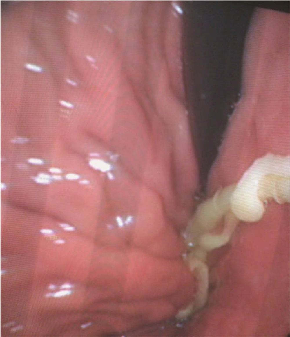
Tapeworm in Stomach shown in Upper Gastrointestinal Endoscopy
Figure 1.
Tapeworm in Stomach shown in Upper Gastrointestinal Endoscopy
Humans are the only definitive hosts for Taenia saginata (T. saginata). Eggs or gravid proglottids are passed through human stool. Cattles become infected by ingesting vegetation, contaminated with eggs or gravid proglottids. In animal’s intestine, the oncospheres hatch, invade the intestinal wall, and migrate hematogenously to striated muscles, where they develop in cysticerci. Humans become infected by ingesting raw or undercooked infected meat containing cysticerci. In human intestine, protoscolices are released from the cysts and attached to the intestinal wall via suckers and hooks (1). The prevalence of taeniasis in Iran has been disregarded in recent years - estimated to be about 0.5% in Mazandaran province - where it appears to have the highest prevalence rate (2). Tapeworm infections are usually asymptomatic, however mild abdominal pain or discomfort, nausea, change in appetite, weakness, and weight loss can also occur (3). It is usually diagnosed by detection of the eggs or proglottids in stool. Only one other report of taeniasis diagnosed on upper gastrointestinal endoscopy was found (4). A single dose of Praziquantel (10 mg/kg) is highly effective for the treatment of taeniasis. The patient was prescribed Praziquantel 600 mg and a month later at the follow-up visit, she was reported to have no symptoms.

Acknowledgments

Footnotes

  • Financial Disclosure:The author declares that there is no conflict of interest.

References

  • 1.
    Leder K, Weller PF. Intestinal tapeworms. In: Basow DS, editor. Up To Date. Waltham, MA; 2013.
  • 2.
    Kia EB, Masoud J, Yalda A, Mahmoudi M, Farahani H. Study on Human Taeniasis by Administring Anti-Taenia Drug. Iran J Public Health. 2005;34(4):47-50.
  • 3.
    White AC, Jr, Weller PF. Cestode Infections. In: Longo DL, editor. Harrison's principles of internal medicine. 18 ed. New York: McGraw Hill; 2012.
  • 4.
    Liao WS, Bair MJ. Images in clinical medicine. Taenia in the gastrointestinal tract. N Engl J Med. 2007;357(10):1028. [PubMed ID: 17804847]. https://doi.org/10.1056/NEJMicm067761.

Copyright

Copyright © 2012, Infectious Diseases and Tropical Medicine Research Center. This is an open-access article distributed under the terms of the Creative Commons Attribution-NonCommercial 4.0 International License (http://creativecommons.org/licenses/by-nc/4.0/) which permits copy and redistribute the material just in noncommercial usages, provided the original work is properly cited.

Similar Articles

25
Feb
2011
Celiac disease in dyspeptic patients

Celiac disease in dyspeptic patients

Mohammad Rostami-Nejad,
Hamid Mahbobipor,
Zeynab Fazeli,
Reza Mashayekhi,
Daryosh MirSatari,
Ehasn NazemolHoseini
,et al.

Rostami-Nejad M, Mahbobipor H, Fazeli Z, Mashayekhi R, MirSatari D, et al. Celiac disease in dyspeptic patients. koomesh. 2011;12(2):e153818. doi:

10
Jun
2013
Comparison the Sensitivity of Stomach Mucus Touching Cytology and Urease Rapid Test in Helicobacter Pylori Diagnosis in Endoscopies Patients with Gastritis or Peptic Ulcer

Comparison the Sensitivity of Stomach Mucus Touching Cytology and Urease Rapid Test in Helicobacter Pylori Diagnosis in Endoscopies Patients with Gastritis or Peptic Ulcer

Mohammad Jafari,
Alireza Khalilian,
Farshad Rostampour

Jafari M, Khalilian A, Rostampour F. Comparison the Sensitivity of Stomach Mucus Touching Cytology and Urease Rapid Test in Helicobacter Pylori Diagnosis in Endoscopies Patients with Gastritis or Peptic Ulcer. Jundishapur J Microbiol. 2013;6(4):e7446. doi: https://doi.org/10.5812/jjm.7446

30
Aug
2007
Atypical Radiographic Manifestations of Duodenal Diverticula: A Pictorial Essay

Atypical Radiographic Manifestations of Duodenal Diverticula: A Pictorial Essay  

Leila Kiani,
S. Rad

Kiani L, Rad S. Atypical Radiographic Manifestations of Duodenal Diverticula: A Pictorial Essay  . I J Radiol. 2007;4(3):e79102. doi:

31
Jan
2011

Helicobacter Pylori Infection: A Review

Nasser Ebrahimi Daryani,
Mohammad Taher,
Samira Shirzad

Ebrahimi Daryani N, Taher M, Shirzad S. Helicobacter Pylori Infection: A Review. Arch Clin Infect Dis. 2011;6(1):. doi:

17
Mar
2011
Assessment clinical features of gastro esophageal reflux diseases in infants

Assessment clinical features of gastro esophageal reflux diseases in infants

Shahla Behramand,
Noshin Jalili,
Esmaeil Mohammadnejad,
Jiran Zebardast,
Azadeh Khatibi

Behramand S, Jalili N, Mohammadnejad E, Zebardast J, Khatibi A. Assessment clinical features of gastro esophageal reflux diseases in infants. Zahedan J Res Med Sci. 2012;13(10):e93887. doi:

More by these authors

Mohsen MasoudiPubMedScholar
Zahedin KheyriPubMedScholar
Ali Ali-AsgariPubMedScholar
Share
Cited by
Metrics