Full Text
Full text is available in PDF
Asian Journal of Sports Medicine
Sports Medicine Research Center, TUMS
Authors
The study of the right ventricular (RV) chamber has been recently improved by use of the 3D method. In young asymptomatic trained athletes, RV is not routinely investigated if not in suspected disease. This study is designed to test if the 3D method compared to 2D, adds information to study of RV morphology and function, in the early stages of myocardial remodeling of a group of young athletes.
The RV chamber function was assessed in 25 young trained athletes (20 soccer and 5 basketball) aged 203 yrs and compared to 20 sedentary controls by 2D-AC and 3D-RV methods (TomTec), measuring RV diastolic (RVDV) and systolic (RVSV) volumes or ejection fraction (EF).
3D RV volumes were slightly higher in athletes than the 2D volumes in presence of lower EF values, but not significantly higher than in sedentary. Significant differences were conversely found comparing 3D systolic and diastolic RV volumes and 2D-AC volumes within each group with higher values in athletes (RVDV:
Although the found EF values were substantially similar, the morphological assessment of the initial modifications of this chamber of the young athlete's heart, results seem to be more accurate using the 3D method than 2D. The clinical implication of this aspect could be of interest in case of difficulty in drawing a clear diagnosis of any RV chamber disease in young athletes.
© 2013, Author(s). This open-access article is available under the Creative Commons Attribution 4.0 (CC BY 4.0) International License (https://creativecommons.org/licenses/by/4.0/), which allows for unrestricted use, distribution, and reproduction in any medium, provided that the original work is properly cited.
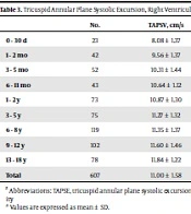
Yılmaz M, Çetin M, Dundar PE, Coşkun Ş. Normal Values of Echocardiographic Parameters Indicating Right Ventricular Systolic Functions in 607 Healthy Children. Inn J Pediatr. 2023;33(2):e134196. doi: https://doi.org/10.5812/ijp-134196
Karaca O, Avcı A, Babur Güler G, Omaygenc O, Cakal B, et al. Predictors of Right Ventricular Systolic Dysfunction in Non-Ischemic Dilated Cardiomyopathy: An Echocardiographic Study. Int Cardiovasc Res J. 2016;10(1):e10307. doi:
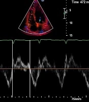
Parsaee M, Amin A, Mombeini H, Khansari N. Right Ventricular Function in Heart Failure Patients with Normal Left Ventricular Size: Analysis by Conventional Echocardiographic Parameters and Magnetic Resonance Imaging. Multidiscip Cardio Annal. 2019;10(2):e95507. doi: https://doi.org/10.5812/mca.95507
Sadeghpour A, Harati H, Kiavar M, Esmaeizadeh M, Maleki M, et al. Correlation of Right Ventricular dP/dt with Functional Capacity and RV Function in Patients with Mitral Stenosis. Int Cardiovasc Res J. 2007;1(4):e69664. doi:
Abtahi F, Bazyar Z, Zarrabi K, Hassanzadeh M, Mehdipour Namdar Z, et al. A Prospective Study of Right Ventricular Function After Coronary Artery Bypass Grafting: Are Usual Echocardiographic Parameters Useful?. Int Cardiovasc Res J. 2024;18(1):e146326. doi: https://doi.org/10.5812/icrj-146326
Last Update: 1 week ago
Last Update: 1 week ago
Last Update: 1 week ago
Last Update: 1 hour ago
Last Update: 8 hours ago
Last Update: 5 days ago