Fulltext
Discussion
Initially, he was referred to our clinic only due to elevation in Alk-Phos and liver aminotransferases from which he had been suffering for more than 3 years. Liver smears showed hepatic granulomas with significant fibrosis. In this case, we will discuss the causes of hepatic granulomas that are associated with significant liver fibrosis and portal hypertension.
Differential Diagnosis:
Primary Biliary Cirrhosis (PBC): PBC is characterized by a T-lymphocytemediated attack on small intralobular bile ducts(1). PBC typically presents in middle life and women are about ten times as commonly affected as men(2). Some of the patients are asymptomatic at the time of diagnosis; however, symptoms will appear with progression of hepatic disease over time (3). Fatigue and pruritis are the most common presenting symptoms of PBC. Jaundice is usually a late symptom that heralds the onset of the advanced disease (4). Physical examination may show hyperpigmentation, hepatosplenomegaly and osteopenia (5, 6). The most typical biochemical abnormality in PBC is an elevated AlkPhos. The finding of AMA seropositivity is highly sensitive (98%) as a diagnostic test (2). The diagnosis of PBC should be confirmed by percutaneous liver biopsy. Also, liver biopsy is needed to determine the stage of the disease.
The early stages of PBC are characterized by portal hepatitis, granulomas and bile-duct lesions. Hepatic granulomas tend to be located in portal tracts. The progressive stage of PBC is dominated by fibrosis spanning between portal tracts and also ductopenia. In addition, the inflammatory features described with early stages are often seen (7, 8, 9). The histological findings of present case can suggest PBC, but pulmonary involvement and mediastinal lymph node enlargement are not seen in PBC. Furthermore, patients with progressive lesions of PBC are usually symptomatic, but our patient was asymptomatic. Also, AMA test for this case was negative. Thus, it seems that PBC is an unlikely diagnosis.
Tuberculosis:
Hepatic granulomas have been described in about 90 percent of patients with miliary tuberculosis, 75 percent of cases with extra pulmonary tuberculosis and 25 percent of patients with pulmonary tuberculosis (10). Also, liver may be primarily involved (11). Most of the patients are symptomatic for one to two years prior to time of diagnosis. Symptoms include fever, night sweats, fatigue, anorexia and weight loss(11). Rarely, may splenomegaly, ascites and symptoms of liver failure be seen (12). In miliary tuberculosis, hepatic granulomas are distributed throughout the liver parenchyma, but in other forms of hepatic tuberculosis, granulomas are characteristically located in periportal and portal areas (13, 14). Hepatic granulomas may or may not caseate and acid-fast organisms can occasionally be found within the granulomas (14). The presence of hepatic granulomas in a patient would suggest tuberculosis. In the current case; however, tuberculosis is an unlikely diagnosis for many reasons.
First, there was no fever, fatigue, weight loss or respiratory symptoms in spite of severe liver damage and pulmonary involvement. Hepatic granulomas and significant liver fibrosis may be found in the end stage of tuberculous hepatitis, but bilateral hilar and mediastinal lymphadenopathy are not consistent with post primary tuberculosis.
Furthermore, there were no caseation or acid bacilli in liver smears and PPD test was negative.
Drugs:
Many drugs have been associated with noncaseating hepatic granulomas (15). These granulomas are usually located in periportal and portal areas. Some of these drugs can be associated with progressive liver injury and subsequent cirrhosis (16). However, this patient had not received any drugs for the past 10 months.
Sarcoidosis:
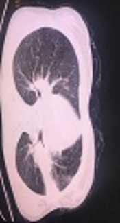
Sarcoidosis is a multisystem granulomatous disorder of unknown etiology and is characterized by the presence of noncaseating granulomas in involved organs (17). Sarcoidosis most frequently involves lungs- in over 90 percent of patients with Sarcoidosis (18). The classic chest roentgenogram reveals bilateral hilar adenopathy. This finding may occur in combination with parenchymal infiltrates, depending upon the stage of the disease. The serum angiotensine converting enzyme level is elevated in 75 percent of untreated patients with Sarcoidosis(19). Hepatic involvement is usually silent. The clinical consequences of the liver involvement are variable ranging from asymptomatic biochemical abnormalities (usually a mildly elevated Alk-Phos and GGT to cholestatic liver disease and cirrhosis(15). These patients may present with FUO. Hepatic granulomas are characteristically well-formed and non-caseating(15). They tend to be in portal and periportal regions. Some of them heal, but leave fibrosis and scarring. A component of lobular hepatitis and portal triaditis may be present. Cirrhosis may develop because of scarring of granulomas. Similar to PBC, destruction of intralobular bile ducts may be seen (20, 21, 22).In the present case, hepatic noncaseating granulomas and significant fibrosis would suggest sarcoidosis. Granulomas of sarcoidosis are predominantly located in portal area but can be seen in lobular region. Our patient had lobular granulomas proliferation of bile ducts that was seen in this case, is not specific for PBC and can be found in sarcoidosis. In addition to this histologic feature, elevated ACE and CT scan findings (bilateral hilar and mediastinal lymph node enlargement and also pulmonary involvement) can confirm the diagnosis of sarcoidosis. Although, hepatic granulomas with bile duct lesions are most often seen in PBC extrahepatic findings exclude this diagnosis.
It is surprising that this case of sarcoidosis was presented to the clinician exclusively with liver enzyme abnormalities, but extensive investigation revealed a severe liver disease with pulmonary and mediastinal lymph node involvement.