


1. Background
2. Objectives
3. Patients and Methods
3.1. General Information and Patients’ Enrollment
3.2. Imaging Information
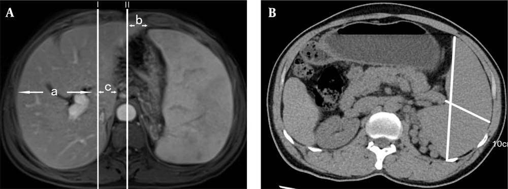
A, Contrast enhanced MRI, measurement of hepatic lobe atrophy and hyperplasia; B, Plain CT scan, measurements of the spleen length and thickness
3.3. Histopathology of the Liver
3.4. Serological Examination
3.5. Statistical Methods
4. Results
4.1. Summary of Patients' Clinical Baseline Data
| Parameter | Liver Cirrhosis (n = 30) | IPH (n = 14) | X2/t/Z | P-Value |
|---|---|---|---|---|
| Gender | 3.727 | 0.054 | ||
| Male | 20 (66.7) | 5 (35.7) | ||
| Female | 10 (33.3) | 9 (64.3) | ||
| Age | 49.47 ± 14.50 | 54.07 ± 8.88 | - 1.092 | 0.281 |
| DBIL (μmol/L) | 5.90 [4.03,7.70] | 4.35 [2.85,6.60] | - 1.55 | 0.121 |
| IBIL (μmol/L) | 14.90 [11.33,20.38] | 12.15 [7.88,25.92] | - 0.655 | 0.512 |
| TP (g/L) | 68.02 ± 7.40 | 63.71 ± 7.32 | 1.802 | 0.079 |
| ALB (g/L) | 38.29 ± 6.02 | 38.57 ± 5.41 | - 0.151 | 0.881 |
| GLB (g/L) | 29.73 ± 5.99 | 24.79 ± 3.72 | 2.836 | 0.007 |
| A/G Ratio | 1.34 ± 0.35 | 1.58 ± 0.28 | - 2.243 | 0.030 |
| ALT (U/L) | 32.50 [21.00,47.75] | 19.00 [17.00,25.75] | - 2.599 | 0.009 |
| AST (U/L) | 35.50 [24.50,44.25] | 24.00 [19.25,30.50] | - 1.992 | 0.046 |
| ALP (U/L) | 122.50 [101.50,147.75] | 98.50 [63.75,157.00] | - 1.222 | 0.222 |
| CHE | 5187.10 ± 2467.29 | 5039.71 ± 1280.86 | 0.261 | 0.796 |
| GGT (U/L) | 50.50 [35.25,112.50] | 31.00 [25.00,43.00] | - 2.521 | 0.012 |
| TBA (U/L) | 18.20 [7.20,53.27] | 6.55 [4.05,9.43] | - 2.797 | 0.005 |
| WBC (109 /L) | 4.30 [3.04,5.73] | 2.48 [2.06,3.45] | - 2.356 | 0.018 |
| ANC (109 /L) | 2.33 [1.94,3.70] | 1.83 [1.40,2.63] | - 1.399 | 0.162 |
| RBC (1012 /L) | 4.21 ± 0.85 | 3.69 ± 0.92 | 1.845 | 0.072 |
| PLT (s) | 80.50 [53.50,173.25] | 57.50 [38.25,125.00] | - 1.084 | 0.279 |
| PT (s) | 12.55 [11.85,13.70] | 12.55 [11.80,13.70] | - 0.088 | 0.930 |
| INR (s) | 1.19 [1.11,1.30] | 1.17 [1.11,1.27] | - 0.479 | 0.632 |
| APTT (s) | 30.30 [28.88,33.62] | 27.80 [26.13,32.75] | - 1.764 | 0.078 |
| TT (s) | 18.45 [17.40,19.48] | 16.25 [15.77,16.78] | - 4.335 | < 0.001 |
| FG (g/L) | 1.85 [1.57,2.37] | 2.40 [2.16,3.04] | - 2.848 | 0.004 |
Abbreviations: DBIL, direct bilirubin; IBIL, indirect bilirubin; TP, total protein; ALB, albumin ; GLB, globulin; A/G ratio, the albumin-globulin ratio; ALT, alanine aminotransferase ; AST, aspartate aminotransferase; ALP, alkaline phosphatase; CHE, cholinesterase; GGT, γ-glutamyl transferase ;TBA, total bile acid ; WBC, white blood cell; ANC, absolute neutrophil counts; RBC, red blood cell; PLT, blood platelet; PT, prothrombin time; INR, international normalized ratio; APTT, activated partial thromboplastin time; TT, thrombin time; FG, fibrinogen; IPH, idiopathic portal hypertension.
a One patient with cirrhosis after splenectomy.
b Values are expressed as No. (%), Mean ± SD or median [IQR].
c Normally or approximately normally distributed variables including: AGE, TP, ALB, GLB, A/G ratio, CHE, and RBC; Non-normally distributed variables including: DBIL, IBIL, ALT, AST, ALP, GGT, TBA, WBC, ANC, PLT, PT, INR, APTT, TT, and FG.
4.2. Summary of IPH Imaging Features
4.2.1. Comparison of the Quantitative Values of Hepatic Lobe Atrophy and Hyperplasia and Spleen Thickness and Length Between the IPH Group and the Cirrhosis Group
| Parameter | Liver Cirrhosis (n = 30) | IPH (n = 14) | Z/X2 | P-Value |
|---|---|---|---|---|
| Abnormal liver morphology | 21 (70) | 14 (100) | 3.597 | 0.058 |
| Portal vein thrombosis | 1 (3.3) | 1 (7.1) | - | 0.540 b |
| Arterial and venous phase perfusion abnormalities | 3 (10) | 5 (35.7) | 2.69 | 0.101 |
| Liver regenerative nodules | 8 (26.7) | 0 (0) | 2.946 | 0.086 |
| Focal nodular Hyperplasia-like Lesions | 0 (0) | 2 (14.3) | - | 0.096 b |
| Abnormal portal vein morphology | 9 (30) | 14 (100) | 18.748 | < 0.001 |
| Spleen-kidney shunt | 4 (13.3) | 1 (7.1) | 0.009 | 0.926 |
| Gastro-renal shunt | 2 (6.7) | 3 (21.4) | 0.86 | 0.354 |
| Spleen length (cm) | 10.20 [9.20,12.90] | 15.75 [12.55,17.15] | - 3.384 | 0.001 |
| Spleen thickness (cm) | 4.94 ± 0.95 | 6.31 ± 1.56 | - 3.042 | 0.007 |
Abbreviation: IPH, idiopathic portal hypertension.
a Values are expressed as No. (%), median (interquartile range) [M(IQR)] or Mean ± SD.
b Fisher's precision probability test; one patient with cirrhosis after splenectomy; Normally or approximately normally.
| Parameter | Liver Cirrhosis (n = 30) | IPH (n = 14) | t/Z | P-Value |
|---|---|---|---|---|
| Coronal diameter of the right lobe of the liver (mm) | 8.84 ± 0.98 | 8.91 ± 0.95 | -0.223 | 0.824 |
| Coronal diameter of the left lobe of the liver (mm) | 5.37 [4.04,7.96] | 3.32 [2.41,6.12] | -2.218 | 0.027 |
| Coronal diameter of the caudate lobe (mm) | 3.69 [3.34,3.80] | 2.90 [2.40,3.26] | -3.077 | 0.002 |
| Caudal right ratio | 0.42 [0.37, 0.44] | 0.31 [0.26, 0.39] | -2.642 | 0.009 |
Abbreviation: IPH, idiopathic portal hypertension.
a Values are expressed as Mean ± SD or median (interquartile range) [M(IQR)].
| Parameters | AUC | Cutoff | Sensitivity | Specificity | PPV | NPV |
|---|---|---|---|---|---|---|
| Coronal diameter of the left lobe of the liver | 0.710 | 3.205 | 0.867 | 0.500 | 0.788 | 0.267 |
| Coronal diameter of the caudate lobe | 0.790 | 3.440 | 0.700 | 0.857 | 0.913 | 0.350 |
| Caudal right ratio | 0.748 | 0.378 | 0.733 | 0.714 | 0.846 | 0.373 |
Abbreviations: AUC, area under the curve; PPV, positive predictive value; NPV, negative predictive value.
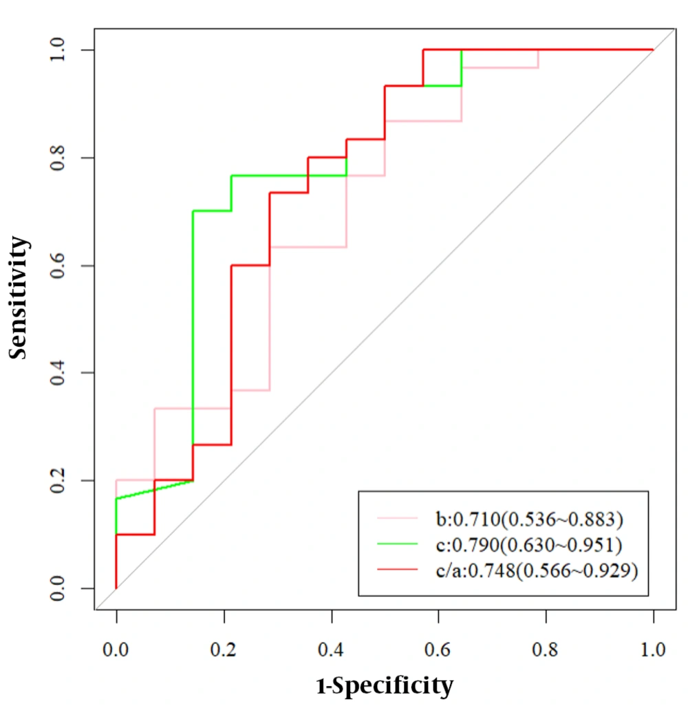
Receiver operating characteristic (ROC) curves for atrophy and hyperplasia of the liver for the diagnosis of Idiopathic portal hypertension (IPH)
4.2.2. Changes in Liver Morphology and Liver Parenchyma in the IPH and Cirrhosis Groups
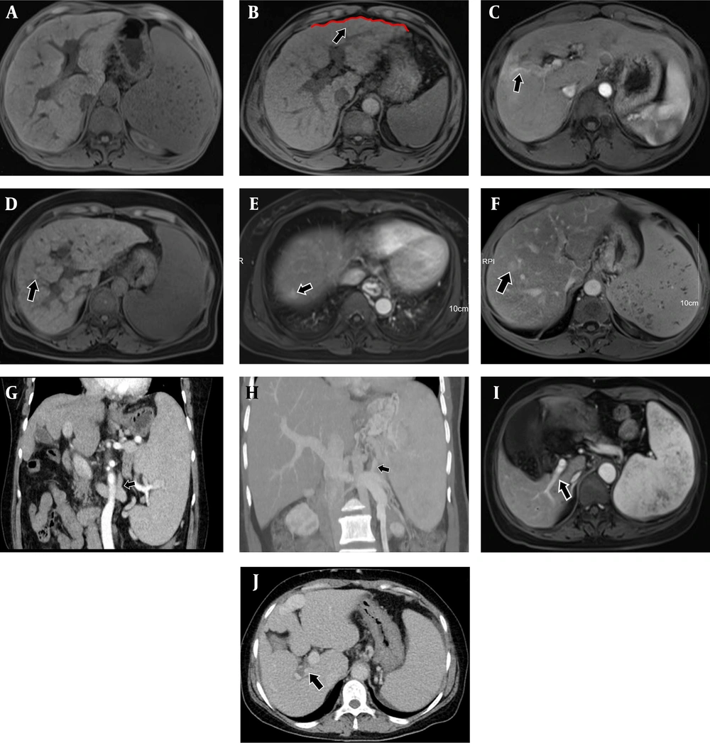
A, Magnetic resonance imaging (MRI)-T1WI.Idiopathic portal hypertension (IPH) patient with giant spleen, caudate lobe hypertrophy, smooth and slightly atrophic peripheral segments, and "fist-like" liver morphology in cross-section; B, Contrast enhanced MRI - arterial phase. Cirrhotic patient with uneven and "serrated" liver surface (black arrow); C, Contrast enhanced MRI-arterial phase. IPH patient with focal perfusion abnormalities in the arterial phase of the periportal liver parenchyma (black arrow). D, MRI-T1WI.cirrhotic patient with diffuse intrahepatic regenerative nodules on T1WI sequence (black arrow); E, F, Contrast enhanced MRI-venous phase. In 2 patients with IPH, FNH-like lesions could be observed in the venous phase (black arrow), a type of benign lesion that was more strongly enhanced in the venous phase than in the adjacent liver parenchyma on imaging; G, Contrast enhanced computed tomography (CT)-delay period. A patient with IPH whose tortuous gastric vein branches into the renal vein figure (black arrow); H, Portal CT angiography. A patient with IPH whose tortuous splenic vein branches converged into the renal vein (black arrow); I, Contrast enhanced MRI - arterial phase. In a patient with IPH, the right posterior branch of the portal vein was stiff and straight, and the distal branch of the portal vein was slender (black arrow); J, Contrast enhanced CT-delay period. A patient with IPH in the portal phase with a localized filling defect in the right secondary branch of the portal vein (black arrow)
4.2.3. Splenic-renal Shunts and Gastrorenal Shunts
4.2.4. Abnormalities of the Portal Venous System
4.3. Pathological Changes in Patients with Idiopathic Portal Hypertension and Cirrhosis
| Parameter | Liver cirrhosis (n = 30) | IPH (n = 14) | X2 | P-Value |
|---|---|---|---|---|
| Pseudolobule formation | 30 (100) | 0 (0) | 39.511 | < 0.001 |
| Fibrosis in the portal areas | 0 (0) | 14 (100) | 39.511 | < 0.001 |
| Septal fibrosis | 30 (100) | 7 (50) | 14.296 | < 0.001 |
| Portal vein occlusion | 0 (0) | 4 (28.6) | 6.288 | 0.012 |
| Lobular inflammation | 26 (86.7) | 3 (21.4) | 17.029 | < 0.001 |
| Cell necrosis | 22 (73.3) | 2 (14.3) | 13.423 | < 0.001 |
Abbreviation: IPH, idiopathic portal hypertension.
a Values are expressed as No. (%).
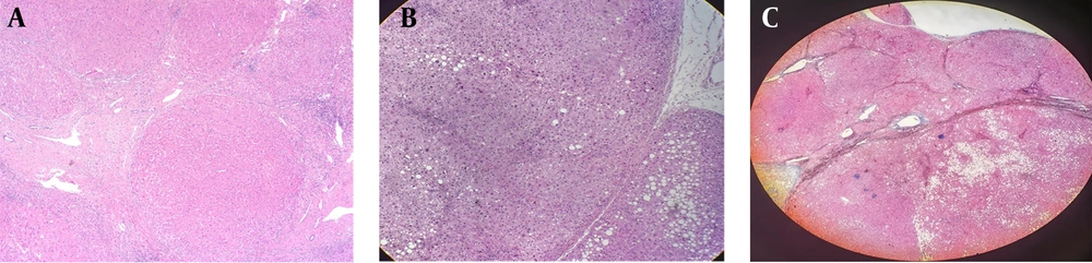
A, Cirrhotic patient, nodular liver cirrhosis in peripheral hepatocytes (upper side, lower right side), hematoxylin and eosin (H&E) staining, × 40; B, Regenerative nodule (left side), H&E staining, × 100. C, Masson stain showing fibrotic nodules
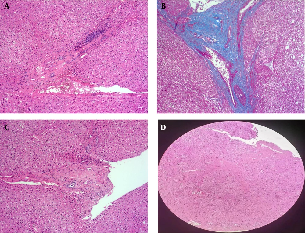
A, A patient with idiopathic portal hypertension (IPH) exhibited portal fibrosis; B, Masson staining showing portal vein fibrosis; C, Portal vein occlusion; D, Unclear structure of portal vein branches, hematoxylin and eosin (H&E) staining, × 100.


