Fulltext
Full-text is available in PDF file.
Authors
Introduction: Atrial septal defect (ASD) is a common congenital heart disease and causes left-to-right shunting and significant right ventricular (RV) volume overload. The goal of this study was to evaluate the influence of these hemodynamic changes on pulmonary venous flow pattern in young adults.
Methods: Complete echocardiographic examination was performed in a group of 40 young adults (aged < 40 years) who had secundum type ASD and was compared to 40 age-matched individuals in control group who had no cardiac abnormality. Systolic and diastolic flow velocities in pulmonary veins (PV), superior vena cava (SVC), inferior vena cava (IVC) and RV functional parameters were recorded and evaluated.
Results: As opposed to healthy young individuals who showed distinct S and D waves with diastolic predominance in pulmonary vein Doppler, in patients with ASD a continuous flow with increased systolic peak that began in systole and continued to the late diastole was observed. The RV systolic function increased compared to the control group.
Conclusions: In patients with ASD, the pattern of pulmonary veins flow transforms into a single continuous antegrade wave with systolic dominance due to persistent shunting of left atrial blood in to right heart chambers as well as increased RV pump function on pulmonary vein (by means of ASD), SVC and IVC, and could be used as a screening method for the presence of secundum type ASDs in young adults.
© 2016, International Journal of Cardiovascular Practice. This open-access article is available under the Creative Commons Attribution-NonCommercial 4.0 (CC BY-NC 4.0) International License (https://creativecommons.org/licenses/by-nc/4.0/), which allows for the copying and redistribution of the material only for noncommercial purposes, provided that the original work is properly cited.
Mirdamadi A, Nejati M, Behjati M. A Case Report of Total Anomalous Pulmonary Vein Connection in Adult, through application of multi-modality imaging. Int Cardiovasc Res J. 2018;12(2):e11287. doi:
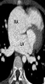
Choi B, Joo Jeong Y, Lee G, Song S, Cho M, et al. Drainage of the Inferior Vena Cava Into the Left Atrium with Atrial Septal Defect and Partial Anomalous Pulmonary Venous Return: Initial and Postoperative Computed Tomography (CT) Findings. I J Radiol. 2018;15(2):e13878. doi: https://doi.org/10.5812/iranjradiol.13878
Hosseinzadeh maleki M, Alireza M. Absent Right Superior Vena Cava with Atrial Septal Defect and Unroofed coronary sinus. Multidiscip Cardio Annal. 2011;3(3):e8754. doi:
Amoozgar H, Fallahi M, Ajami G, Borzouee M. The Relationship of Coronary Sinus Dilation with Pulmonary Artery Pressure in Pediatric Patients. Int Cardiovasc Res J. 2017;6(2):e12818. doi:
Ranjan R, Gucuk Ipek E, Mostafavi Toroghi H, Schiller N. Continuous Flow from Inferior Vena Cava into the Right Atrium Predicts Normal Central Venous pressure. Int Cardiovasc Res J. 2018;12(4):e67163. doi:
Last Update: 4 days ago
Last Update: 4 days ago
Last Update: 4 days ago
Last Update: 6 days ago
Last Update: 6 hours ago
Last Update: 4 days ago