
1. Introduction
Gastric adenocarcinoma is extremely rare in children, and its median age at diagnosis is over 50 years. The pediatric gastrointestinal malignancies and gastric adenocarcinoma account for 5 and 0.05% of all pediatric malignancies, respectively (1). Although infectious causes and lifestyle factors have a role in the etiology of this malignancy in adults, the role of these factors in children continues to remain unknown. The gastric adenocarcinoma prognosis in children is very poor, and the mean survival rate of these patients is about five months (2). Early clinical manifestations in these children, such as epigastric pain, nausea, vomiting, dyspepsia, gastrointestinal bleeding, and weight loss, are typically nonspecific (3). Since this malignancy is rare in children and has nonspecific symptoms, it is often diagnosed with a delay, and the patients are diagnosed with metastasis (4). Here, we describe a 12-year-old girl with gastric adenocarcinoma, who was presented with upper abdominal pain, anorexia, abdominal distension, and nausea.
2. Case Presentation
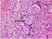
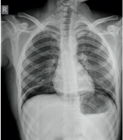
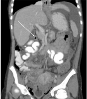
The patient was a 12-year-old Afghan girl, who was presented with abdominal pain and distention since two months ago. She complained of anorexia and nausea and had 6 kg weight loss. There was no evidence of hematemesis or melena. However, she had normal growth and development. She was the third child of a family with no history of gastric cancer. Physical examination revealed pallor, as well as abdominal distention and tenderness. No sign of lymphadenopathy or organomegaly was observed. Abdominal ultrasound showed a large volume of ascites in the abdomen and pelvis. In addition to a large volume of ascites, abdominal computed tomography (CT) scan findings revealed increased thickness of the proximal gastric mucosa, irregular thickening of anterior wall of antrum region of stomach, peritoneal involvement by metastatic cells, left side pleural effusion, and lymphadenopathy adjacent to the superior mesenteric artery and aorto-caval area (Figure 1). The results of the electrolytes, stool for occult blood, liver function test, bilirubin, albumin, PT, PTT, and alpha fetoprotein (AFP) tests were normal. The stool occult blood test was negative. Complete blood count showed WBC: 9200/mm3; PMN: 70%; HB: 10.1 g/dL; MCV: 80 fL; and Platelet: 584000/mm3. Erythrocyte sedimentation rate (ESR) was 57 mm/hr. The level of tumor marker was significantly increased in CA125 (120 u/mL, normal range: < 35 u/mL) but was normal in CA19-9 (1.6 u/mL, normal range: < 27 u/mL) and CEA (2.5 ng/mL, normal range: < 4 ng/mL). Upper gastrointestinal endoscopy showed gastric mucosa thickness, severe nodularity, and thickening of antrum. Six biopsy specimens were obtained from the antrum region and gastric mucosa. The histopathologic examination results were positive for diffuse-type adenocarcinoma (Figure 2) and negative for Helicobacter pylori. Cytology of ascites fluid also indicated the presence of malignant cells. The patient received two cycles of chemotherapy with capecitabine, oxaliplatin, and epirubicin. Unfortunately, she did not respond to the treatment, and the patient died two months later.

Figure 1.
Contrast-enhanced computed tomography of the abdomen shows increased thickness of the proximal gastric mucosa, irregular thickening of anterior wall of antrum region of stomach (arrow), and moderate to severe ascites.

Figure 2.
The biopsy of gastric mucosa shows cells with large cytoplasmic vacuoles and peripherally displaced, and crescent shaped nuclei (signet ring cells) consistent with gastric adenocarcinoma.
3. Discussion
Gastric adenocarcinoma is often seen in the elderly, rarely in individuals under the age of 30 years, and extremely rare in children (2). The clinical manifestations and biological behavior of tumor are similar in children and adults; however, the etiology of pediatric gastric cancer is still unclear and may be associated with gene mutations (5). Although various risk factors, such as Helicobacter pylori infection, high salt intake, smoked foods, nitrates and carbohydrates, alcohol consumption, tobacco smoking, and family history of gastric cancer, in adults have been suggested for this malignancy, the role of these factors in children is not clear yet (6, 7). The most common gastric adenocarcinoma symptoms in these patients are epigastric pain, nausea, anorexia, weight loss, and hematemesis. The most prevalent symptoms are epigastric mass and tenderness, as well as the symptoms associated with anemia and cachexia (8). Given that it is a rare malignancy in children and has nonspecific symptoms, along with insufficient physicians’ knowledge, it is often diagnosed with a delay and associated with metastases at the time of diagnosis. Therefore, most of the patients are in the advanced stage, which leads to their poor prognosis. The mean survival rate in these patients is about five months after diagnosis. Moreover, the patients with diffuse type adenocarcinoma, which accounts for a high percentage of pediatric gastric adenocarcinoma, are at more advanced stages than the patients with non-diffuse type adenocarcinoma when they are presented (7, 8). In a study by Tessler RA, 0.10% of gastric adenocarcinoma cases occurred in children (≤ 21 years). Pediatric cases presented with more advanced disease and signet ring adenocarcinoma comprised 45% of cases in children as compared to 20% of cases in the adults (9).
Given that it is a rare health problem in children, management of this cancer in this group of patients is not clear and is often based on the same principles used for adults. Depending on the disease stage and condition, surgical, chemotherapy, and radiotherapy treatments are used for these patients. The only curative treatment for localized form of gastric adenocarcinoma is radical gastrectomy with lymph node dissection; however, recurrence is relatively common within two years after the treatment (7, 10, 11).
We presented this case due to the rarity of gastric adenocarcinoma in children under 18 and its nonspecific symptoms. The lack of physicians' knowledge also leads to delayed diagnosis, and consequently the referral of patients with metastasis and in advanced stages of the disease, for whom the treatments are not effective and cause a poor prognosis. Therefore, this malignancy should be considered in any child with abdominal pain, nausea, and weight loss, who does not respond to routine treatments. This can be helpful for early and timely diagnosis.