Drugs and Chemicals
PEITC, >98% purity was purchased from Abcam (ab142645, Cambridge, UK). Doxorubicin HCl (Adricin) was purchased from EIMC United Pharmaceuticals (Cairo, Egypt). 3-(4,5-dimethyl-2-thiazolyl) 2,5diphenyl-2H-tetrazolium-bromide (MTT) was purchased from SERVA electrophoresis GmbH (Heidelberg, Germany). Trypan Blue and Dimethyl sulfoxide (DMSO) were purchased from Sigma–Aldrich (St. Louis, MO, USA). Roswell Park Memorial Institute (RPMI) - 1640 medium was purchased from Gibco Laboratories (Life Technologies Inc., Grand Island, NY, USA). Fetal bovine serum (FBS) and penicillin-streptomycin antibiotic were purchased from Lonza (Basel, Switzerland). All other chemicals are of high purity and analytical grade.
Animals
Adult Swiss female albino mice (25–30 g) were purchased from Theodore Bilharz Institute, Giza, Egypt and kept in animal house of Faculty of Pharmacy, Mansoura University, Egypt. Animals were kept in standard size polycarbonate cages under standard laboratory conditions (26 ± 1 °C, 12-h light:12-h dark cycle) and had free access to food and water ad libitum. Ehrlich ascites carcinoma (EAC) induction started 10 days following animals’ accommodation under these standard conditions. All animal experiments carried out in this study were complied with ″Research Ethics Committee″ Faculty of Pharmacy, Mansoura University, Mansoura, Egypt, in accordance with ″Principles of Laboratory Animal Care″ (NIH publication No. 85-23, revised 1985).
Ehrlich ascites carcinoma cells
EAC cell line was obtained from the National Cancer Institute, Cairo University, Cairo, Egypt. EAC cells were maintained by intraperitoneal (IP) passage of 1 X 10
6 cells in adult female Swiss albino mice (
17). Seven days later, the ascetic fluid was collected from mice peritoneal cavity under sterile condition by needle aspiration. Viable tumor cells were counted in a Neubauer hemocytometer using the trypan blue dye (0.4%) exclusion method. EAC cells were used for both
in-vivo and
in-vitro experiments.
Ehrlich ascites carcinoma model
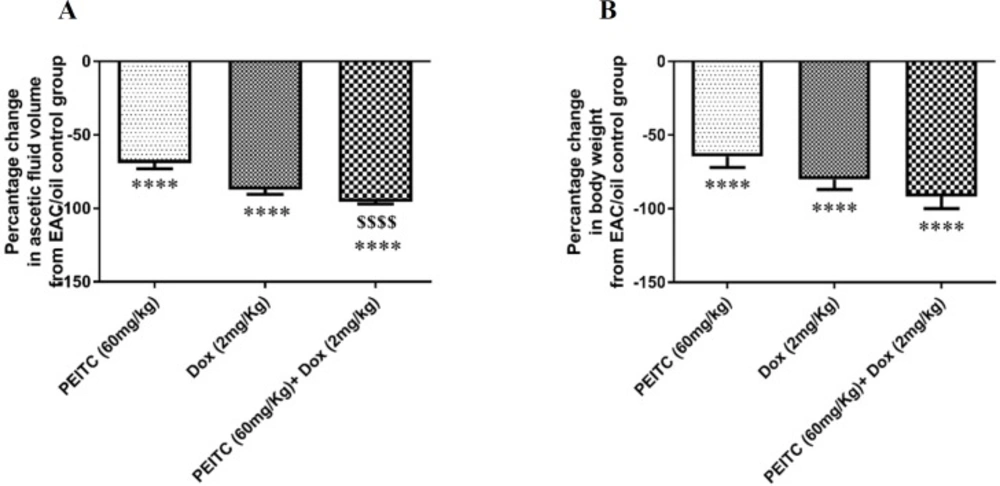
On day zero, mice were injected intraperitoneally (IP) with 1 X 10⁶ EAC cells (0.1 mL/mouse) for tumor induction. On the next day, mice were weighted then randomly divided into 4 groups; each group consists of 10 animals. Treatments were administered for 14 days as follows:
EAC/oil control group that received 0.1 mL oil/mouse/day orally,
PEITC-treated group that received
PEITC at a dose of 60 mg/kg/0.1 mL oil/mouse/day orally (
18), Dox-treated group that received IP injection of Dox at a dose of 2 mg/kg/0.1 mL/mouse/day (
19) and
PEITC/Dox combination treated group that received Dox (2 mg/kg/0.1 mL/mouse/ day, IP) 2 h following receiving
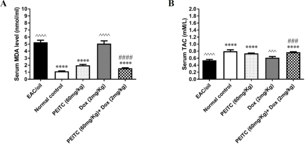
PEITC (60 mg/kg/0.1 mL oil/mouse/day, orally). A fifth group of eight mice that received 0.1 mL oil/day/mouse orally was used as normal control group. On day 15, animals were weighted, blood was collected by retro-orbital puncture and then sacrificed. Serum samples were prepared by centrifugation of blood samples that was withdrawn via retro-orbital puncture at 4000 rpm for 10 min at 4 °C. Ascetic fluid was aspirated from all animals for measuring tumor volume as an indication for the antitumor effect. Ascetic fluid samples were centrifuged at 1800 rpm for 5 min at 4 °C. The pellet was flash-frozen in liquid nitrogen then stored in -80 °C till used.
Malondialdehyde and total antioxidant capacity assay
Commercially available malondialdehyde (MDA) and total antioxidant capacity (TAC) colorimetric assay kits from Biodiagnostic Company (Giza, Egypt) were used for spectrophotometric determination of serum MDA levels and serum TAC according to the methods reported by Satoh (
20) and Koracevic
et al. (
21), respectively.
Cell culture
EAC cells were cultured in RPMI-1640 medium supplied with 10% FBS and 1% (v/v) penicillin-streptomycin antibiotic. They were maintained at 37 °C in a humidified atmosphere with 5% CO2 (Binder, C-series, Germany).
Cell viability assay
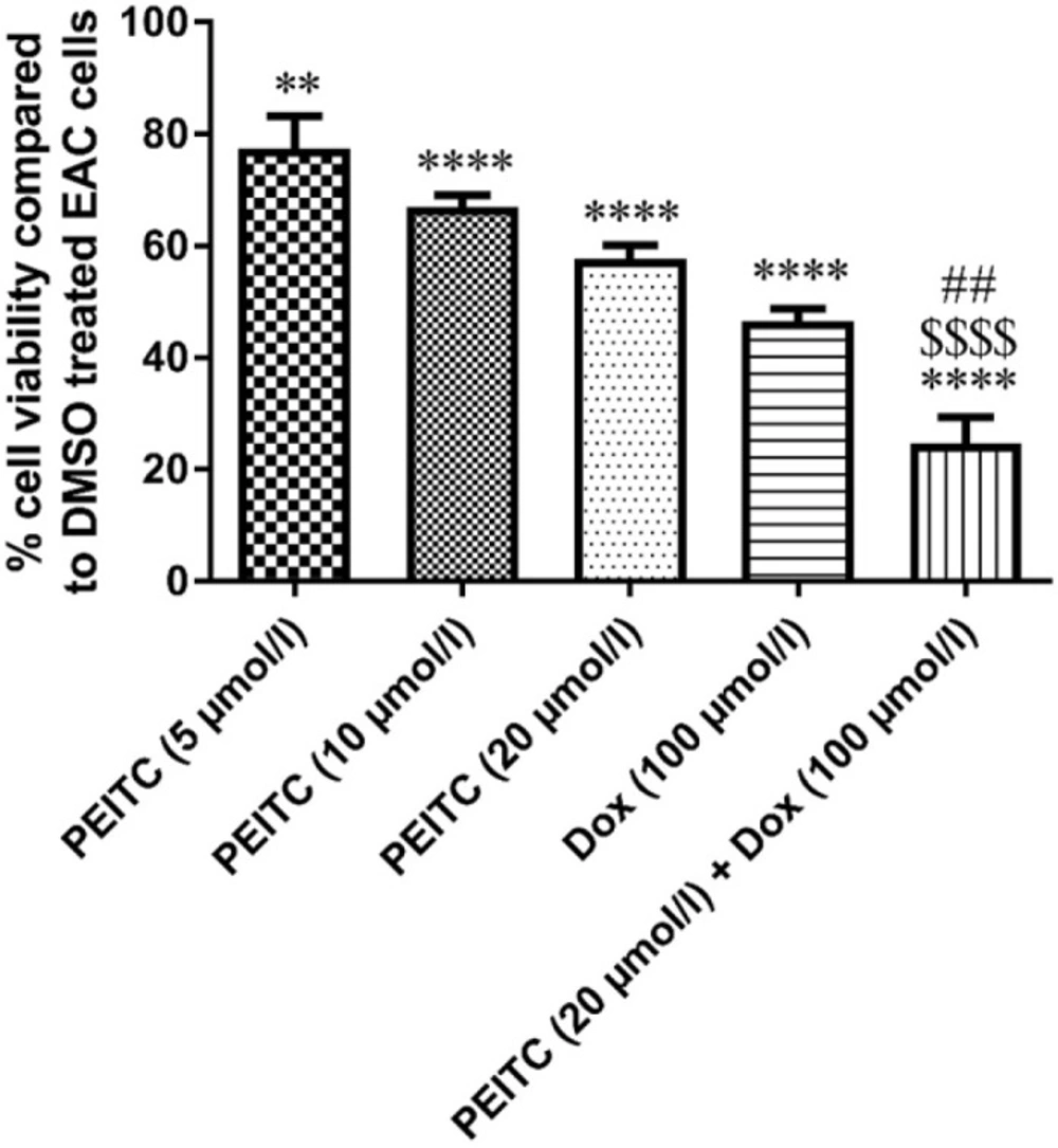
MTT assay was used for the assessment of cell viability. EAC cells (1 X 10⁵ cells/well) were inoculated in 96-well flat bottom tissue culture plate (Griener, Germany). One day after, cells were treated with three different concentrations of PEITC (5, 10 and 20 µmol/L), Dox (100 µmol/L), a combination of PEITC 20 µmol/L + Dox 100 µmol/L and DMSO with a final concentration of 0.1% that have been used as a control. Cells were incubated for 24 h and incubation period continued for additional 4 h at 37 °C following the addition of 20 μL/well of MTT solution (5 mg/mL in PBS). The formazan crystals formed as a result of MTT reduction were solubilized by treating cells with 100 µL of 0.04 N acidic isopropanol. Absorbance was measured at 540 nm using a microplate reader, (Bio-Tek ELX800, USA). The experiment was repeated three times. Viable cells were calculated as percentage relative to DMSO treated control cells.
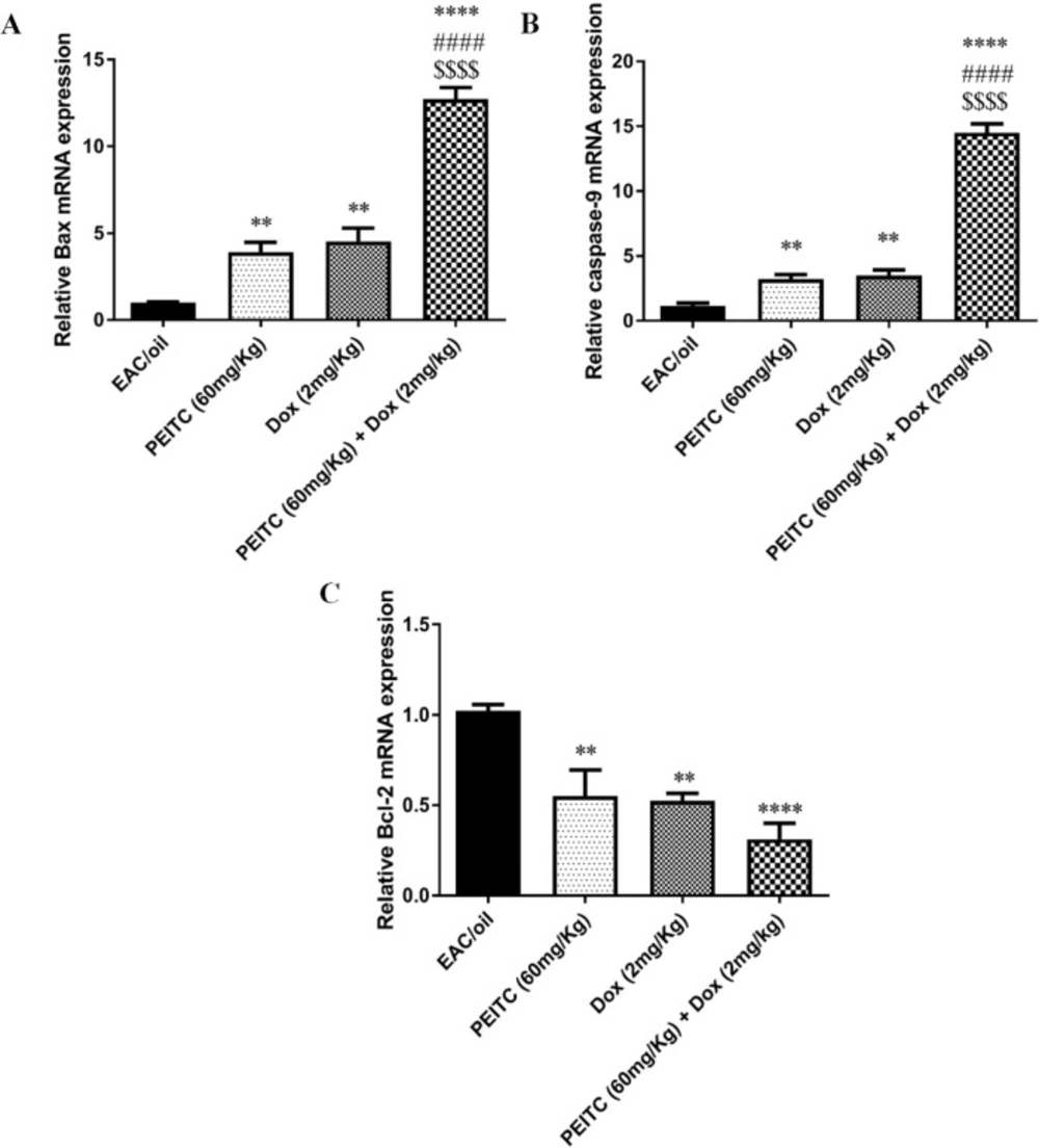
Quantitative, Real-Time Polymerase Chain Reaction (RT-PCR) for Bax, caspase-9 and Bcl-2 gene expression
Total RNA was isolated from EAC cells using TRIzol
® Reagent (Ambion, Life Technologies, USA) according to the manufacturer’s instructions. The quantity and quality of the isolated RNA was assessed spectrophotometrically at 260 nm and 260/280 nm ratio, respectively, using NanoPhotometer
® (Implen, GmbH, Germany). One microgram of the total RNA was reverse transcribed into single-stranded complementary DNA using QuantiTect Reverse Transcription Kit (Qiagen, USA) according to the manufacturer’s instructions. The mRNA expression level of apoptosis genes Bax, caspase-9 and Bcl-2 was determined in different EAC cells. For normalization of gene expression, mouse Glyceraldehyde 3-phosphate dehydrogenase (GAPDH) was quantified in parallel with target genes. Reactions were performed using HOT FIREPol EvaGreen qPCR Mix (Solis BioDyne, Tartu, Estonia) in Rotor-Gene Q (Qiagen, USA). Gene specific primers are summarized in
Table 1. The primers were designed using PREMIER Biosoft (USA) according to gene sequence obtained from PubMed (Entrez Gene), blasted on NCBI/Blast and purchased from Invitrogen-Life Technologies. Thermal cycling program was as follows: initial activation cycle at 95 °C for 15 min followed by 40 cycles at 95 °C for 15 sec for denaturation, 65 °C for 20 sec for annealing and finally 72 °C for 20 sec for elongation. Relative expression of studied genes was determined using 2
−ΔΔCT method relative to GAPDH. The specificity of the designed primers and the size of amplified PCR products were confirmed by melt curve analysis and 2% agarose gel electrophoresis, respectively.
| Gene of Interest | Primer Sequence | Reference Sequence | Product size (bp) |
|---|
| GAPDH Forward | 5`-ATGGTGAAGGTCGGTGTGAAC-3` | NM_008084.3 NM_001289726.1 | 251 |
| GAPDH Reverse | 5`-TTGATGTTAGTGGGGTCTCGC-3` | | |
| Bax Forward | 5`-CCACCAGCTCTGAACAGATC-3` | NM_007527.3 | 140 |
| Bax Reverse | 5`-CAGCTTCTTGGTGGACGCAT-3` | | |
| Caspase-9 Forward | 5`-TGGACATTGGTTCTGGCG-3` | NM_015733.5 NM_001277932.1 | 117 |
| Caspase-9 Reverse | 5`-TGTTGATGATGAGGCAGTGG-3` | | |
| Bcl-2 Forward | 5`-GGATGACTTCTCTCGTCGCTAC-3` | NM_009741.4 NM_177410.2 | 199 |
| Bcl-2 Reverse | 5`-TGACATCTCCCTGTTGACGCT-3` | | |
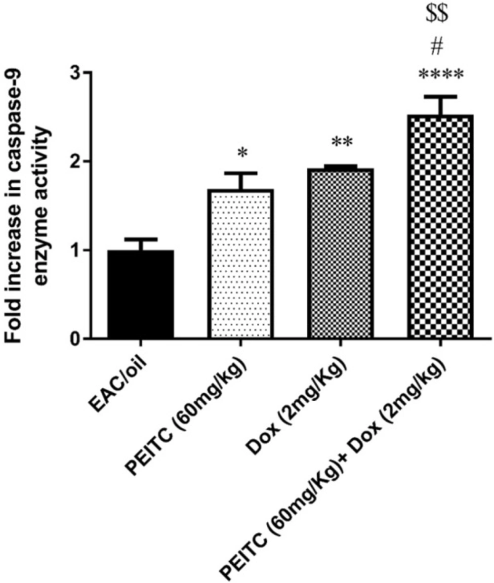
Caspase-9 enzyme activity colorimetric assay
EAC cells collected from control and treated groups were lysed using chilled lysis buffer provided by Caspase-9 Colorimetric Assay Kit (BioVision, CA, USA) and then caspase-9 enzyme activity was determined spectrophotometrically according to the manufacturer’s instructions.
Statistical analysis
Results are presented as mean ± standard error of mean (SEM). One-way analysis of variance (ANOVA) followed by Tukey’s post-hoc test using GraphPad Prism 6.01 (GraphPad Software, SanDiego, CA, USA) was used to find out statistically significant results. P-value of less than 0.05 was considered statistically significant.