Cover image of the article: Recent Progress in PET and SPECT Myocardial Perfusion Imaging: From Radiopharmaceuticals to Imaging Innovations

Image Credit:Iran J Pharm Res

Recent Progress in PET and SPECT Myocardial Perfusion Imaging: From Radiopharmaceuticals to Imaging Innovations

Authors

Elham Sattarzadeh KhamenehElham Sattarzadeh Khameneh ORCID1, Saeed KakaeiSaeed Kakaei ORCID1,*, Yashar Noshadi2, Behrouz AlirezapourBehrouz Alirezapour ORCID1, Fariba Johari Daha1
1Radiation Application Research School, Nuclear Science and Technology Research Institute (NSTRI), Tehran, 11365 - 3486, Iran
2Medical Radiation, Nuclear Engineering, Shahid Beheshti University, Tehran, Iran
*Corresponding Author: Radiation Application Research School, Nuclear Science and Technology Research Institute, Tehran, Iran. Email: ken1.kke@gmail.com

Iranian Journal of Pharmaceutical Research:Vol. 25, issue 1; e171684
Published online:Sep 14, 2026
Article type:Review Article
Received:May 05, 2026
Accepted:Sep 05, 2026
How to Cite:Sattarzadeh Khameneh E, Kakaei S, Noshadi Y, Alirezapour B, Johari Daha F. Recent Progress in PET and SPECT Myocardial Perfusion Imaging: From Radiopharmaceuticals to Imaging Innovations. Iran J Pharm Res. 2026;25(1):e171684. doi: https://doi.org/10.5812/ijpr-171684

Abstract

Context:

Cardiovascular diseases (CVDs) remain a leading cause of morbidity and mortality worldwide, underscoring the need for accurate, noninvasive diagnostic methods to support clinical decision-making and treatment planning. Myocardial perfusion imaging (MPI) with positron emission tomography (PET) and single-photon emission computed tomography (SPECT) plays a key role in the evaluation of myocardial ischemia and coronary artery disease. This review provides a comparative overview of PET and SPECT MPI, with particular emphasis on radiopharmaceuticals, technical advances, clinical applications, and the limitations of each modality.

Evidence Acquisition:

A systematic literature search was conducted in PubMed/MEDLINE, Scopus, Web of Science, and Google Scholar for studies published between 1990 and July 2026. Search terms included myocardial perfusion imaging, PET radiotracers, SPECT radiotracers, rubidium-82, nitrogen-13 ammonia, technetium-99m sestamibi, flurpiridaz, and relevant combinations of these terms using Boolean operators. Original research articles, clinical trials, and review articles addressing radiopharmaceutical development, image reconstruction techniques, quantitative imaging, and hybrid PET/CT and SPECT/CT technologies were included. Non-English publications, case reports, and studies lacking relevant original or comparative data were excluded. The reference lists of eligible articles were also manually reviewed. Radiotracers were compared with respect to pharmacokinetics, myocardial extraction fraction, target-to-background ratio, image quality, radiation dosimetry, and clinical utility.

Results:

SPECT MPI, particularly with 99mTc-sestamibi and 99mTc-tetrofosmin, remains widely used because of its established clinical infrastructure, relatively low equipment costs, broad availability, and ability to assess both myocardial perfusion and left ventricular function using gated imaging. Its principal limitations include lower spatial resolution, susceptibility to photon attenuation artifacts, and a limited capacity for absolute quantification of myocardial blood flow. PET MPI offers superior spatial and temporal resolution, routine attenuation correction, and quantitative measurement of myocardial blood flow and myocardial flow reserve. Established PET tracers, including 82Rb and 13N-ammonia, generally provide higher diagnostic accuracy than conventional SPECT; however, their short physical half-lives require generator-based or on-site cyclotron production and may limit the feasibility of exercise stress protocols. Emerging 18F-labeled tracers, particularly flurpiridaz, offer several advantages, including a longer half-life suitable for centralized production and distribution, improved image contrast, exercise stress imaging, rapid extracardiac clearance, and a more linear relationship between myocardial tracer uptake and blood flow over a broad flow range. Recent phase III studies have further supported the diagnostic performance of flurpiridaz.

Conclusions:

PET and SPECT play important and complementary roles in noninvasive myocardial perfusion imaging. SPECT remains the most widely available and cost-effective technique, whereas PET provides superior image quality, attenuation correction, and quantitative assessment of myocardial blood flow. Ongoing advances in radiopharmaceutical development, hybrid imaging systems, reconstruction methods, and quantitative software are expected to further enhance the diagnostic and prognostic value of MPI and support increasingly personalized approaches to cardiovascular care.

1. Context

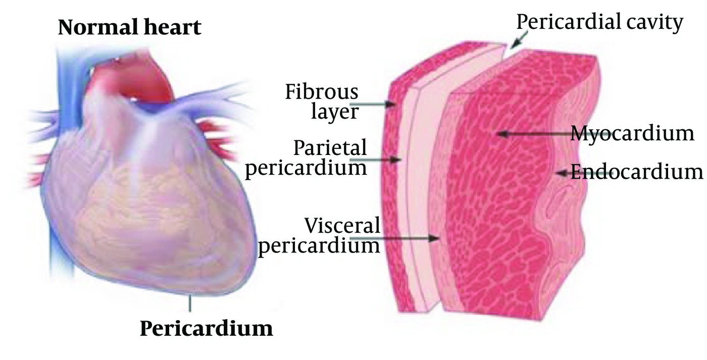
The heart is a vital muscular organ that serves as the central pump of the circulatory system. It is enclosed within a fibrous sac called the pericardium (Figure 1). The heart has four chambers, including two atria and two ventricles, separated by septal walls. The right atrium receives deoxygenated blood from the body and pumps it to the right ventricle, which then pumps blood to the lungs. The left atrium receives oxygenated blood from the lungs and pumps it to the left ventricle, which distributes blood throughout the body (1-3).
Pericardial sac and different parts of the pericardium
Figure 1.
Pericardial sac and different parts of the pericardium
The coronary arteries supply blood to the heart muscle, and blockages in these arteries are a leading cause of myocardial infarction and heart failure. Cardiovascular diseases (CVDs) are disorders that affect the heart and blood vessels. Coronary artery disease (CAD) is the most common type and occurs when the coronary arteries become narrowed or blocked by atherosclerotic plaque buildup, potentially leading to myocardial infarction, heart failure, or arrhythmias (4, 5). Additional CVDs include angina pectoris (chest pain due to reduced myocardial blood flow), heart failure (impaired pumping function), hypertension, cardiomyopathy, cardiac arrhythmias, valvular heart disease, and peripheral artery disease. Despite medical advances, CAD remains the leading cause of death worldwide (6), and accurate diagnostic techniques are essential for disease identification and management (7). Myocardial perfusion imaging (MPI) has evolved substantially over the past four decades (8, 9) as a noninvasive method for assessing coronary blood flow and identifying perfusion abnormalities (10). Currently, two major molecular imaging techniques are widely used for cardiovascular assessment: single-photon emission computed tomography (SPECT) and positron emission tomography (PET) (8, 11). These modalities provide noninvasive assessment of perfusion and myocardial function (12) and support disease staging and treatment planning (13).
SPECT is the standard and most widely used nuclear imaging method and has been broadly available in most hospitals since the early 1990s (14). Nuclear cardiology has been a clinical specialty for approximately four decades, with the first use of radioisotopes to study circulatory processes reported in 1927 (15). Labeled molecules enable visualization, measurement, and tracking of metabolic processes, thereby distinguishing between healthy and diseased tissue (15). Recent advances in physics and related technologies have improved all aspects of medical imaging, particularly nuclear cardiology. Nuclear medicine imaging is now essential for diagnosing and managing cardiac disease, assessing blood supply during exercise and rest, identifying regions of poor perfusion, and detecting viable tissue in patients with prior myocardial infarction (16). The development of 2D and 3D imaging devices has created new opportunities for medical imaging research (16).

1.1. Scope and Objective

This review provides a comprehensive comparative overview of PET and SPECT myocardial perfusion imaging, with a detailed focus on radiopharmaceuticals, technical innovations, clinical applications, and limitations. The review addresses the following questions: 1) what are the key characteristics of current SPECT and PET perfusion tracers, and how do they compare? 2) what are the advantages and limitations of each modality in clinical practice? 3) what emerging tracers and technologies may overcome current limitations? The comparative analysis emphasizes radiopharmaceutical performance, clinical utility, and practical considerations for implementation in cardiovascular imaging.

2. Evidence Acquisition

A systematic literature search was performed in PubMed/MEDLINE, Scopus, Web of Science, and Google Scholar to identify studies published between 1990 and July 2026. Search terms included myocardial perfusion imaging, PET radiotracers, SPECT radiotracers, rubidium-82, nitrogen-13 ammonia, technetium-99m sestamibi, flurpiridaz, and relevant Boolean combinations. Original research articles, clinical trials, and review articles addressing radiopharmaceutical development, image reconstruction techniques, quantitative imaging, and hybrid PET/CT and SPECT/CT technologies were included. Non-English publications, case reports, and studies lacking relevant original or comparative data were excluded. Reference lists of eligible articles were also reviewed manually. Radiotracers were compared with respect to pharmacokinetics, myocardial extraction fraction, target-to-background ratio, image quality, radiation dosimetry, and clinical utility.

3. Results

3.1. Myocardial Perfusion SPECT Radiopharmaceuticals

Selection of an appropriate radiopharmaceutical is critical in nuclear cardiology, as the radionuclide’s physical half-life should align with the duration of the physiological process under investigation (7, 17).

3.1.1. Thallium-201 Chloride (201Tl)

Thallium-201 (201Tl, half-life 73 hours) was the first clinically adopted SPECT MPI tracer, introduced in the 1970s. It is a potassium analog transported across cell membranes via the Na+/K+ ATPase pump, with rapid myocardial extraction and subsequent redistribution reflecting myocardial viability (18-20). 201Tl is cyclotron-produced and has a long physical half-life, requiring lower administered activities to minimize radiation exposure. However, its low-energy photons (71 - 80 keV) result in longer imaging times and limited image quality due to photon attenuation and scatter, particularly in obese patients (21, 22). 201Tl distribution is proportional to organ blood flow. Following intravenous injection, normal myocardium rapidly extracts the tracer (5 - 8% remains in blood at 5 minutes), with slow excretion via feces and urine (21, 22).
As a monovalent cation, 201Tl behaves as a potassium analog and is actively transported into viable myocytes. Injection occurs at peak exercise or during pharmacological stress. Initial perfusion defects may indicate ischemia or non-viable myocardium. After cessation of stress, 201Tl redistributes, and ischemic areas may demonstrate reversible defects (filling in on delayed images) (23).

3.1.2. Thallium-201 Disadvantages

201Tl provides good contrast between normal and ischemic regions. However, its 73-hour physical half-life and approximately 10-day biological half-life result in a long effective half-life of about 56 hours. Significant accumulation occurs in the liver, kidneys, and skeleton, contributing to an effective dose of 34.5 mSv per 150 MBq (Table 1). The relatively high absorbed dose limits administered radioactivity to less than 150 MBq, potentially compromising image quality (23). 201Tl decays by electron capture to 201Hg, emitting low-energy X-rays (75 - 80 kV) that are captured during imaging (10).
Table 1.
201Tl-Cl Tracer Features for Myocardial Perfusion Blood Flow
Tracer201Tl
Chemical structure201Tl+
Type of tracerMetal cation
ProductionCyclotron
Half-life73 h
Scan Duration (rest and stress)4 h
Intravenously administered activity (MBq)74 - 148
Effective Dose (mSv/MBq)0.23
Approval year (US FDA / EU)1977 / 1980

3.1.3. Technetium-99m-Based Tracers

99mTc-tetrofosmin is a cationic technetium-99m complex in the +5 oxidation state, formulated as [99mTc(tetrofosmin)2O2]+ (24, 25). It is commercially available as a lyophilized kit (Myoview, GE Healthcare) containing 0.23 mg tetrofosmin and excipients. Labeling is performed by adding sodium pertechnetate (99mTc) from a 99Mo/99mTc generator to the vial and incubating for 15 minutes at room temperature. Radiochemical purity must exceed 90% (24, 25).
Cardiac uptake of 99mTc-tetrofosmin occurs via passive diffusion without redistribution. Following intravenous injection, myocardial uptake reaches a maximum within 5 minutes. Approximately 66% of the injected dose is excreted within 48 hours (40% urine, 26% feces). 99mTc-tetrofosmin is used for myocardial perfusion imaging under stress and rest conditions to identify reversible ischemia and infarcted tissue (Figure 2). It effectively detects perfusion changes due to pharmacological stress (adenosine) in CAD patients. 99mTc-tetrofosmin rapidly clears from blood (5% remains at 10 minutes), with myocardial uptake of approximately 1.2% at peak, minimal redistribution (~1% at 2 hours), and rapid hepatic and pulmonary clearance (26-28). It is also used to evaluate left ventricular function.
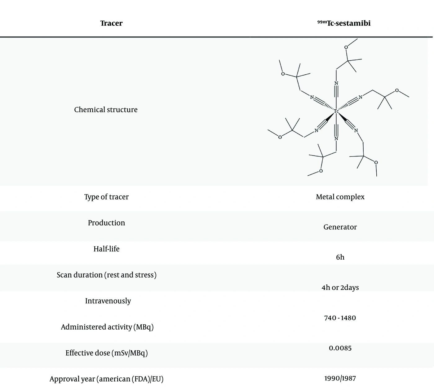
<sup>99</sup>mTc-tetrofosmin tracer features for myocardial perfusion blood flow
Figure 2.
99mTc-tetrofosmin tracer features for myocardial perfusion blood flow

3.1.4. 99mTc-Sestamibi (MIBI)

99mTc-Methoxyisobutylisonitrile (99mTc-MIBI, sestamibi) is a cationic, lipophilic complex with a half-life of 6.01 hours. It consists of a 99mTc atom in the 1+ oxidation state coordinated to six MIBI ligands. The isonitrile groups form a complex with reduced 99mTc. Due to the volatility and instability of isonitrile, MIBI is stabilized as [Cu(MIBI)4]BF4, which decomposes during high-temperature radiolabeling. Commercial kits (Cardiolite, CardioTop, Technescan Sestamibi) contain [tetrakis (2-methoxy-2-methylpropyl-1-isocyanide) copper (1+)] tetrafluoroborate with stannous chloride (reducing agent), L-cysteine hydrochloride, sodium citrate (buffer), and mannitol. Labeling involves adding sodium pertechnetate (99mTc) and heating in a boiling water bath for 10 minutes (7).
99mTc-sestamibi is primarily used to diagnose myocardial perfusion abnormalities, particularly ischemia and infarction, and to evaluate myocardial function using first-pass radionuclide ventriculography. It enters myocardial cells via passive diffusion, localizes to mitochondria, and accumulates proportionally to myocardial perfusion with slow washout (7). Under stress, > 3% of the injected dose accumulates in the myocardium. 99mTc-sestamibi acts as a calcium analog, is trapped in mitochondria, and does not redistribute, unlike 201Tl. It provides good perfusion imaging with rapid blood clearance, high myocardial extraction, and limited uptake in the thyroid and salivary glands (Figure 3).
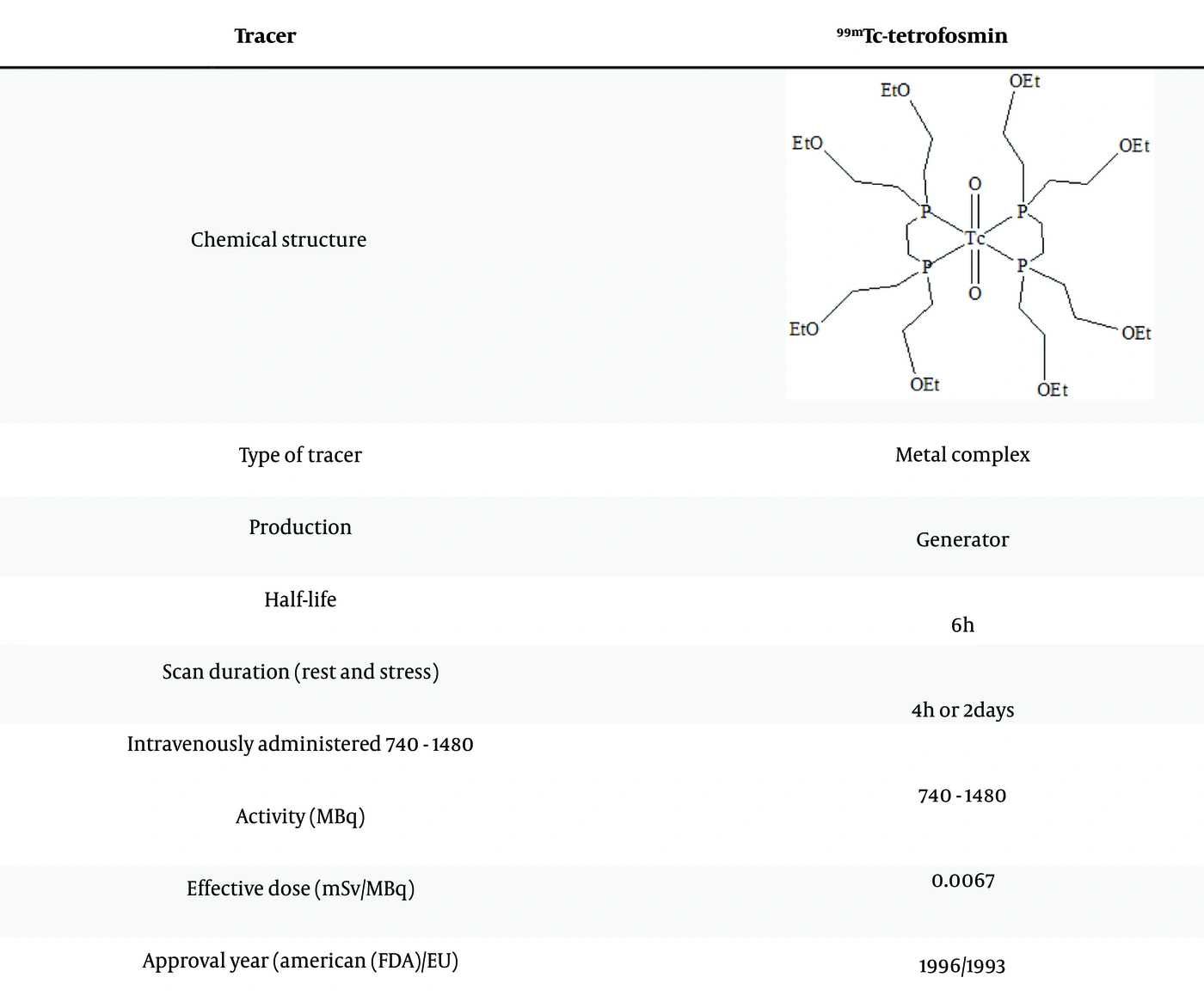
<sup>99</sup>mTc-sestamibi tracer features for myocardial perfusion blood flow
Figure 3.
99mTc-sestamibi tracer features for myocardial perfusion blood flow

3.1.5. 123I-Meta-Iodobenzylguanidine (mIBG)

Heart failure (HF) is characterized by sympathetic nervous system activation (29-31, 32). Metaiodobenzylguanadine (mIBG), or iobenguane, originally developed as a pheochromocytoma imaging agent, accumulates in address an effective dose of 2.4 mSv in adults, with doses up to 55 - 111 MBq/kg possible with new gamma cameras. MIBG is excreted primarily via the kidneys. Distribution in SPECT studies is similar to perfusion tracers (e.g., 123Tc-tetrofosmin).

3.1.6. 99mTc-HYNIC-Annexin V

Apoptosis (programmed cell death) plays a significant role in cardiovascular disease pathogenesis (33, 34). During early apoptosis, phosphatidylserine appears on the outer cell membrane. Annexin V, a 319-amino acid human protein, binds with high affinity to phosphatidylserine (35, 36). 99mTc-Annexin V is the most widely used radiopharmaceutical for apoptosis imaging (37).
Apoptosis imaging with 99mTc-Annexin V, combined with perfusion imaging (99mTc-sestamibi, 99mTc-tetrofosmin, or 201Tl), has been used in patients with myocardial infarction after percutaneous coronary angioplasty (PTCA) (38, 39). Increased 99mTc-Annexin V uptake at infarct sites correlates with myocardial hypoperfusion, indicating apoptosis. The absence of 99mTc-Annexin V uptake at 4 days post-infarction, corresponding to reduced 99mTc-sestamibi defect size, suggests reversible myocardial damage rather than necrosis (Table 2) (39, 40).
Table 2.
Comparison of the Main Blood-Flow SPECT Tracers
TracerPhysical Half-lifeMyocardial Extraction (%)Clearance Half-Life
Thallium-20173 h85slow
99mTc-sestamibi6 h65Very slow
99mTc-tetrofosmin6 h60Very slow

3.1.7. Summary of SPECT Tracer Clinical Status

Currently, 99mTc-sestamibi and 99mTc-tetrofosmin are the most widely used SPECT MPI tracers, with 201Tl used less frequently due to its radiation dose and imaging limitations. 123I-mIBG and 99mTc-Annexin V are used for specialized applications (sympathetic innervation and apoptosis imaging, respectively) rather than routine perfusion assessment.

3.1.8. Extracardiac Activity and Artifact Reduction in SPECT

A significant limitation of SPECT MPI is interference from extracardiac activity, particularly from the liver, bowel, and stomach (41, 42). Hepatobiliary excretion of 99mTc-based agents often results in liver and bowel activity, while gastric activity may arise from duodenal reflux or uptake of free 99mTc-pertechnetate by the gastric mucosa. These artifacts can mimic or obscure inferior wall perfusion defects, reducing diagnostic accuracy. Strategies to minimize extracardiac activity include: 1) patient hydration and fatty meal consumption to promote hepatobiliary clearance; 2) prone imaging to shift extracardiac activity away from the inferior wall; 3) carbonated beverage ingestion to displace gastric activity; and 4) delayed imaging when necessary (43, 44).

3.1.9. CZT Detector Technology in Modern SPECT

Cadmium-zinc-telluride (CZT) semiconductor detectors represent a significant technological advance in SPECT imaging. Unlike conventional NaI (Tl) scintillation detectors with photomultiplier tubes, CZT detectors directly convert photons to electrical signals, providing: 1) superior energy resolution (approximately 5 - 6% at 140 keV vs. 9 - 10% for NaI (Tl)); 2) markedly improved sensitivity (up to 10-fold higher); 3) reduced acquisition times (down to 5 - 10 minutes); and 4) potential for significant dose reduction (up to 50%). CZT-based SPECT systems have demonstrated improved diagnostic accuracy for CAD detection, particularly in challenging patient populations such as obese individuals and women with breast attenuation. The technology enables dynamic SPECT imaging and has the potential to facilitate absolute myocardial blood flow (MBF) quantification, although this remains an area of active investigation.

3.2. Myocardial Perfusion PET Tracers

Positron emission tomography (PET) offers significant advantages for MPI, including superior spatial and temporal resolution, routine attenuation correction, and the capacity for absolute quantification of MBF. The field is evolving through the integration of hybrid PET systems, advanced computational techniques, and the development of novel, mechanism-specific radiopharmaceuticals aimed at overcoming longstanding limitations in cardiac imaging (45).

3.2.1. Positron Range and Its Impact on Spatial Resolution

The effective spatial resolution of PET imaging is limited by the positron range, the distance a positron travels before annihilation. This physical phenomenon causes misregistration of annihilation events relative to the original tracer location. Positron range varies substantially among radionuclides based on their maximum positron energy: 18F (0.64 MeV, mean range 1.03 mm) provides the highest spatial resolution; 13N (1.19 MeV, mean range 2.53 mm) offers intermediate resolution; 15O (1.73 MeV, mean range 4.14 mm) has moderate resolution; and 82Rb (3.38 MeV, mean range 8.6 mm) has the lowest resolution. This range-dependent degradation means that 82Rb PET images have inherent blurring that can limit detection of small perfusion defects, whereas 18F-labeled tracers achieve near-optimal spatial resolution. Modern PET/CT systems with time-of-flight capability partially compensate for these effects, but the fundamental limitations remain.
Accurate attenuation correction (AC) is essential for quantitative PET and SPECT imaging. PET offers routine, robust AC using transmission scans or CT-based attenuation maps. In PET/CT, the CT image provides a high-quality attenuation map that is co-registered with the PET emission data, enabling accurate correction for photon attenuation in heterogeneous tissues. This is particularly important in obese patients and those with breast or diaphragm attenuation. In contrast, SPECT AC is more challenging; conventional methods use external radioisotope sources (e.g., 153Gd line sources) or CT-based maps on SPECT/CT systems. However, SPECT AC is limited by the non-overlapping energies of the emission and transmission photons, potential misregistration, and the need for correction of scatter and tissue heterogeneity. The inability to routinely perform accurate AC has historically limited SPECT’s diagnostic specificity compared with PET.

3.2.2. Absolute Quantification of Myocardial Blood Flow With PET

A unique advantage of PET MPI is the ability to quantify absolute MBF (mL/g/min) using dynamic imaging and tracer kinetic modeling. This contrasts with SPECT, which provides only semi-quantitative indices such as the summed stress score (SSS), summed rest score (SRS), and summed difference score (SDS). PET MBF quantification uses compartmental models that account for tracer delivery, extraction, and retention. For 13N-ammonia, a two-tissue compartment model is typically employed:
dCm(t)/dt = K1 · Ca(t) – k2 · Cm(t) – k3 · Cm(t) + k4 · Ct(t)
dCt(t)/dt = k3 · Cm(t) – k4 · Ct(t)
where Ca is the arterial input function, Cm is free ammonia in myocardium, Ct is metabolically trapped ammonia, and K1, k2, k3, k4 are rate constants. For 82Rb, a simpler one-compartment model is often sufficient:
dCm(t)/dt = K1 · Ca(t) – k2 · Cm(t)
Myocardial flow reserve (MFR = MBFstress/ MBFrest) provides a measure of the hemodynamic significance of coronary stenosis. Quantitative MBF has demonstrated superior diagnostic performance compared with relative perfusion assessment, particularly for detecting balanced ischemia and multivessel disease. However, accurate quantification requires careful attention to: 1) arterial input function extraction; 2) correction for partial volume effects; 3) spillover between the blood pool and myocardium; and 4) appropriate model selection and parameter estimation.

3.2.3. Partial Volume Effects and Spillover in PET MPI

Partial volume effects (PVE) arise from the limited spatial resolution of PET, causing underestimation of activity in small structures and spillover between adjacent regions. In cardiac PET, this is particularly important for the left ventricular cavity (blood pool) and myocardial wall. PVE and spillover affect tracer kinetic modeling and MBF quantification. Different tracers have different PVE characteristics: 15O-water (long positron range) shows significant spillover between blood and myocardium, requiring complex correction models; 82Rb (intermediate range) has moderate PVE; and 18F-labeled tracers (short range) minimize PVE. Correction methods include recovery coefficient approaches, geometric transfer matrix methods, and partial volume correction algorithms integrated into kinetic models. Failure to account for PVE and spillover can lead to systematic errors in MBF estimates, particularly in thin-walled myocardial segments.

3.2.4. Dynamic Framing Challenges for Short-Lived Tracers

Generating the arterial input function for MBF quantification requires dynamic PET acquisition with short frame durations. For 82Rb (76-second half-life), this is challenging because: 1) early frames must be very short (1 - 3 seconds) to capture the rapid peak; 2) count statistics are limited, increasing noise; 3) rapid decay requires precise timing; and 4) the input function is highly dependent on accurate bolus characterization. Typical framing protocols for 82Rb include 30 frames: 12 × 1 - 3 seconds, 6 × 5 seconds, 4 × 10 seconds, 3 × 20 seconds, and 5 × 60 seconds. For 13N-ammonia (10-minute half-life), framing is less demanding, with protocols including 6 × 5 seconds, 4 × 15 seconds, 4 × 30 seconds, and 6 × 60 seconds. The quality of the arterial input function directly affects MBF quantification accuracy; poor count statistics or improper framing can introduce significant errors, particularly at high flow rates during pharmacological stress.

3.2.5. Rubidium-82 (82Rb) and Nitrogen-13-Ammonia (13N-Ammonia)

Rubidium-82 (82Rb) and nitrogen-13-ammonia (13N-ammonia) are the only PET radiotracers currently FDA-approved for MPI, with 15O-water limited to research applications (7, 46). These tracers have short half-lives: 76 seconds (82Rb), 2 minutes (15O-water), and 10 minutes (13N-ammonia), requiring either on-site cyclotrons or generators. 82Rb, a potassium analog, is actively transported into the myocardium via the Na+/K+ ATPase pump. Its short half-life and generator availability enable rapid imaging protocols with favorable dosimetry (47). Despite a relatively low myocardial extraction fraction (approximately 65%), 82Rb has been validated for MBF measurement using single-tissue compartment modeling (48).
13N-ammonia diffuses across cell membranes as NH3, equilibrates with NH4+, and is trapped in cells via conversion to 13N-glutamine, glutamic acid, and carbamoyl phosphate (49). It has a high first-pass extraction fraction of approximately 80% and minimal redistribution, offering advantages over 82Rb for MBF quantification. Its small positron range (2.53 mm) provides good spatial resolution and defect detection (7, 46). However, the need for an on-site cyclotron and short half-life (10 minutes) make exercise stress impractical in clinical settings. Different techniques for absolute quantification of dynamic 13N-ammonia PET data have been validated, with single-tissue or two-tissue compartment models used (48, 50, 51). Model selection affects MBF estimates, highlighting the importance of standardized protocols.

3.2.6. Oxygen-15-Water (15O-Water)

15O-water is considered the reference standard for quantitative MBF measurement due to its freely diffusible nature and linear relationship between first-pass extraction and perfusion (7, 46, 49). Its short half-life (2.04 minutes) requires on-site cyclotron synthesis, limiting clinical use. Additionally, the long positron range (4.14 mm) and poor myocardial-to-background ratio make visual image interpretation challenging. Despite recent data supporting its clinical use (52), 15O-water is not FDA-approved for routine clinical use, being primarily restricted to research applications.
15O-water PET has been used as a reference standard for MBF quantification, against which other methods have been validated (53-58). The rest and stress protocol can be completed in approximately 30 minutes using modern PET/CT scanners (59). A parametric perfusable tissue index, derived from a single 15O-water PET/CT scan, has been developed as a marker of myocardial viability (60), potentially increasing clinical utility. However, the short half-life precludes exercise stress imaging, and the low myocardial accumulation limits visual assessment capabilities. The US FDA and Japanese Ministry of Health, Labour, and Welfare have not approved 15O-water for clinical use (61).

3.2.7. Rubidium-82 (82Rb): Detailed Discussion

82Rb, generated by the 82Sr/82Rb generator (CardioGen-82), has practical advantages, including independence from an on-site cyclotron and a generator lifespan of 4 - 5 weeks (62, 63). The ultra-short half-life (76 seconds) enables rapid sequential studies, and radiation exposure is lower than 99mTc-based SPECT (effective dose 2 - 3.7 mSv vs. 5 - 10 mSv) (64, 65). 82Rb can show subdiaphragmatic activity in the stomach and pancreas due to Na+/K+-ATPase-mediated uptake, which can interfere with inferior wall interpretation (42-44, 66). Strategies to reduce this artifact include water ingestion, prone imaging, and fatty meals. The availability of daily generator elutions and rapid acquisition times (10-minute intervals) allow imaging of up to 10 - 15 patients per day (67). The 82Sr/82Rb generator can be operated for 28 - 42 days, with potential extension to 60 days to reduce per-patient cost.
82Rb PET offers superior count density and uniformity compared with SPECT, with higher interpretive certainty and reader agreement (68). Because PET cameras do not require collimators, they provide better sensitivity and resolution. The primary advantage of 82Rb PET is accurate MBF and flow reserve quantification, which remains a challenge for SPECT (Table 3).
Table 3.
Characteristics of Various Cardiac PET Perfusion Tracers
Characteristic18F-Flurpiridaz13N-Ammonia15O-Water82Rb
Half-life (min)1099.962.061.25
ProductionRegional cyclotronOnsite or nearby cyclotronOnsite cyclotronGenerator
Positron range (mm)1.032.534.148.60
Image resolutionHighestIntermediate-highIntermediateLowest
Myocardial extraction fraction (%)948010065
Perfusion defect contrastHighestIntermediateIntermediate aLowest
Pharmacologic stress imaging protocolFeasibleFeasibleFeasibleFeasible
Treadmill exercise imaging protocolFeasibleFeasible but not practicalNot feasibleNot feasible
a Theoretically, a 100 percent myocardial extraction fraction of 15O-water should produce the most incredible perfusion defect contrast. On the other hand, a weak myocardial-to-background ratio reduces the contrast defect.

3.3. Stress Protocols for Myocardial Perfusion Imaging

Stress MPI can be performed using either exercise (treadmill or bicycle) or pharmacological agents. The choice of stress protocol significantly affects hemodynamic parameters and MBF quantification:
Exercise produces physiological hyperemia through increased myocardial oxygen demand, with peak MBF typically reaching 3 - 5 mL/g/min. It provides additional functional information (symptoms, ECG changes, exercise capacity) and patient satisfaction. Limitations include the inability to achieve target heart rate in deconditioned patients and the lack of a steady-state MBF during imaging. Exercise stress is only feasible with tracers that allow post-exercise injection (201Tl, 99mTc-agents, and potentially 18F-labeled agents).
A non-selective vasodilator that produces potent coronary hyperemia with minimal hemodynamic changes, achieving MBF of 4 - 6 mL/g/min. Side effects include chest pain, dyspnea, flushing, and atrioventricular block. Adenosine requires continuous infusion and is contraindicated in reactive airway disease and advanced heart block. Regadenoson: A selective A2A adenosine receptor agonist with rapid bolus administration and fewer side effects, providing comparable hyperemia to adenosine. Regadenoson has largely replaced adenosine in clinical practice because of ease of use and improved patient tolerability.
A β-1 agonist that increases heart rate and contractility, producing flow reserve comparable to exercise. It is used in patients unable to exercise and with contraindications to vasodilators. Dobutamine increases blood pressure, which may affect MBF quantification.
Stress protocol selection influences MBF quantification: vasodilators produce more uniform hyperemia and are better suited for MBF reserve assessment, whereas dobutamine stress provides functional information similar to exercise. For PET MBF quantification, vasodilators are typically preferred because of the steady-state MBF required for accurate measurement. Current guidelines recommend pharmacologic stress for PET MPI, although 18F-labeled tracers may enable exercise protocols.
Despite these advantages, PET is not universally used for clinical myocardial perfusion imaging for several reasons. Financial factors include higher equipment costs for PET/CT scanners, continuous generator expenses for 82Rb, and the need for cyclotron facilities for 13N-ammonia. Clinical limitations include short tracer half-lives that preclude exercise stress, gastrointestinal background activity, and variable myocardial extraction characteristics. However, emerging 18F-labeled tracers aim to address these limitations.

3.4. The Emergence of Fluorine-18-Labeled Tracers

Over the past several decades, substantial effort has been devoted to developing 18F-labeled PET MPI tracers. 18F (half-life 110 minutes, positron range 1 mm) offers advantages that may overcome current limitations (69-71):
Long half-life enabling centralized production and distribution, comparable to 18F-FDG logistics (72, 73).
Lowest positron range among MPI PET tracers, providing the highest spatial resolution (72, 74).
Lower positron energy and higher positron fraction allowing reduced administered activities (75).
Half-life suitable for delayed imaging protocols (76).
Potential for exercise stress protocols, unlike short-lived PET agents (69).
18F-labeled phosphonium cations are classified by mitochondrial targeting mechanisms: 1) voltage-dependent cations that penetrate mitochondria based on membrane potential; and 2) mitochondrial complex-1 (MC1) inhibitors (7, 77).
Phosphonium analogues, including 18F-FBnTP, 18F-FTPP, and 18F-FPTP, have shown rapid myocardial uptake and retention (78-80).
18F-FBnTP, a lipophilic cation, accumulates in cardiac mitochondria (81). In canine studies, left ventricular activity plateaued at 5 minutes and remained detectable at 90 minutes, with minimal metabolite formation and high heart-to-lung ratios (12:1 at 5 minutes) compared with SPECT agents. 18F-FBnTP demonstrated superior diagnostic performance to 99mTc-tetrofosmin in detecting coronary stenoses in canine models (82-84). In Wistar rat studies, 18F-FBnTP showed no washout or redistribution after transient coronary occlusion, with defects matching histology (85). 18F-FBnTP has been used as a reference perfusion marker in myocarditis studies (86), although human data remain limited.
Additional 18F-fluoroalkylphosphonium derivatives (18F-FATPs: 18F-FPTP, 18F-FHTP, 18F-FETP) demonstrated superior image quality and rapid liver/lung clearance compared with 13N-ammonia in rat coronary occlusion models (87). These tracers had higher first-pass extraction fractions than 13N-ammonia at flow rates exceeding 4.0 mL/min, suggesting improved detection of mild stenosis (88).

3.4.1. 18F-Flurpiridaz

Flurpiridaz (formerly [18F]BMS-747158 - 02), a structural analog of the pyridazinone insecticide pyridaben (Figure 4), is a mitochondrial complex-1 (MC1) inhibitor that competitively binds the ubiquinone site without affecting cell viability (89). It demonstrates rapid myocardial uptake (time to half-maximal uptake approximately 35 seconds), high retention (9.5% injected dose/g at 60 minutes), slow washout (efflux half-time > 120 minutes), flow-independent first-pass extraction, minimal lung uptake, and rapid hepatic clearance (washout between 5 and 15 minutes) (49, 73, 89-91).
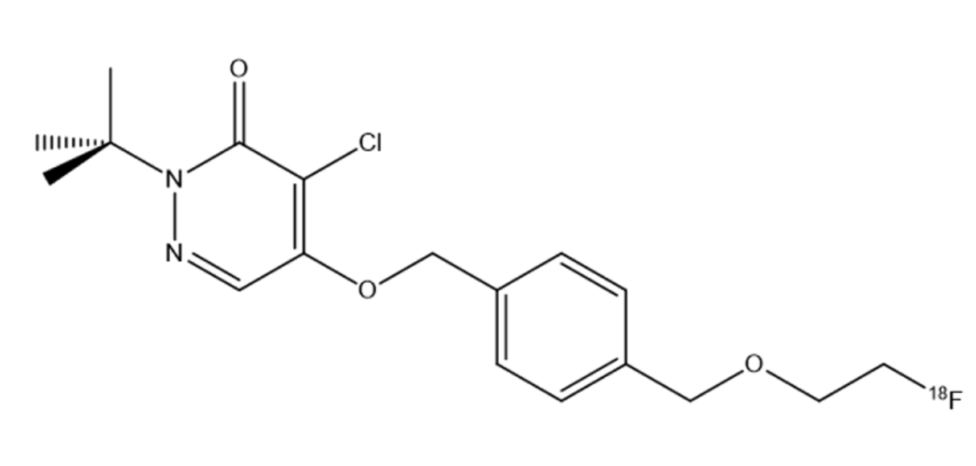
Chemical structure of <sup>18</sup>F-flurpiridaz
Figure 4.
Chemical structure of 18F-flurpiridaz
Electrocardiographic safety has been confirmed in both control and myocardial infarction rabbit models (92). Flurpiridaz demonstrates flow-independent first-pass extraction and a linear uptake-flow relationship across a wide flow range (93-99).

3.4.2. Phase I-III Clinical Data

Phase I studies established injected activities of 1.9 mCi (70 MBq) for rest and 6.5 mCi (240 MBq) for exercise stress, or 2.9 mCi (105 MBq) for rest and 6.8 mCi (250 MBq) for pharmacologic stress. Mean effective radiation doses were approximately 4.8 mSv (exercise) and 6.4 mSv (pharmacologic), higher than short-lived tracers 82Rb or 13N-ammonia (94, 100, 101). The phase II trial demonstrated non-inferiority to 99mTc-SPECT and superiority in obese patients. The phase III trial (101, 102) showed significantly higher diagnostic accuracy than SPECT (90.4% vs. 85.6%, P < 0.001), with better sensitivity and specificity. As of 2026, flurpiridaz remains under FDA review and is not yet available for routine clinical use (Table 4) (73, 100-107).
Table 4.
Advantages and Limitations of 18F-Flurpiridaz as a PET Radiotracer for MPI
AdvantagesLimitations
High first-pass extraction of > 90%Limited to academic centers
Almost linear correlation between tracer uptake and myocardial blood flow in isolated perfused rat heartCyclotron production
Radiotracer redistribution after reperfusionNo FDA approval yet
Phase I: radiotracer present up to 5 h after injection, i.e., injection at peak treadmill exercise is feasibleCost-effectiveness data are lacking
Phase II: compared to stress-rest SPECT MPI, PET MPI with superior performance characteristics for overall CAD diagnosis-
First Phase III study: superior perfusion defect detection of Flurpiridaz relative to SPECT MPI for the assessment of CAD in obese subjects; efficacy and high sensitivity of Flurpiridaz PET MPI in detecting significant CAD compared to SPECT MPI in patients scheduled for invasive coronary angiography-

3.5. Comprehensive Comparison of SPECT and PET MPI Tracers

The following table provides a comprehensive comparison of all SPECT and PET MPI tracers using consistent evaluation criteria (Table 5) (93):
Table 5.
Comprehensive Comparison of SPECT and PET Myocardial Perfusion Tracers
Characteristic201Tl99mTc-Sestamibi99mTc-Tetrofosmin82Rb13N-Ammonia15O-Water18F-Flurpiridaz
Half-life73 h6 h6 h76 s10 min2 min110 min
Positron Range (mm)N/AN/AN/A8.602.534.141.03
Myocardial Extraction (%)856560658010094
ProductionCyclotronGeneratorGeneratorGeneratorCyclotronCyclotronCyclotron
Exercise StressYesYesYesNoNoNoYes
Absolute MBF QuantificationNoNoNoYesYesYesYes
Effective Dose (mSv)34.56.35.02.0 - 3.71.3 - 2.01.0 - 1.54.8 - 6.4
Spatial ResolutionPoorModerateModerateModerateGoodGoodExcellent
Attenuation CorrectionLimitedLimitedLimitedRoutineRoutineRoutineRoutine
AvailabilityWidelyWidelyWidelyModerateLimitedResearchInvestigational
Regulatory StatusFDA/EUFDA/EUFDA/EUFDA/EUFDA/EUResearchPhase III
CostLowModerateModerateHighHighVery HighModerate

4. Conclusions

The updated evidence supports a complementary model of PET and SPECT MPI. 99mTc-sestamibi and 99mTc-tetrofosmin remain clinically established SPECT agents with major advantages in availability, workflow, and cost. CZT detector technology and modern attenuation correction have improved the performance of contemporary SPECT and can reduce acquisition time or administered activity. PET offers higher sensitivity, routine attenuation correction, and quantitative MBF/MFR; however, these benefits depend on appropriate dynamic acquisition, kinetic modeling, motion correction, and tracer availability.
The major recent radiopharmaceutical development is the clinical translation of 18F-flurpiridaz. Its 2024 U.S. approval changes the previous regulatory landscape and establishes a longer-lived 18F perfusion tracer as a clinical option for rest and stress PET MPI, including exercise stress. Its clinical value should nevertheless be considered in the context of local regulatory status, reimbursement, production and distribution capabilities, and the availability of validated quantitative analysis.
Several technical issues remain important. Positron range contributes to the lower intrinsic spatial resolution of high-energy emitters such as 82Rb compared with 18F. PET quantitative accuracy can be affected by partial-volume and blood-pool spillover, dynamic framing, attenuation misregistration, and motion. SPECT remains vulnerable to photon attenuation and extracardiac activity, although CT attenuation correction, prone imaging, CZT detectors, iterative reconstruction, and emerging machine-learning approaches can mitigate these limitations.

4.1. Limitations

This review has limitations. It is a narrative rather than a systematic review; no formal risk-of-bias assessment or meta-analysis was performed, and study selection and synthesis were author-directed. Regulatory availability and reimbursement are time- and jurisdiction-dependent and should be verified locally before clinical implementation. Future comparative research should emphasize standardized MBF/MFR protocols, multicenter diagnostic validation, health-economic analysis, patient-specific acquisition, and integration of quantitative PET with anatomic and clinical risk information. The evidence therefore supports a role for both modalities rather than a universal replacement of SPECT by PET.

Acknowledgments

Footnotes

  • AI Use Disclosure:The authors declare that no generative AI tools were used in the creation of this article.

  • Authors' Contribution:Conceptualization: S. K. and E. S. K.; Methodology: S. K. and E. S. K.; Investigation: Y. N.; Formal Analysis:Y. N.; Resources: S. K. and E. S. K.; Writing – Original Draft: S. K. and F. J. D.; Writing – Review & Editing: S. K. and E. S. K.; Visualization: Y. N.; Supervision: S. K. All authors contributed to writing the article.

  • Conflict of Interests Statement:The authors do not declare any conflicts of interests for this study.

  • Data Availability:The dataset presented in the study is available on request from the corresponding author during submission or after publication.

  • Funding/Support:No funding was received for this study.

References

  • 1.
    Gavaghan M. Cardiac anatomy and physiology: a review. AORN journal. 1998;67(4):800-22. [PubMed ID: 9561274]. https://doi.org/10.1016/S0001-2092(06)62644-6.
  • 2.
    Farb A, Burke AP, Virmani R. Anatomy and pathology of the right ventricle (including acquired tricuspid and pulmonic valve disease). Cardiology clinics. 1992;10(1):1-21. [PubMed ID: 1739953]. https://doi.org/10.1016/S0733-8651(18)30252-2.
  • 3.
    Flanigan M, Gaskell SM. A review of cardiac anatomy and physiology. Home Healthcare Now. 2004;22(1):45-51. [PubMed ID: 14734995]. https://doi.org/10.1097/00004045-200401000-00011.
  • 4.
    Anderson RH, Razavi R, Taylor AM. Cardiac anatomy revisited. Journal of anatomy. 2004;205(3):159-77. [PubMed ID: 15379923]. [PubMed Central ID: PMC1571338]. https://doi.org/10.1111/j.0021-8782.2004.00330.x.
  • 5.
    Lee FA. Hemodynamics of the right ventricle in normal and disease states. Cardiology clinics. 1992;10(1):59-67. [PubMed ID: 1739960]. https://doi.org/10.1016/S0733-8651(18)30255-8.
  • 6.
    Triposkiadis F, Xanthopoulos A, Parissis J, Butler J, Farmakis D. Pathogenesis of chronic heart failure: cardiovascular aging, risk factors, comorbidities, and disease modifiers. Heart Failure Reviews. 2022;27(1):337-44. [PubMed ID: 32524327]. https://doi.org/10.1007/s10741-020-09987-z.
  • 7.
    Dilsizian V, Bacharach SL, Beanlands RS, Bergmann SR, Delbeke D, Dorbala S, et al. ASNC imaging guidelines/SNMMI procedure standard for positron emission tomography (PET) nuclear cardiology procedures. Journal of Nuclear Cardiology. 2016;23(5):1187-226. [PubMed ID: 27392702]. https://doi.org/10.1007/s12350-016-0522-3.
  • 8.
    Baggish AL, Boucher CA. Radiopharmaceutical agents for myocardial perfusion imaging. Circulation. 2008;118(16):1668-74. [PubMed ID: 18852377]. https://doi.org/10.1161/CIRCULATIONAHA.108.778860.
  • 9.
    Adak S, Bhalla R, Vijaya Raj KK, Mandal S, Pickett R, Luthra SK. Radiotracers for SPECT imaging: current scenario and future prospects. Radiochimica Acta. 2012:111024035950006. https://doi.org/10.1524/ract.2012.1891.
  • 10.
    Mikołajczak R, Garnuszek P. Radiopharmaceuticals in cardiology. Nuclear Medicine Review. 2012;15(1):39-45. [PubMed ID: 23047572]. https://doi.org/10.5603/NMR.2012.0008.
  • 11.
    Bateman TM, Maddahi J, Udelson J, Beanlands R, Knuuti J, Heller G, et al. Improved assessment of CAD in obese subjects with flurpiridaz F18 PET myocardial perfusion imaging: A subset analysis of the flurpiridaz F18 301 phase 3 study. Journal of the American College of Cardiology. 2016;67(13):1578. https://doi.org/10.1016/S0735-1097(16)31579-0.
  • 12.
    Masuda A, Takeishi Y. Choosing the appropriate examination for diagnosis of stable ischemic heart disease. Annals of Nuclear Cardiology. 2016;2(1):167-73. https://doi.org/10.17996/ANC.02.01.167.
  • 13.
    Chua SC, Ganatra RH, Green DJ, Groves AM. Nuclear cardiology: myocardial perfusion imaging with SPECT and PET. Imaging. 2006;18(3):166-77. https://doi.org/10.1259/imaging/20803801.
  • 14.
    Taillefer R, Harel F. Radiopharmaceuticals for cardiac imaging: Current status and future trends. 25(4). Radiopharmaceuticals for cardiac imaging: Springer; 2018. p. 1242-1246. [PubMed ID: 29417412]. https://doi.org/10.1007/s12350-018-1194-y.
  • 15.
    Zanzonico P. Physics in Nuclear Medicine. Medical Physics. 2014;41(3). 37303. https://doi.org/10.1118/1.4865807.
  • 16.
    Rao Chervu L. Radiopharmaceuticals in cardiovascular nuclear medicine. 9(4). Seminars in nuclear medicine: Elsevier; 1979. p. 241-256. [PubMed ID: 231327]. https://doi.org/10.1016/S0001-2998(79)80012-4.
  • 17.
    John Wiley & Sons. Handbook of Radiopharmaceuticals. Chichester, UK: John Wiley & Sons; 2003. https://doi.org/10.1002/0470846380.
  • 18.
    Wackers FJ, Schoot JB, Sokole EB, Samson G, Niftrik GJ, Lie KI, et al. Noninvasive visualization of acute myocardial infarction in man with thallium-201. Heart. 1975;37(7):741-4. [PubMed ID: 1156481]. [PubMed Central ID: PMC482866]. https://doi.org/10.1136/hrt.37.7.741.
  • 19.
    Beller GA, Zaret BL. Contributions of nuclear cardiology to diagnosis and prognosis of patients with coronary artery disease. Circulation. 2000;101(12):1465-78. [PubMed ID: 10736294]. https://doi.org/10.1161/01.CIR.101.12.1465.
  • 20.
    Atkins HL, Budinger TF, Lebowitz E, Ansari AN, Greene MW, Fairchild RG, et al. Thallium-201 for medical use. Part 3: Human distribution and physical imaging properties. J Nucl Med. 1977;18(2):133-140. [PubMed ID: 833658].
  • 21.
    Krahwinkel W, Herzog H, Feinendegen LE. Pharmacokinetics of thallium-201 in normal individuals after routine myocardial scintigraphy. J Nucl Med. 1988;29(9):1582-1586. [PubMed ID: 3411375].
  • 22.
    Strauss HW, Harrison K, Langan JK, Lebowitz E, Pitt B. Thallium-201 for myocardial imaging. Relation of thallium-201 to regional myocardial perfusion. Circulation. 1975;51(4):641-5. [PubMed ID: 1116254]. https://doi.org/10.1161/01.CIR.51.4.641.
  • 23.
    Amin KC, Patel S, Doke A, Saha GB. An alternative method for determining the radiochemical purity of 99mTc-tetrofosmin. Journal of nuclear medicine technology. 2011;39(1):51-4. [PubMed ID: 21349827]. https://doi.org/10.2967/jnmt.110.080788.
  • 24.
    Kelly JD, Forster AM, Higley B, Archer CM, Booker FS, Canning LR, et al. Technetium-99m-tetrofosmin as a new radiopharmaceutical for myocardial perfusion imaging. J Nucl Med. 1993;34(2):222-227. [PubMed ID: 8429340].
  • 25.
    Higley B, Smith FW, Smith T, Gemmell HG, Das Gupta P, Gvozdanovic DV, et al. Technetium-99m-1,2-bis[bis(2-ethoxyethyl)phosphino]ethane: human biodistribution, dosimetry and safety of a new myocardial perfusion imaging agent. J Nucl Med. 1993;34(1):30-38. [PubMed ID: 8418268].
  • 26.
    Zaret BL, Rigo P, Wackers FJT, Hendel RC, Braat SH, Iskandrian AS, et al. Myocardial perfusion imaging with 99mTc tetrofosmin. Comparison to 201Tl imaging and coronary angiography in a phase III multicenter trial. Tetrofosmin International Trial Study Group. Circulation. 1995;91(2):313-9. [PubMed ID: 7805233]. https://doi.org/10.1161/01.cir.91.2.313.
  • 27.
    Stabin MG. Radiopharmaceuticals for nuclear cardiology: radiation dosimetry, uncertainties, and risk. Journal of Nuclear Medicine. 2008;49(9):1555-63. [PubMed ID: 18765586]. https://doi.org/10.2967/jnumed.108.052241.
  • 28.
    Flamen P, Rosseel M, Everaert H, Bossuyt A, Franken PR. Technetium-99m-tetrofosmin in dipyridamole-stress myocardial SPECT imaging: intraindividual comparison with technetium-99m-sestamibi. Journal of nuclear medicine: official publication, Society of Nuclear Medicine. 1995;36(2):2009-15. https://doi.org/10.1016/S1071-3581(05)80208-9.
  • 29.
    Ashraf M, Venugopal R. Management and role of pharmacist in chronic heart failure. Am J PharmTech Res. 2019;9(1):1-19. https://doi.org/10.46624/ajptr.2019.v9.i1.018.
  • 30.
    Bauersachs J. Heart failure drug treatment: the fantastic four. European Heart Journal. 2021;42(6):681-683. [PubMed ID: 33447845]. [PubMed Central ID: PMC7878007]. https://doi.org/10.1093/eurheartj/ehaa1012.
  • 31.
    Levy D, Kenchaiah S, Larson MG, Benjamin EJ, Kupka MJ, Ho KKL, et al. Long-term trends in the incidence of and survival with heart failure. New England Journal of Medicine. 2002;347(18):1397-402. [PubMed ID: 12409541]. https://doi.org/10.1056/NEJMoa020265.
  • 32.
    Dimitriu-Leen AC, Scholte AJHA, Jacobson AF. 123I-MIBG SPECT for evaluation of patients with heart failure. Journal of Nuclear Medicine. 2015;56(Supplement 4):25S-30S. [PubMed ID: 26033900]. https://doi.org/10.2967/jnumed.115.157503.
  • 33.
    Shekhar A, Heeger P, Reutelingsperger C, Arbustini E, Narula N, Hofstra L, et al. Targeted imaging for cell death in cardiovascular disorders. JACC: Cardiovascular Imaging. 2018;11(3):476-93. [PubMed ID: 29361478]. https://doi.org/10.1016/j.jcmg.2017.11.018.
  • 34.
    Boersma HH, Kietselaer BL, Stolk LM, Bennaghmouch A, Hofstra L, Narula J, et al. Past, present, and future of annexin A5: from protein discovery to clinical applications. J Nucl Med. 2005;46(12):2035-2050. [PubMed ID: 16330568].
  • 35.
    Schutters K, Reutelingsperger C. Phosphatidylserine targeting for diagnosis and treatment of human diseases. Apoptosis. 2010;15(9):1072-82. [PubMed ID: 20440562]. [PubMed Central ID: PMC2929432]. https://doi.org/10.1007/s10495-010-0503-y.
  • 36.
    Tait JF, Engelhardt S, Smith C, Fujikawa K. Prourokinase-annexin V chimeras. Construction, expression, and characterization of recombinant proteins. The Journal of biological chemistry. 1995;270(37):21594-9. [PubMed ID: 7665573]. https://doi.org/10.1074/jbc.270.37.21594.
  • 37.
    Thimister PW, Hofstra L, Liem IH, Boersma HH, Kemerink G, Reutelingsperger CP, et al. in vivo detection of cell death in the area at risk in acute myocardial infarction. J Nucl Med. 2003;44(3):391-396. [PubMed ID: 12621005].
  • 38.
    Hofstra L, Liem IH, Dumont EA, Boersma HH, van Heerde WL, Doevendans PA, et al. Visualisation of cell death in vivo in patients with acute myocardial infarction. The Lancet. 2000;356(9225):209-12. [PubMed ID: 10963199]. https://doi.org/10.1016/S0140-6736(00)02482-X.
  • 39.
    Wang X, Feng H, Zhao S, Xu J, Wu X, Cui J, et al. SPECT and PET radiopharmaceuticals for molecular imaging of apoptosis: from bench to clinic. Oncotarget. 2017;8(12):20476-95. [PubMed ID: 28108738]. [PubMed Central ID: PMC5386778]. https://doi.org/10.18632/oncotarget.14730.
  • 40.
    Sogbein OO, Pelletier-Galarneau M, Schindler TH, Wei L, Wells RG, Ruddy TD. New SPECT and PET radiopharmaceuticals for imaging cardiovascular disease. Biomed Res Int. 2014;2014:942960-24. [PubMed ID: 24901002]. [PubMed Central ID: PMC4034657]. https://doi.org/10.1155/2014/942960.
  • 41.
    Rehm PK, Atkins FB, Ziessman HA, Green SE, Akin EA, Fox LM, et al. Frequency of extra-cardiac activity and its effect on 99Tcm-MIBI cardiac SPET interpretation. Nuclear medicine communications. 1996;17(10):851-6. [PubMed ID: 8951905]. https://doi.org/10.1097/00006231-199610000-00005.
  • 42.
    van Dongen AJ, van Rijk PP. Minimizing liver, bowel, and gastric activity in myocardial perfusion SPECT. J Nucl Med. 2000;41(8):1315-1317. [PubMed ID: 10945520].
  • 43.
    Vermeltfoort IAC, van Dijk AB, de Jong JAF, Teule GJJ, Gevers M, Verhoeven B, et al. A randomized study of the effect of carbonated water prior to myocardial SPECT. Annals of nuclear medicine. 2014;28(7):669-73. [PubMed ID: 24912581]. https://doi.org/10.1007/s12149-014-0863-4.
  • 44.
    Flotats A, Bravo PE, Fukushima K, Chaudhry MA, Merrill J, Bengel FM. ⁸²Rb PET myocardial perfusion imaging is superior to ⁹⁹mTc-labelled agent SPECT in patients with known or suspected coronary artery disease. European journal of nuclear medicine and molecular imaging. 2012;39(8):1233-9. [PubMed ID: 22648514]. https://doi.org/10.1007/s00259-012-2140-x.
  • 45.
    Murthy VL, Bateman TM, Beanlands RS, Berman DS, Borges-Neto S, Chareonthaitawee P, et al. Clinical Quantification of Myocardial Blood Flow Using PET: Joint Position Paper of the SNMMI Cardiovascular Council and the ASNC. J Nucl Med. 2018;59(2):273-93. [PubMed ID: 29242396]. https://doi.org/10.2967/jnumed.117.201368.
  • 46.
    Senthamizhchelvan S, Bravo PE, Esaias C, Lodge MA, Merrill J, Hobbs RF, et al. Human biodistribution and radiation dosimetry of 82Rb. Journal of Nuclear Medicine. 2010;51(10):1592-9. [PubMed ID: 20847168]. [PubMed Central ID: PMC3172678]. https://doi.org/10.2967/jnumed.110.077669.
  • 47.
    Berti V, Sciagrà R, Neglia D, Pietilä M, Scholte AJ, Nekolla S, et al. Segmental quantitative myocardial perfusion with PET for the detection of significant coronary artery disease in patients with stable angina. European journal of nuclear medicine and molecular imaging. 2016;43(8):1522-9. [PubMed ID: 26993310]. https://doi.org/10.1007/s00259-016-3362-0.
  • 48.
    Maddahi J, Packard RRS. Cardiac PET perfusion tracers: current status and future directions. 44(5). Seminars in nuclear medicine: Elsevier; 2014. p. 333-343. [PubMed ID: 25234078]. [PubMed Central ID: PMC4333146]. https://doi.org/10.1053/j.semnuclmed.2014.06.011.
  • 49.
    Bellina CR, Parodi O, Camici P, Salvadori PA, Taddei L, Fusani L, et al. Simultaneous in vitro and in vivo validation of nitrogen-13-ammonia for the assessment of regional myocardial blood flow. J Nucl Med. 1990;31(8):1335-1343. [PubMed ID: 2384801].
  • 50.
    Muzik O, Duvernoy C, Beanlands RSB, Sawada S, Dayanikli F, Wolfe ER, et al. Assessment of diagnostic performance of quantitative flow measurements in normal subjects and patients with angiographically documented coronary artery disease by means of nitrogen-13 ammonia and positron emission tomography. Journal of the American College of Cardiology. 1998;31(3):534-40. [PubMed ID: 9502631]. https://doi.org/10.1016/S0735-1097(97)00526-3.
  • 51.
    Danad I, Raijmakers PG, Driessen RS, Leipsic J, Raju R, Naoum C, et al. Comparison of coronary CT angiography, SPECT, PET, and hybrid imaging for diagnosis of ischemic heart disease determined by fractional flow reserve. JAMA cardiology. 2017;2(10):1100-7. [PubMed ID: 28813561]. [PubMed Central ID: PMC5710451]. https://doi.org/10.1001/jamacardio.2017.2471.
  • 52.
    Werner RA, Chen X, Rowe SP, Lapa C, Javadi MS, Higuchi T. Moving into the next era of PET myocardial perfusion imaging: introduction of novel 18 F-labeled tracers. The international journal of cardiovascular imaging. 2019;35(3):569-77. [PubMed ID: 30334228]. [PubMed Central ID: PMC6454078]. https://doi.org/10.1007/s10554-018-1469-z.
  • 53.
    Mori Y, Manabe O, Naya M, Tomiyama Y, Yoshinaga K, Magota K, et al. Improved spillover correction model to quantify myocardial blood flow by 11 C-acetate PET: comparison with 15 OH 2 O PET. Annals of nuclear medicine. 2015;29(1):15-20. [PubMed ID: 25209232]. https://doi.org/10.1007/s12149-014-0904-z.
  • 54.
    Yoshinaga K, Manabe O, Katoh C, Chen L, Klein R, Naya M, et al. Quantitative analysis of coronary endothelial function with generator-produced 82 Rb PET: comparison with 15 O-labelled water PET. European journal of nuclear medicine and molecular imaging. 2010;37(12):2233-41. [PubMed ID: 20625721]. [PubMed Central ID: PMC2975915]. https://doi.org/10.1007/s00259-010-1541-y.
  • 55.
    Tsukamoto T, Ito Y, Noriyasu K, Morita K, Katoh C, Okamoto H, et al. Quantitative assessment of regional myocardial flow reserve using tc-99m-sestamibi imaging. Circulation Journal. 2005;69(2):188-93. [PubMed ID: 15671611]. https://doi.org/10.1253/circj.69.188.
  • 56.
    Tomiyama Y, Manabe O, Oyama‐Manabe N, Naya M, Sugimori H, Hirata K, et al. Quantification of myocardial blood flow with dynamic perfusion 3.0 Tesla MRI: Validation with 15o‐water PET. Journal of Magnetic Resonance Imaging. 2015;42(3):754-62. [PubMed ID: 25557072]. https://doi.org/10.1002/jmri.24834.
  • 57.
    Kikuchi Y, Oyama-Manabe N, Naya M, Manabe O, Tomiyama Y, Sasaki T, et al. Quantification of myocardial blood flow using dynamic 320-row multi-detector CT as compared with 15 OH 2 O PET. European radiology. 2014;24(7):1547-56. [PubMed ID: 24744200]. https://doi.org/10.1007/s00330-014-3164-3.
  • 58.
    Yoshinaga K, Manabe O, Tamaki N. Absolute quantification of myocardial blood flow. Journal of Nuclear Cardiology. 2018;25(2):635-51. [PubMed ID: 27444500]. https://doi.org/10.1007/s12350-016-0591-3.
  • 59.
    Harms HJ, de Haan S, Knaapen P, Allaart CP, Lammertsma AA, Lubberink M. Parametric images of myocardial viability using a single 15O-H2O PET/CT scan. Journal of Nuclear Medicine. 2011;52(5):745-9. [PubMed ID: 21498530]. https://doi.org/10.2967/jnumed.110.085431.
  • 60.
    Manabe O, Kikuchi T, Scholte AJHA, El Mahdiui M, Nishii R, Zhang MR, et al. Radiopharmaceutical tracers for cardiac imaging. Journal of Nuclear Cardiology. 2018;25(4):1204-36. [PubMed ID: 29196910]. [PubMed Central ID: PMC6133155]. https://doi.org/10.1007/s12350-018-1184-0.
  • 61.
    Rasmussen T, Kjær A, Hasbak P. Stomach interference in 82 Rb-PET myocardial perfusion imaging. Journal of Nuclear Cardiology. 2019;26(6):1934-42. [PubMed ID: 29968157]. https://doi.org/10.1007/s12350-018-1359-8.
  • 62.
    Di Carli MF, Dorbala S, Meserve J, El Fakhri G, Sitek A, Moore SC. Clinical myocardial perfusion PET/CT. Journal of Nuclear Medicine. 2007;48(5):783-93. [PubMed ID: 17475968]. https://doi.org/10.2967/jnumed.106.032789.
  • 63.
    Hunter CRRN, Hill J, Ziadi MC, Beanlands RSB, deKemp RA. Biodistribution and radiation dosimetry of 82 Rb at rest and during peak pharmacological stress in patients referred for myocardial perfusion imaging. European journal of nuclear medicine and molecular imaging. 2015;42(7):1032-42. [PubMed ID: 25820674]. https://doi.org/10.1007/s00259-015-3028-3.
  • 64.
    Senthamizhchelvan S, Bravo PE, Lodge MA, Merrill J, Bengel FM, Sgouros G. Radiation dosimetry of 82Rb in humans under pharmacologic stress. Journal of Nuclear Medicine. 2011;52(3):485-91. [PubMed ID: 21321275]. [PubMed Central ID: PMC3172687]. https://doi.org/10.2967/jnumed.110.083477.
  • 65.
    Burrell S, MacDonald A. Artifacts and pitfalls in myocardial perfusion imaging. J Nucl Med Technol. 2006;34(4):193-211. [PubMed ID: 17146108].
  • 66.
    Rehm PK, Atkins FB, Ziessman HA, Green SE, Akin EA, Fox LM, et al. FREQUENCY OF EXTRACARDIAC ACTIVITY ON SPECT Tc-99m SESTAMIBI (MIBI) AND ITS EFFECT ON INTERPRETATION. Clinical Nuclear Medicine. 1996;21(2):165. https://doi.org/10.1097/00003072-199602000-00043.
  • 67.
    Valenta I, Schindler TH. 82 Rb PET/CT: entering a new area of myocardial perfusion imaging? European journal of nuclear medicine and molecular imaging. European Journal of Nuclear Medicine and Molecular Imaging. 2012;39(8):1231-2. [PubMed ID: 22652982]. https://doi.org/10.1007/s00259-012-2154-4.
  • 68.
    Chatal JF, Rouzet F, Haddad F, Bourdeau C, Mathieu C, Le Guludec D. Story of rubidium-82 and advantages for myocardial perfusion PET imaging. Frontiers in medicine. 2015;2:65. [PubMed ID: 26442267]. [PubMed Central ID: PMC4566054]. https://doi.org/10.3389/fmed.2015.00065.
  • 69.
    Nekolla SG, Reder S, Saraste A, Higuchi T, Dzewas G, Preissel A, et al. Evaluation of the novel myocardial perfusion positron-emission tomography tracer 18F-BMS-747158 - 02: comparison to 13N-ammonia and validation with microspheres in a pig model. Circulation. 2009;119(17):2333-42. [PubMed ID: 19380625]. https://doi.org/10.1161/CIRCULATIONAHA.108.797761.
  • 70.
    Promislow SJ, Ruddy TD. The evolving landscape of nuclear imaging in cardiac amyloidosis. J Nucl Cardiol. 2020;27(1):210-214. [PubMed ID: 29777480]. https://doi.org/10.1007/s12350-018-1295-7.
  • 71.
    Ducharme J, Goertzen AL, Patterson J, Demeter S. Practical aspects of 18F-FDG PET when receiving 18F-FDG from a distant supplier. Journal of nuclear medicine technology. 2009;37(3):164-9. [PubMed ID: 19692456]. https://doi.org/10.2967/jnmt.109.062950.
  • 72.
    Li Y, Zhang W, Wu H, Liu G. Advanced tracers in PET imaging of cardiovascular disease. Biomed Res Int. 2014;2014:504532-13. [PubMed ID: 25389529]. [PubMed Central ID: PMC4214169]. https://doi.org/10.1155/2014/504532.
  • 73.
    Liga R, Neglia D. Emerging F-18-Labelled PET Myocardial Perfusion Tracers. Current cardiology reports. 2020;22(10). 116. [PubMed ID: 32772178]. https://doi.org/10.1007/s11886-020-01368-0.
  • 74.
    Werner RA, Wakabayashi H, Chen X, Hirano M, Shinaji T, Lapa C, et al. Functional renal imaging with 2-deoxy-2 - 18F-fluorosorbitol PET in rat models of renal disorders. Journal of Nuclear Medicine. 2018;59(5):828-32. [PubMed ID: 29242399]. https://doi.org/10.2967/jnumed.117.203828.
  • 75.
    Werner RA, Rischpler C, Onthank D, Lapa C, Robinson S, Samnick S, et al. Retention kinetics of the 18F-labeled sympathetic nerve PET tracer LMI1195: comparison with 11C-hydroxyephedrine and 123I-MIBG. Journal of Nuclear Medicine. 2015;56(9):1429-33. [PubMed ID: 26182969]. https://doi.org/10.2967/jnumed.115.158493.
  • 76.
    Li J, Lu J, Zhou Y. Mitochondrial-Targeted Molecular Imaging in Cardiac Disease. BioMed Research International. 2017;2017:5246853-11. [PubMed ID: 28638829]. [PubMed Central ID: PMC5468586]. https://doi.org/10.1155/2017/5246853.
  • 77.
    Klein R, Celiker-Guler E, Rotstein BH, deKemp RA. PET and SPECT tracers for myocardial perfusion imaging. 50(3). Seminars in nuclear medicine: Elsevier; 2020. p. 208-218. [PubMed ID: 32284107]. https://doi.org/10.1053/j.semnuclmed.2020.02.016.
  • 78.
    Cheng Z, Subbarayan M, Chen X, Gambhir SS. Synthesis of (4‐[18F] fluorophenyl) triphenylphosphonium as a potential imaging agent for mitochondrial dysfunction. Journal of Labelled Compounds and Radiopharmaceuticals: The Official Journal of the International Isotope Society. 2005;48(2):131-7. https://doi.org/10.1002/jlcr.906.
  • 79.
    Kim DY, Kim HS, Le UN, Jiang SN, Kim HJ, Lee KC, et al. Evaluation of a mitochondrial voltage sensor,(18F-fluoropentyl) triphenylphosphonium cation, in a rat myocardial infarction model. Journal of Nuclear Medicine. 2012;53(11):1779-85. [PubMed ID: 23038748]. https://doi.org/10.2967/jnumed.111.102657.
  • 80.
    Madar I, Ravert H, Nelkin B, Abro M, Pomper M, Dannals R, et al. Characterization of membrane potential-dependent uptake of the novel PET tracer 18F-fluorobenzyl triphenylphosphonium cation. European journal of nuclear medicine and molecular imaging. 2007;34(12):2057-65. [PubMed ID: 17786439]. https://doi.org/10.1007/s00259-007-0500-8.
  • 81.
    Madar I, Ravert HT, Du Y, Hilton J, Volokh L, Dannals RF, et al. Characterization of uptake of the new PET imaging compound 18F-fluorobenzyl triphenyl phosphonium in dog myocardium. J Nucl Med. 2006;47(8):1359-1366. [PubMed ID: 16883017].
  • 82.
    Sinusas AJ, Shi Q, Saltzberg MT, Vitols P, Jain D, Wackers FJ, et al. Technetium-99m-tetrofosmin to assess myocardial blood flow: experimental validation in an intact canine model of ischemia. J Nucl Med. 1994;35(4):664-671. [PubMed ID: 8151391].
  • 83.
    Higuchi T, Fukushima K, Rischpler C, Isoda T, Javadi MS, Ravert H, et al. Stable delineation of the ischemic area by the PET perfusion tracer 18F-fluorobenzyl triphenyl phosphonium after transient coronary occlusion. Journal of Nuclear Medicine. 2011;52(6):965-9. [PubMed ID: 21571789]. https://doi.org/10.2967/jnumed.110.085993.
  • 84.
    Madar I, Ravert H, DiPaula A, Du Y, Dannals RF, Becker L. Assessment of severity of coronary artery stenosis in a canine model using the PET agent 18F-fluorobenzyl triphenyl phosphonium: comparison with 99mTc-tetrofosmin. Journal of Nuclear Medicine. 2007;48(6):1021-30. [PubMed ID: 17504876]. https://doi.org/10.2967/jnumed.106.038778.
  • 85.
    Werner RA, Wakabayashi H, Bauer J, Schütz C, Zechmeister C, Hayakawa N, et al. Longitudinal 18F-FDG PET imaging in a rat model of autoimmune myocarditis. European Heart Journal-Cardiovascular Imaging. 2019;20(4):467-74. [PubMed ID: 30102319]. [PubMed Central ID: PMC6429237]. https://doi.org/10.1093/ehjci/jey119.
  • 86.
    Kim DY, Kim HS, Reder S, Zheng JH, Herz M, Higuchi T, et al. Comparison of 18F-labeled fluoroalkylphosphonium cations with 13N-NH3 for PET myocardial perfusion imaging. Journal of Nuclear Medicine. 2015;56(10):1581-6. [PubMed ID: 26069304]. https://doi.org/10.2967/jnumed.115.156794.
  • 87.
    Brunken RC. Promising new 18F-labeled tracers for PET myocardial perfusion imaging. Journal of Nuclear Medicine. 2015;56(10):1478-9. [PubMed ID: 26229147]. https://doi.org/10.2967/jnumed.115.161661.
  • 88.
    de Jong RM, Blanksma PK, Willemsen AT, Anthonio RL, Meeder JG, Pruim J, et al. Posterolateral defect of the normal human heart investigated with nitrogen-13-ammonia and dynamic PET. J Nucl Med. 1995;36(4):581-585. [PubMed ID: 7699445].
  • 89.
    Iskandrian AE, Bateman TM, Belardinelli L, Blackburn B, Cerqueira MD, Hendel RC, et al. Adenosine versus regadenoson comparative evaluation in myocardial perfusion imaging: results of the ADVANCE phase 3 multicenter international trial. Journal of Nuclear Cardiology. 2007;14(5):645-58. [PubMed ID: 17826318]. https://doi.org/10.1016/j.nuclcard.2007.06.114.
  • 90.
    Ahmed H, Haider A, Gisler L, Schibli R, Gebhard C, Ametamey SM. [18F]Flurpiridaz: facile and improved precursor synthesis for this next-generation cardiac positron emission tomography imaging agent. ChemMedChem. 2020;15(12):1040-1043. [PubMed ID: 32324949]. https://doi.org/10.1002/cmdc.202000085.
  • 91.
    Yu M, Bozek J, Guaraldi M, Kagan M, Azure M, Robinson SP. Cardiac imaging and safety evaluation of BMS747158, a novel PET myocardial perfusion imaging agent, in chronic myocardial compromised rabbits. Journal of nuclear cardiology. 2010;17(4):631-6. [PubMed ID: 20339964]. https://doi.org/10.1007/s12350-010-9221-7.
  • 92.
    Huisman MC, Higuchi T, Reder S, Nekolla SG, Poethko T, Wester HJ, et al. Initial characterization of an 18F-labeled myocardial perfusion tracer. Journal of Nuclear Medicine. 2008;49(4):630-6. [PubMed ID: 18344426]. https://doi.org/10.2967/jnumed.107.044727.
  • 93.
    Yahiro DS, Leite LF, Azevedo GL, Al-Mallah MH, Mesquita CT. Comparison of PET-CT and CZT-SPECT on myocardial blood flow and flow reserve measurement: A systematic review and meta-analysis. Journal of nuclear cardiology: official publication of the American Society of Nuclear Cardiology. 2025;52. 102279. [PubMed ID: 40527443]. https://doi.org/10.1016/j.nuclcard.2025.102279.
  • 94.
    Berman DS, Maddahi J, Tamarappoo BK, Czernin J, Taillefer R, Udelson JE, et al. Flurpiridaz F 18 PET: phase II safety and clinical comparison with SPECT myocardial perfusion imaging for detection of coronary artery disease. Journal of the American College of Cardiology. 2012;61(4):469-477. [PubMed ID: 23265345]. [PubMed Central ID: PMC4316725]. https://doi.org/10.1016/j.jacc.2012.11.022.
  • 95.
    Moody JB, Ficaro EP, Murthy VL. Simplified quantification of PET myocardial blood flow: The need for technical standardization. Journal of nuclear cardiology: official publication of the American Society of Nuclear Cardiology. 2020;27(3):829-32. [PubMed ID: 30397868]. [PubMed Central ID: PMC6500765]. https://doi.org/10.1007/s12350-018-01497-8.
  • 96.
    Otaki Y, Manabe O, Miller RJH, Manrique A, Nganoa C, Roth N, et al. Quantification of myocardial blood flow by CZT-SPECT with motion correction and comparison with (15)O-water PET. Journal of nuclear cardiology: official publication of the American Society of Nuclear Cardiology. 2021;28(4):1477-86. [PubMed ID: 31452085]. [PubMed Central ID: PMC7042031]. https://doi.org/10.1007/s12350-019-01854-1.
  • 97.
    Packard RRS, Maddahi J, Pelletier-Galarneau M, Al-Mallah MH, Coelho M, Dorbala S, et al. SNMMI/EANM/ASNC/ACNM procedure standard/practice guideline for 18F-Flurpiridaz PET myocardial perfusion imaging and blood flow quantitation. Journal of Nuclear Cardiology. 2025;53. 102473. [PubMed ID: 41086188]. https://doi.org/10.1016/j.nuclcard.2025.102473.
  • 98.
    Shiraishi S, Tsuda N, Sakamoto F, Ogasawara K, Tomiguchi S, Tsujita K, et al. Clinical usefulness of quantification of myocardial blood flow and flow reserve using CZT-SPECT for detecting coronary artery disease in patients with normal stress perfusion imaging. Journal of Cardiology. 2020;75(4):400-9. [PubMed ID: 31753710]. https://doi.org/10.1016/j.jjcc.2019.09.006.
  • 99.
    Zhang J, Xie J, Li M, Fang W, Hsu B. SPECT myocardial blood flow quantitation for the detection of angiographic stenoses with cardiac-dedicated CZT SPECT. Journal of nuclear cardiology: official publication of the American Society of Nuclear Cardiology. 2023;30(6):2618-32. [PubMed ID: 37491508]. https://doi.org/10.1007/s12350-023-03334-z.
  • 100.
    Aljizeeri A, Badarin FA, Al-Mallah MH. Automation in Nuclear Cardiology: Time for Flurpiridaz to Join the Club: Aljizeeri et al. Journal of Nuclear Cardiology. 2022;29(2):709-11. [PubMed ID: 33205327]. https://doi.org/10.1007/s12350-020-02421-9.
  • 101.
    Maddahi J, Lazewatsky J, Udelson JE, Berman DS, Beanlands RSB, Heller GV, et al. Phase-III clinical trial of fluorine-18 flurpiridaz positron emission tomography for evaluation of coronary artery disease. Journal of the American College of Cardiology. 2020;76(4):391-401. [PubMed ID: 32703509]. https://doi.org/10.1016/j.jacc.2020.05.063.
  • 102.
    Horgan SJ, Heller GV, Maddahi J, Bateman TM, Bax JJ, Beanlands RSB, et al. Diagnostic performance of F-18 flurpiridaz in women: A sub-study of the Aurora Phase 3 trial. Journal of Nuclear Cardiology. 2026;58. 102640. [PubMed ID: 41577167]. https://doi.org/10.1016/j.nuclcard.2026.102640.
  • 103.
    Maddahi J, Czernin J, Lazewatsky J, Huang SC, Dahlbom M, Schelbert H, et al. Phase I, first-in-human study of BMS747158, a novel 18F-labeled tracer for myocardial perfusion PET: dosimetry, biodistribution, safety, and imaging characteristics after a single injection at rest. Journal of Nuclear Medicine. 2011;52(9):1490-8. [PubMed ID: 21849402]. https://doi.org/10.2967/jnumed.111.092528.
  • 104.
    Schelbert HR, Phelps ME, Huang SC, MacDonald NS, Hansen H, Selin C, et al. N-13 ammonia as an indicator of myocardial blood flow. Circulation. 1981;63(6):1259-72. [PubMed ID: 7226473]. https://doi.org/10.1161/01.CIR.63.6.1259.
  • 105.
    Baggiano A, Mushtaq S, Fusini L, Muratori M, Pontone G, Pepi M. Artificial Intelligence in Cardiovascular Imaging: Current Applications and New Horizons. Journal of cardiovascular echography. 2025;35(2):97-107. [PubMed ID: 40950368]. [PubMed Central ID: PMC12425272]. https://doi.org/10.4103/jcecho.jcecho_62_25.
  • 106.
    Bulusu S, Uddin AS, Oderinde OM, Zheng QH. (18)F-Flurpiridaz PET in myocardial perfusion imaging: comparing effectiveness against traditional tracers. American journal of nuclear medicine and molecular imaging. 2026;16(2):67-76. [PubMed ID: 42183166]. [PubMed Central ID: PMC13191561]. https://doi.org/10.62347/jych8034.
  • 107.
    Maddahi J. (18)F flurpiridaz PET MPI: Imaging characteristics and clinical trials. Journal of nuclear cardiology: official publication of the American Society of Nuclear Cardiology. 2025;45s. 102175. [PubMed ID: 40180504]. https://doi.org/10.1016/j.nuclcard.2025.102175.

Copyright

Copyright © 2026, Sattarzadeh Khameneh et al. This open-access article is available under the Creative Commons Attribution 4.0 (CC BY 4.0) International License (https://creativecommons.org/licenses/by/4.0/), which allows for unrestricted use, distribution, and reproduction in any medium, provided that the original work is properly cited.

Similar Articles

10
Aug
2016
Comparison of Reconstruction Methods in a Small Animal Cardiac Positron Emission Tomography Study Using a 18F-Labeled Myocardial Agent, [18F] FPTP

Comparison of Reconstruction Methods in a Small Animal Cardiac Positron Emission Tomography Study Using a 18F-Labeled Myocardial Agent, [18F] FPTP

Hyeon-Sik Kim,
Byeong-il Lee,
Ju Han Kim,
Hee-Seung Bom,
Dong-Yeon Kim,
Jung-Joon Min

Kim H, Lee B, Han Kim J, Bom H, Kim D, et al. Comparison of Reconstruction Methods in a Small Animal Cardiac Positron Emission Tomography Study Using a 18F-Labeled Myocardial Agent, [18F] FPTP. I J Radiol. 2017;14(1):e13463. doi: https://doi.org/10.5812/iranjradiol.26806

28
Feb
2017
Evaluation of the Correlation Between Myocardial Perfusion Scan Findings and Invasive Coronary Angiography Results

Evaluation of the Correlation Between Myocardial Perfusion Scan Findings and Invasive Coronary Angiography Results

Rahimeh Eskandarian,
Mohammad Reza Razavi,
Somayeh Safaei,
Raheb Ghorbani,
Mohammad Forozeshfard

Eskandarian R, Razavi MR, Safaei S, Ghorbani R, Forozeshfard M. Evaluation of the Correlation Between Myocardial Perfusion Scan Findings and Invasive Coronary Angiography Results. Middle East J Rehabil Health Stud. 2017;4(2):e45216. doi: https://doi.org/10.5812/mejrh.45216

16
Jun
2022
Investigation of the Clinical Efficacy of 99mTc-Sestamibi Washout in Patients with Acute Myocardial Infarction and Comparison with Stress Myocardial Imaging with 99mTc-Sestamibi Using a Two-Day Protocol

Investigation of the Clinical Efficacy of 99mTc-Sestamibi Washout in Patients with Acute Myocardial Infarction and Comparison with Stress Myocardial Imaging with 99mTc-Sestamibi Using a Two-Day Protocol

Mieko Ota,
Fuminori Hyodo,
Shinro Matsuo,
Takashi Kato,
Nobuyuki Kawai,
Fumihiko Nakamura
,et al.

Ota M, Hyodo F, Matsuo S, Kato T, Kawai N, et al. Investigation of the Clinical Efficacy of 99mTc-Sestamibi Washout in Patients with Acute Myocardial Infarction and Comparison with Stress Myocardial Imaging with 99mTc-Sestamibi Using a Two-Day Protocol. I J Radiol. 2022;19(2):e122423. doi: https://doi.org/10.5812/iranjradiol-122423

1
Jun
2024
Evaluating the Predictive Value of a Cardiac Perfusion Scan for Cardiac Events in Chronic Kidney Disease

Evaluating the Predictive Value of a Cardiac Perfusion Scan for Cardiac Events in Chronic Kidney Disease

Samaneh Hoseinzadeh,
Mehrdad Jafari Fesharaki,
Shiva Samavat,
Hossein Amini,
Vahid Eslami,
Masoumeh Hakiminejad
,et al.

Hoseinzadeh S, Jafari Fesharaki M, Samavat S, Amini H, Eslami V, et al. Evaluating the Predictive Value of a Cardiac Perfusion Scan for Cardiac Events in Chronic Kidney Disease. Int J Cardiovasc Pract. 2024;9(1):e143902. doi: https://doi.org/10.5812/intjcardiovascpract-143902

6
Mar
2017
A Narrative-Review on Transient Ischemic Dilatation from Single Photon Emission Computed Tomography Myocardial Perfusion Imaging

A Narrative-Review on Transient Ischemic Dilatation from Single Photon Emission Computed Tomography Myocardial Perfusion Imaging

Salih Sinan Gultekin,
Mehmet Erdogan

Sinan Gultekin S, Erdogan M. A Narrative-Review on Transient Ischemic Dilatation from Single Photon Emission Computed Tomography Myocardial Perfusion Imaging. Thrita J Neu. 2017;6(1):e45175. doi: https://doi.org/10.5812/thrita.45175

More by these authors

Elham Sattarzadeh KhamenehPubMedScholar
Saeed KakaeiPubMedScholar
Yashar NoshadiPubMedScholar
Behrouz AlirezapourPubMedScholar
Fariba Johari DahaPubMedScholar
Share
Cited by
Metrics