
Idiopathic Right Atrial Enlargement (Volume: 700 cc) in a Young Man: A Case Report

Author(s):
Zahra KhajaliZahra KhajaliZahra Khajali ORCID1, Fateme JorfiFateme JorfiFateme Jorfi ORCID1,*, Mozhghan ParsaeeMozhghan ParsaeeMozhghan Parsaee ORCID1, Mohammad KasaeiMohammad Kasaei1, Mehrdad JafarifesharakiMehrdad JafarifesharakiMehrdad Jafarifesharaki ORCID2
1Rajaei Heart Medical and Research Center, Iran University of Medical Sciences, Tehran, IR Iran
2School of Medicine, Shahid Beheshti University of Medical Sciences, Tehran, IR Iran
*Corresponding Author: Heart Medical and Research Center, Iran University of Medical Sciences, Tehran, Iran. Cellphone: +98-09163082294, postal code: 1995614331, Email: [email protected].[email protected]

International Cardiovascular Research Journal:Vol. 13, issue 4; e92367
Published online:Dec 01, 2019
Article type:Case Report
Received:Apr 16, 2019
Accepted:Oct 05, 2019
How to Cite:Khajali Z, Jorfi F, Parsaee M, Kasaei M, Jafarifesharaki M. Idiopathic Right Atrial Enlargement (Volume: 700 cc) in a Young Man: A Case Report. Int Cardiovasc Res J. 2019;13(4):e92367. doi:

Abstract

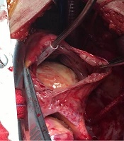
Idiopathic right atrial dilation is known as a congenital anomaly in the absence of obvious causes that produce right ventricular pressure or volume overload. Congenital giant right atrium can cause functional tricuspid regurgitation, arrhythmia, and right-sided heart failure. A massive Idiopathic Dilation of Right Atrium (IDRA) was reported in a 30-year-old man presented with intermittent palpitation and dyspnea on effort. Transthoracic echocardiography revealed huge right atrium leading to severe tricuspid regurgitation, moderate right ventricular enlargement with moderate dysfunction, and no evidence of other valvular heart diseases increasing the right ventricular pressure or volume, left to right shunt, and Ebstein anomaly. Cardiac Magnetic Resonance Imaging (MRI) confirmed the right atrial enlargement. The estimated right atrium volume by MRI was about 700 mL. With regard to the patient’s symptoms, he was scheduled for surgical treatment. The patient underwent partial surgical resection of the dilated right atrium wall and mechanical tricuspid valve replacement because the tricuspid valve was unrepairable on the operation table. Idiopathic right atrial enlargement is a very rare disease and may be easily confused with other anomalies that lead to right atrial enlargement, such as Ebstein anomaly. Hence, the probability of this pathology should be kept in mind when the usual etiologies of right atrial enlargement are excluded. Partial surgical resection of the dilated right atrium is an effective therapeutic option in symptomatic patients.

Fulltext

The full-text is available in PDF.
 
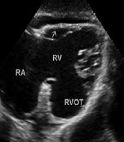
Video 1. The Huge Inappropriate Right Atrial Enlargement Seen in Apical Four-Chamber Echocardiographic View
 
Video 2. Axial Cine MR Showing Severe Right Atrial Enlargement and Severe Tricuspid Regurgitation with Partial Distortion due to Severe Right Atrial Dilation without Apical Displacement of Any of Its Leaflets
References

  • 1.
    The references are available in PDF.

Copyright

© 2019, Author(s). This open-access article is available under the Creative Commons Attribution 4.0 (CC BY 4.0) International License (https://creativecommons.org/licenses/by/4.0/), which allows for unrestricted use, distribution, and reproduction in any medium, provided that the original work is properly cited.

Similar Articles

31
Jul
2016

Vital Importance of Delineation of Coronary Artery Anatomy in Atypical Congenital Giant Right Atrial Aneurysm

Elaheh Malakan Rad,
Hassan Redmehr

Malakan Rad E, Redmehr H. Vital Importance of Delineation of Coronary Artery Anatomy in Atypical Congenital Giant Right Atrial Aneurysm. J Compr Ped. 2016;7(3):e37586. doi: https://doi.org/10.17795/compreped-37586

15
Dec
2014

Intermittent Prolapse of Right Atrial Myxoma in a Four-Year-Old Girl

Hager Barakizou,
Fethi Bayoudh

Barakizou H, Bayoudh F. Intermittent Prolapse of Right Atrial Myxoma in a Four-Year-Old Girl. J Compr Ped. 2014;5(4):e22931. doi: https://doi.org/10.17795/compreped-22931

23
Nov
2019
Journal of Comprehensive Pediatrics

An Affected Child with Arrhythmogenic Right Ventricular Dysplasia and Severe Left Ventricle Dysfunction: A Rare Case Report

Ali Akbar Zeinaloo,
Ehsan Aghaei Moghadam,
Amir Salimi,
Azin Ghamari,
Helia Mojtabavi,
Amir Bahador Golchin
,et al.

Zeinaloo AA, Aghaei Moghadam E, Salimi A, Ghamari A, Mojtabavi H, et al. An Affected Child with Arrhythmogenic Right Ventricular Dysplasia and Severe Left Ventricle Dysfunction: A Rare Case Report. J Compr Ped. 2020;11(1):e96077. doi: https://doi.org/10.5812/compreped.96077

4
Dec
2019
Iranian Journal of Radiology

Calcified Ball Thrombus in the Right Atrium: A Case Report

Bo Zhang,
Zhong Wei Tian,
Ji Zhang,
Kun Mao Yuan,
Hong Yu

Zhang B, Tian ZW, Zhang J, Yuan KM, Yu H. Calcified Ball Thrombus in the Right Atrium: A Case Report. I J Radiol. 2020;17(1):e95169. doi: https://doi.org/10.5812/iranjradiol.95169

30
Sep
2007

Comparison of transesophageal echocardiographic assessment of right and left atrial appendage functions

Maryam Esmaeilzadeh,
A Sadeghpour Sadeghpour,
A Fazlinezhad,
A Mohebbi,
M Maleki,
F Noohi
,et al.

Esmaeilzadeh M, Sadeghpour AS, Fazlinezhad A, Mohebbi A, Maleki M, et al. Comparison of transesophageal echocardiographic assessment of right and left atrial appendage functions. Int Cardiovasc Res J. 2018;1(3):e74123. doi:

Download PDF1.08 MB
Indexed in
Share on
Cited by
Metrics

Ordering Reprints

Articles are published under the Creative Commons license stated on each article. No permission or royalty fee is required for uses permitted by that license. CCC handles optional bulk and customized reprint orders. Any quotation covers production and delivery services only, not copyright permission. > Request Reprints from CCC 

Search Relations

Author(s):

Related Articles