Journal of Inflammatory Diseases
The Official Journal of Qazvin University of Medical Sciences
In Vitro Enzymatic Virulence Factors of Dermatophytes Species Isolated From Clinical Specimens
Authors

Abstract
Background: Dermatophytes are keratinophilic fungi that affect the stratum corneum of the skin and keratinous structures. Violent factors play a vital role in the pathogenesis and antifungal resistance of dermatophytes. Objective: This study aims to evaluate the activity of extracellular enzymatic and biofilm formation as virulence factors of dermatophyte isolates. Methods: Fifty-eight dermatophyte isolates belonged to 27 Trichophyton. rubrum (46.6%), 19 T. mentagrophytes (32.8%), and 12 Microsporum. canis (20.7%) for evaluating the activity of phospholipase, hemolysin, proteinase, and biofilm formation were examined. The biofilm formed was analyzed by scanning electron microscopy (SEM). Findings: Evaluation of extracellular enzymes production revealed that 86.2%, 77.6 %, and 57% of dermatophyte strains were shown to be phospholipase, hemolysin, and proteinase producers, respectively. Furthermore, all isolates of T. rubrum and M. canis can produce phospholipase and hemolysin, respectively. There was a statistically significant difference between phospholipase activity and dermatophyte strains (P<0.05). In addition, biofilm formation ability was observed in 41.5% of dermatophyte isolates. The highest level of biofilm production was found in 93% of dermatophytes isolated from nail chips. A significant difference between biofilm formation with dermatophyte isolates and different body sites was observed (P <0.05). Conclusion: The activity of hydrolytic enzymes and biofilm formation as important pathogenic factors may play a role in the persistence of dermatophytosis infections. Our results showed that dermatophyte isolates have enzymatic activity and biofilm production at different levels. Therefore, understanding the function of these factors is essential to controlling the spread of dermatophytosis infection.
Highlights
Copyright
© 2024, Journal of Inflammatory Diseases. This open-access article is available under the Creative Commons Attribution-NonCommercial 4.0 (CC BY-NC 4.0) International License (https://creativecommons.org/licenses/by-nc/4.0/), which allows for the copying and redistribution of the material only for noncommercial purposes, provided that the original work is properly cited.
Similar Articles
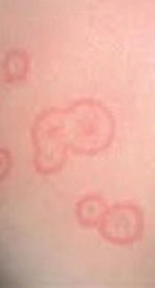
Specific Identification and Antifungal Susceptibility Pattern of Clinically Important Dermatophyte Species Isolated from Patients with Dermatophytosis in Tehran, Iran
Falahati M, Fateh R, Nasiri A, Zaini F, Fattahi A, et al. Specific Identification and Antifungal Susceptibility Pattern of Clinically Important Dermatophyte Species Isolated from Patients with Dermatophytosis in Tehran, Iran. Arch Clin Infect Dis. 2018;13(3):e63104. doi: https://doi.org/10.5812/archcid.63104
In vitro activity of six antifungal drugs against clinically important dermatophytes
Pakshir K, Bahaedinie L, Rezaei Z, Sodaifi M, Zomorodian K, et al. In vitro activity of six antifungal drugs against clinically important dermatophytes. Jundishapur J Microbiol. 2009;2(4):. doi:
Survey of keratinophilic fungi from soils in Ahvaz, Iran
Zarrin M, Haghgoo R. Survey of keratinophilic fungi from soils in Ahvaz, Iran. Jundishapur J Microbiol. 2011;4(3):. doi:
Staphylococcus epidermidis virulence factor and ability of macroscopic biofilm production
najar-peerayeh S, Jazayeri Moghadas A, bakhshi B. Staphylococcus epidermidis virulence factor and ability of macroscopic biofilm production. koomesh. 2016;17(4):e151185. doi:
Some Virulence Factors of Staphylococci Isolated From Wound and Skin Infections in Shahrekord, IR Iran
Ebrahimi A, Ghasemi M, Ghasemi B. Some Virulence Factors of Staphylococci Isolated From Wound and Skin Infections in Shahrekord, IR Iran. Jundishapur J Microbiol. 2014;7(4):e9225. doi: https://doi.org/10.5812/jjm.9225
- Scopus by DOI: 0
Last Update: 1 month ago
- Scopus by Title: 0
Last Update: 1 month ago
- Scopus by Title (Ref): 0
Last Update: 1 month ago
- CrossRef: 0
Last Update: 8 hours ago
- OpenAlex: 0
Last Update: 12 hours ago
- OpenCitations: 0
Last Update: 4 days ago