
1. Background
Modern linear accelerators, such as the Elekta Versa HD, integrate high-definition multileaf collimators (0.5-mm leaf width) and kilovoltage (kV) cone-beam computed tomography (CBCT) to achieve submillimeter positioning accuracy (1-3). Daily CBCT verification reduces planning target volume (PTV) margins from historical standards of 10 - 15 mm to 3 - 5 mm, significantly decreasing the radiation dose to critical structures such as the bladder, rectum, and femoral heads (4). This margin reduction directly translates to 30% - 40% lower complication risks for genitourinary and gastrointestinal toxicities, representing a critical advancement for long-term survivorship (5). However, these clinical benefits depend on rigorous quality assurance (QA). Scatter radiation, beam-hardening artifacts, and suboptimal reconstruction parameters degrade Hounsfield unit (HU) stability and geometric fidelity, thereby compromising adaptive replanning decisions (6, 7). Concurrently, repeated CBCT imaging results in a cumulative dose of 1 - 20 cGy over a standard fractionated course. Pediatric patients face a 2 - 3-fold higher lifetime attributable risk of radiation-induced secondary malignancies because of their greater tissue radiosensitivity and longer life expectancy (8, 9).
Current QA frameworks from the American Association of Physicists in Medicine (AAPM), Task Group (TG)-142 and TG-179, provide essential foundational guidance but have important limitations: they lack standardized longitudinal monitoring protocols, patient-specific tolerance thresholds, and quantitative correlations between image quality metrics and clinical positioning errors (10, 11). Without QA metrics anchored to baseline measurements, geometric inaccuracies may remain undetected, risking target underdosing or excessive irradiation of radiosensitive organs. Retrospective analyses indicate that 15% - 20% of image-guided radiotherapy (IGRT) failures stem from uncorrected CBCT calibration drift or HU instability, particularly in institutions lacking systematic performance monitoring (12). This gap is exacerbated in adaptive radiotherapy workflows, in which image quality directly determines the need for replanning, a vulnerability that is poorly addressed in current vendor guidelines.
2. Objectives
This study provides a comprehensive evaluation based on the existing QA protocol for the Elekta Versa HD XVI CBCT system, linking technical performance with clinical outcomes. We measured quantitative image quality parameters, including uniformity, noise, low- and high-contrast resolution, and HU stability, using the CatPhan 503 CTP404, CTP528, and CTP486 modules. The methodology follows current standards, with uncertainty propagation incorporated into all measurements.
3. Methods
This descriptive-analytical study used a longitudinal, phantom-based experimental design to evaluate the image quality and noise performance of the Elekta Versa HD XVI CBCT system. The protocol was implemented over six months using standardized QA measurements.
3.1. Quality Assurance Framework
Quality assurance for the Elekta Versa HD XVI CBCT system is essential to maintain consistent diagnostic image quality while keeping patient radiation dose as low as possible. To ensure compliance with manufacturer specifications and regulatory standards, specifically AAPM TG-142 and TG-179, we implemented a systematic monthly QA program. The program used comprehensive testing procedures with defined tolerance thresholds. Data acquisition included measurements from a single Elekta Versa HD linear accelerator over six months. No significant recalibrations were applied to the XVI platform during the six analyzed measurements; detector gain calibration was performed afterward in response to the detected noise exceedance. Baseline performance metrics were established during commissioning to support longitudinal stability assessment and to trigger action when required.
3.2. CBCT System Specifications
The Elekta XVI system employs an amorphous silicon flat-panel detector with an active area of 42.5 × 42.5 cm. A standardized clinical Pelvis QA protocol available on the Elekta XVI system was selected for image acquisition. Images were acquired using a full 360° gantry rotation at a tube voltage of 120 kVp. An M20 collimator and an F1 bowtie filter were used. The reconstructed images had a matrix size of 512 × 512 (Supplementary Table S1). A 10-minute tube-cooling interval was maintained between acquisitions, according to the manufacturer's protocol.
3.3. Image Quality Assessment
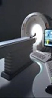
We evaluated five critical parameters using the CatPhan 503 phantom (The Phantom Laboratory, NY; diameter, 20 cm), as shown in Figure 1.
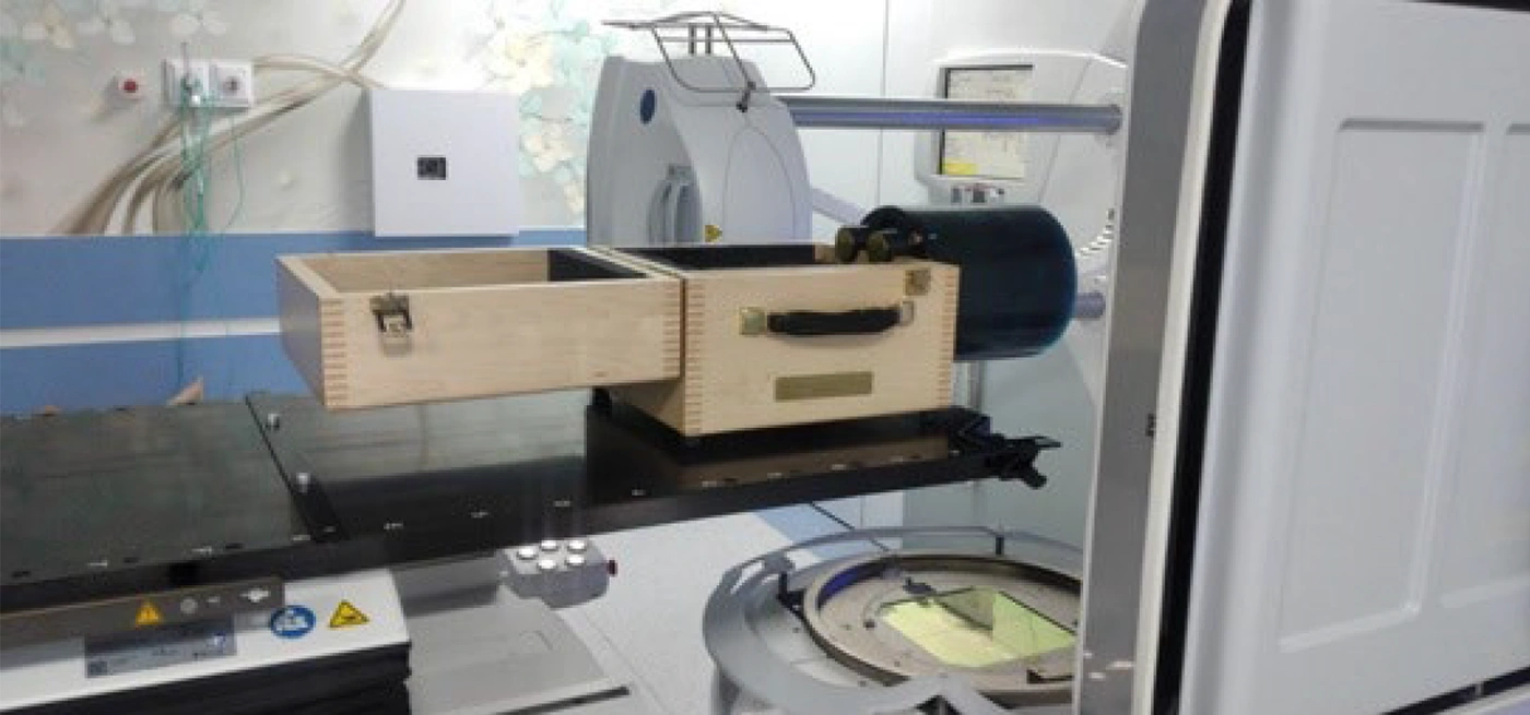
Figure 1.
CatPhan 503 phantom set up on the table of the Elekta Versa HD.
Specifically, we assessed:
1) Uniformity and noise using the CTP486 module.
2) Low-contrast resolution using the CTP404 module.
3) Spatial resolution using the CTP528 module.
4) HU stability using the CTP404 module.
5) Geometric accuracy using the CTP404 module.
All tests followed the AAPM TG-142 and TG-179 recommendations and the manufacturer's guidelines.
3.4. Phantom Setup and Imaging Protocol
The CatPhan 503 was positioned at the isocenter using room lasers and mechanical alignment pins. Before scanning, the X-ray tube underwent a standardized warm-up procedure. The phantom was immobilized on the couch top, and rotational alignment was verified along the G-T axis. DICOM volumes were exported for analysis in PIPSpro software (version 6.0+), and quantitative metrics were compared with institutional tolerance thresholds. Imaging parameters are detailed in Table S1 in the Supplementary File.
3.5. Image Quality Parameters
Uniformity was evaluated monthly for clinical kVp protocols using the CatPhan 503 CTP486 module. Five 1-cm-diameter circular regions of interest (ROIs) were placed on axial slices at the phantom center and at the cardinal points (3, 6, 9, and 12 o'clock) (Figure 2).
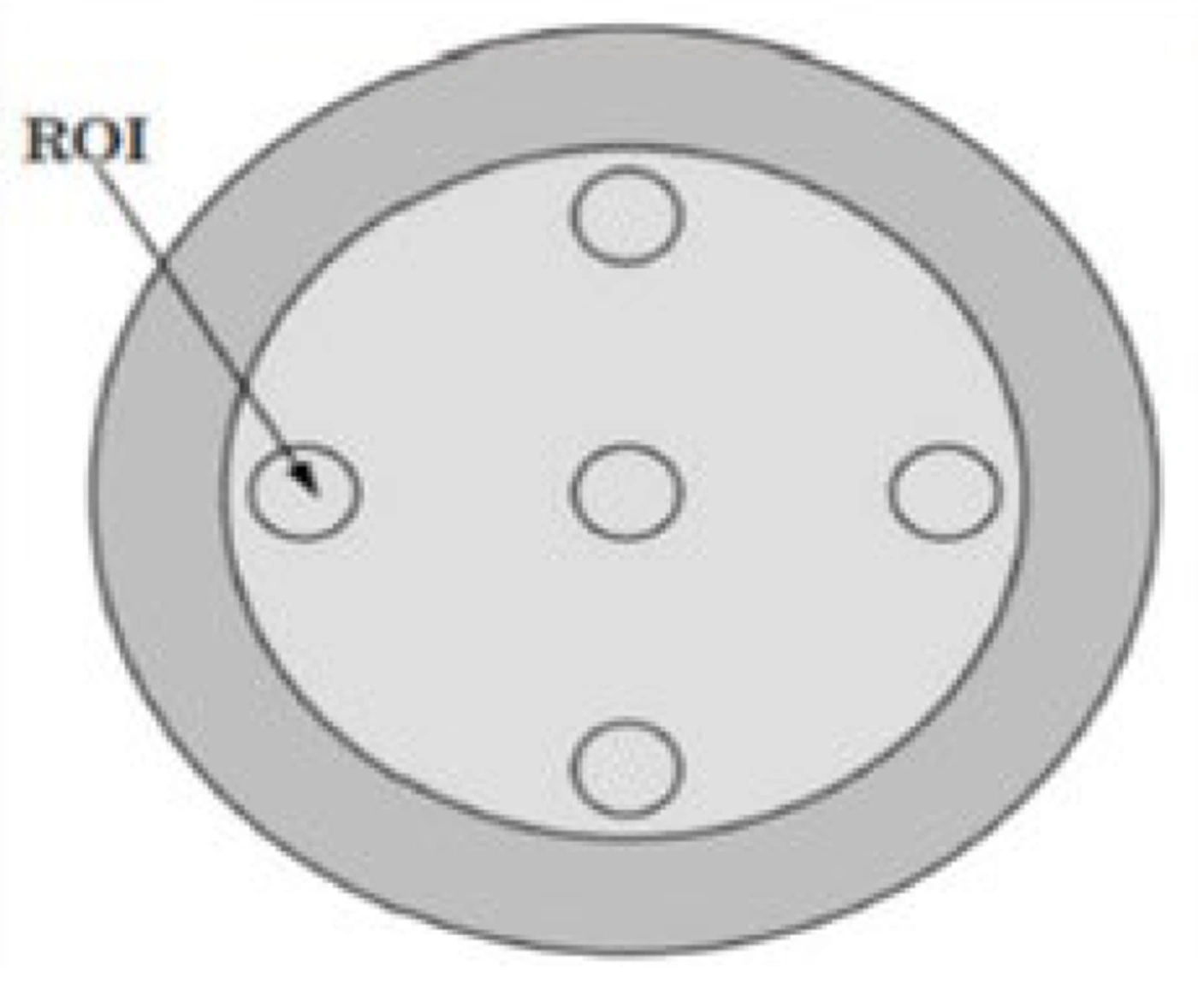
Figure 2.
CTP486 module of the CatPhan 503.
The Uniformity Index (UI) was calculated using the following formula:
The acceptance criterion followed the Elekta Versa HD XVI kV-CBCT guideline: UI ≤ 2.0%.
Noise was quantified as the standard deviation (SD) of HU values within a single 1-cm-diameter circular ROI at the phantom center in the CTP486 module. Images were reconstructed using 1.5-mm slice averaging to minimize random fluctuations. Noise values were reported in HU without normalization. In image quality control calculations, tolerance is usually defined as an absolute value in the measurement unit, here, HU, allowing deviation from a baseline value according to manufacturer specifications. For this imaging device, the allowable deviation from baseline was 2%.
To quantitatively evaluate image noise characteristics, the noise performance descriptor (NPD) was calculated from the HU values as the ratio of the SD of pixel values to the mean pixel value within the central ROI, expressed as a percentage. The calculation used the following formula:
Based on institutional QA guidelines and manufacturer specifications, the selected acceptance threshold for NPD was ≤ 2.0%.
3.6. Low-Contrast Resolution
Low-contrast visibility (LCV) was calculated using the CatPhan 503 CTP404 module (Table S2 in the Supplementary File) and the following formula:
In this formula, the CT value is the HU value obtained for materials within the phantom. Based on institutional QA guidelines and manufacturer specifications, the acceptance criterion for LCV was ≤ 3.0%.
Low-contrast detectability was quantified using the contrast-to-noise ratio (CNR) in the CatPhan 503 CTP404 module. After precise isocenter alignment, CBCT scans were acquired using the "CAT-Image Quality Contrast" preset. Three consecutive 0.5-mm slices were averaged (1.5 mm in total) to reduce noise. The CNR was calculated from two 3.5-mm-diameter ROIs centered on polystyrene (−35 HU) and LDPE (−100 HU) inserts as follows:
A CNR ≥ 1.0 was considered acceptable.
3.7. Spatial Resolution
High-contrast spatial resolution was assessed visually using the CTP528 module's aluminum line-pair patterns, which ranged from 5 - 21 lp/cm. Scans used the "CAT-Image Quality Spatial" preset with 1.5-mm slice averaging. The central slice within the module's axial range was evaluated. Resolution was defined as the highest line-pair group with ≥ 50% visible bars. The manufacturer's acceptance threshold was ≥ 10 lp/cm.
3.8. Hounsfield Unit Stability
HU stability was evaluated using the CTP404 module's eight reference materials, ranging from air to Teflon. Scans used the "CAT-Image Quality HU" preset. Measured HU values were compared with reference values in the phantom documentation. The maximum allowable deviations were ± 40 HU for water-equivalent materials and ± 150 HU for high-density inserts. Deviations exceeding these thresholds triggered system recalibration (Table S2 in the Supplementary File).
3.9. Statistical Analysis
Linear regression analyses were performed to evaluate temporal trends in monthly CBCT image QA parameters over the 6-month observation period. Separate models were fitted for intensity, uniformity, resolution, low-contrast variability (LCV), and relative noise, with month as the independent variable and each QA parameter as the dependent variable. Unstandardized regression coefficients (B), 95% confidence intervals (CIs), P values, and coefficients of determination (R2) were reported. The analyses were intended to characterize linear changes in the measured parameters over time. Statistical analyses were performed using Stata version 17, with statistical significance set at P < 0.05.
4. Results
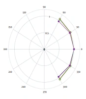
Monthly QA assessments of the Elekta Versa HD XVI CBCT system using the CatPhan 503 phantom demonstrated consistent performance across critical imaging metrics, with one notable exception. Scans used a 360° acquisition protocol with a small field of view (15 cm) and an S20 filter. The UI, calculated across five three-dimensional ROIs at the center and periphery of the CTP486 module, averaged 1.2% ± 0.3% (Figure 3), meeting the acceptance criterion of UI ≤ 2.0%. However, noise measurements in the phantom’s central ROI showed an SD of 22.21 HU and a mean pixel value of 1017 HU (Table S3 in the Supplementary File), yielding an NPD of 2.18%, which exceeded the acceptance threshold (≤ 2.0%).
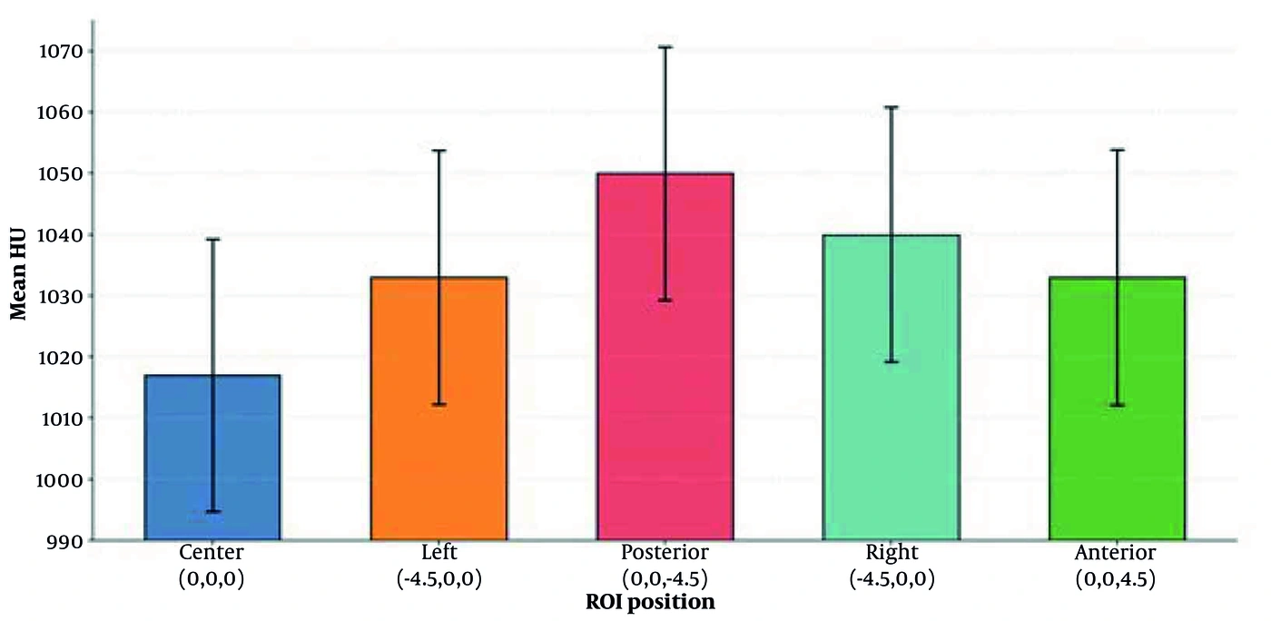
Figure 3.
Spatial distribution of mean HU values across phantom coordinates.
Low-contrast resolution, evaluated using the CTP404 module for eight distinct materials (Figure 4A), yielded a low-contrast variability (LCV) of 0.39%, within the acceptable limit of ≤ 3.0%. High-spatial-resolution testing with the CTP528 module resolved 14 lp/cm, exceeding the minimum requirement of ≥ 10 lp/cm by 40%. The CNR was approximately 2.1 and met the acceptance criterion. HU stability measurements across eight tissue-equivalent materials in the CTP404 module (Table S2 in the Supplementary File) showed strong agreement with reference values: air (−996 ± 4 HU), water (4 ± 2 HU), and acrylic (118 ± 3 HU). The maximum deviation of 18 HU was observed for Teflon and remained within the ± 40 HU tolerance band (Figure 4B).
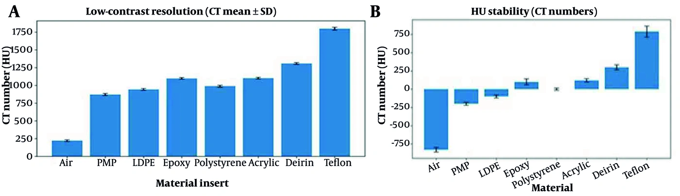
Figure 4.
Comprehensive evaluation of image quality metrics. Mean CT values across eight regions of interest (ROIs) representing diverse material compositions (A) and Hounsfield unit (HU) stability across eight ROIs (B).
After the NPD exceedance (2.18%) was detected, the CBCT system was immediately flagged at the institutional QA action level. An immediate repeat scan was performed to rule out phantom setup errors or transient fluctuations; this confirmed the elevated noise level. The system was subsequently removed from clinical use for IGRT until the issue was resolved. A service engineer was contacted, and detector gain calibration was performed. Following corrective maintenance, the system was returned to clinical operation after meeting standard institutional QA criteria.
Linear regression analyses assessed temporal trends in the five QA parameters over the 6-month observation period. As shown in Table 1, relative noise exhibited a statistically significant increasing linear trend over time (B = 26.778; 95% CI, 24.323 - 29.232; P < 0.001), with month accounting for 98.34% of the observed variance in relative noise (R2 = 0.983). In contrast, no statistically significant linear trends were observed for intensity (B = 0.016, P = 0.874), uniformity (B = 0.118, P = 0.612), resolution (B = −0.163, P = 0.467), or low-contrast variability (LCV; B = −0.020, P = 0.839). The corresponding 95% CIs included zero. These results indicate an increasing temporal trend in relative noise, with no statistically significant linear trends in the other four parameters during the observation period.
Table 1.
Linear Regression Analysis of Temporal Trends in Quality Assurance Parameters Over 6 Months
| Parameters | Regression Coefficient (β) | Standard Error | t-Statistic | P-Value | 95% CI | R2 |
|---|---|---|---|---|---|---|
| Intensity | 0.016 | 0.097 | 0.16 | 0.874 | -0.200, 0.232 | 0.0026 |
| Uniformity | 0.118 | 0.226 | 0.52 | 0.612 | -0.385, 0.622 | 0.0266 |
| Resolution | -0.163 | 0.215 | -0.76 | 0.467 | -0.643, 0.317 | 0.0541 |
| LCV | -0.020 | 0.096 | -0.21 | 0.839 | -0.233, 0.193 | 0.0043 |
| Relative Noise | 26.778 | 1.10 | 24.31 | < 0.001 | 24.32, 29.23 | 0.9834 |
5. Discussion
This study establishes a physics-driven QA framework for the Elekta Versa HD XVI CBCT system using a systematic, phantom-based approach.
The observed UI of 1.2% ± 0.3% highlights the system’s ability to maintain spatial signal homogeneity. In IGRT, signal uniformity is essential for robust automated image registration and accurate dose calculation in adaptive workflows (11). Consistent with the study by Taneja et al. (7), our results suggest that the beam-shaping and reconstruction algorithms remain well calibrated, effectively mitigating artifacts such as cupping or shading under standard acquisition conditions.
The system’s performance in low-contrast and high-spatial-resolution assessments further supports its diagnostic fidelity. A low-contrast variability (LCV) of 0.39% demonstrates sensitivity to soft-tissue density variations, a parameter critical for target volume localization (13). Similarly, the high spatial resolution of 14 lp/cm, which exceeds the threshold by 40%, indicates that the system maintains excellent edge-spread performance. This resolution margin is particularly advantageous for precise bony anatomy alignment, for which visualization of trabecular structures is essential to minimize setup uncertainties (14). Furthermore, the maximum HU deviation of 18 HU from reference standards supports the system’s quantitative consistency, which is foundational for clinical applications requiring reliable voxel intensities (15, 16).
Noise measurements in the phantom’s central ROI showed an SD of 22.21 HU and a mean pixel value of 1017 HU, resulting in an NPD of 2.18%, which exceeded the acceptance threshold (≤ 2.0%).
The NPD exceedance (2.18%) is of particular clinical concern. Although previous studies have emphasized that moderate noise is inherent to CBCT, excessive stochastic noise significantly degrades low-contrast detectability (LCD) (17).
Clinically, this degradation has direct consequences for contouring accuracy (18). Increased noise acts as a stochastic barrier, obscuring anatomical gradients. According to Li et al., high noise-to-signal ratios correlate with increased interobserver variability in contouring soft-tissue structures (13), such as the prostate or pelvic lymph nodes. Furthermore, noise-induced pixel fluctuations could theoretically cause local optimization failures in registration algorithms, potentially resulting in geometric inaccuracies in deformed contours (19). In synthetic CT generation workflows, increased noise propagates into dose recalculation. Even when mean HU values are preserved, increased voxel-intensity variance can introduce uncertainty into dose distributions. This may necessitate larger PTV margins to ensure target coverage, undermining the purpose of margin-reduced adaptive treatments.
The primary finding of this investigation, an NPD of 2.18% exceeding the acceptance threshold of ≤ 2.0%, warrants careful interpretation. Although local contrast variability (LCV) remained within acceptable limits, the NPD exceeded its acceptance threshold. This discrepancy suggests that, although global contrast discrimination was maintained, local signal-to-noise ratios (SNRs) were deteriorating. The elevated noise may result from a combination of factors, including detector aging, inaccuracies in scatter correction, or suboptimal projection data acquisition. From a clinical safety perspective, elevated noise could compromise observer performance and automated segmentation accuracy. This exceedance prompted withdrawal of the system from clinical use, detector gain recalibration, and return to operation only after successful institutional QA testing.
5.1. Study Limitations
This study has inherent limitations. First, although phantom assessments are reproducible, they represent an idealized imaging scenario that may not fully capture the complex scatter environments encountered in larger patients. Second, the analysis used a static protocol; therefore, the effects of varying dose-reduction strategies or field-of-view sizes on noise performance remain to be quantified. Additionally, no noise power spectrum (NPS) analysis was conducted in this study. Future research should incorporate NPS analysis and power-spectrum decomposition to correlate phantom-derived noise metrics with clinical image quality assessments and distinguish the contributions of detector-specific electronic noise from patient-scattered radiation.
5.2. Conclusions
This longitudinal, phantom-based analysis confirms that the Elekta Versa HD XVI CBCT system delivers reliable performance in spatial resolution, uniformity, and HU accuracy. However, the observed noise deviations indicate that reliance on simple descriptors such as NPD may be insufficient. Incorporating advanced metrics such as NPS is essential to fully characterize system performance. Integrating these sensitive assessments into routine QA can support predictive maintenance and help ensure that IGRT workflows remain within optimal technical parameters. Rigorous QA is crucial for adaptive radiotherapy. Although this phantom study did not directly assess patient outcomes, stable, high-quality imaging is necessary to support the broader objectives outlined in AAPM TG-142 and TG-179. Maintaining these technical thresholds may support the safe future use of CBCT data for adaptive replanning, potentially extending CBCT from a positioning tool to a quantitative imaging resource, particularly in resource-constrained settings.