
1. Context
Neurotoxicity induced by CNS agents—such as hallucinogens, stimulants, and opioids—poses a significant threat to neurological function and has been associated with the onset and progression of neurodegenerative diseases, including Alzheimer's disease, which affects over 50 million people worldwide; Parkinson's disease, which affects more than 10 million individuals; and multiple sclerosis, with a global prevalence of approximately 2.8 million (1-3). These conditions are characterized by the deterioration of neuronal structure and function, leading to cognitive and motor deficits (1). Neurotoxic agents, including chemicals, drugs, environmental toxins, and biological substances, can cause extensive CNS damage through mechanisms such as oxidative stress, inflammation, excitotoxicity, and apoptosis (4-7). Natural compounds are chemical substances produced by living organisms, most commonly plants, but also including certain animal-derived, dietary, and endogenous compounds. Chemically, they are classified according to structural features and biosynthetic origin, including alkaloids, terpenoids, phenolics, glycosides, lipids, proteins/peptides, and polysaccharides, many of which exhibit pharmacological activity relevant to neuroprotection.
Oxidative stress is caused by an imbalance between reactive oxygen species (ROS) production and the brain's antioxidant defense system, ultimately resulting in cellular damage. Inflammation, which is often chronic in neurodegenerative conditions, exacerbates neuronal damage through the release of pro-inflammatory cytokines and chemokines (8, 9). Excitotoxicity, primarily mediated by excessive glutamate release and subsequent overstimulation of glutamate receptors, results in calcium overload and neuronal damage (10, 11). Apoptosis, or programmed cell death, is another critical pathway that causes neuronal loss in neurotoxic conditions (12).
In recent years, interest in natural products as neuroprotective agents has increased, particularly given the growing global burden of neurodegenerative diseases (13, 14). These products, derived from plants, animals, and microorganisms, have traditionally been used in medicine for their therapeutic properties and are now being studied extensively for their potential in modern clinical applications (15, 16). Natural compounds exhibit a wide range of pharmacological effects, including antioxidant, anti-inflammatory, anti-excitotoxic, and anti-apoptotic activities, making them promising candidates for mitigating neurotoxic damage (17-19).
Many of the natural products discussed in this review, including curcumin, resveratrol, flavonoids, and other polyphenols, are naturally present in commonly consumed foods and are also widely available as dietary supplements (20). Curcumin is the principal bioactive component of turmeric (Curcuma longa), a spice extensively used in Asian cuisine, whereas resveratrol is found in grapes, berries, and red wine (21). Flavonoids such as quercetin and luteolin occur in onions, apples, citrus fruits, and green tea, whereas catechins are abundant in green tea and related nutraceutical preparations. Ginsenosides are the major active constituents of ginseng and are available in teas, powders, and standardized supplement formulations (22). Similarly, sulforaphane from broccoli sprouts, alpha-lipoic acid from vegetables such as spinach and broccoli, and coenzyme Q10, which is present in meat and fish and is also marketed as a supplement, can be obtained through both dietary intake and commercial products (23).
However, the concentration, formulation, and bioavailability of these compounds differ considerably between natural food sources and manufactured supplements. Consequently, the amounts achieved through a normal diet are often substantially lower than the doses used in experimental studies. Therefore, although regular dietary consumption may contribute to antioxidant and neuroprotective support, controlled supplementation is frequently required to achieve the pharmacologically relevant exposures evaluated in preclinical models.
Mechanistically, these compounds exert protective effects by scavenging ROS, enhancing endogenous antioxidant systems, modulating inflammatory pathways, and inhibiting apoptotic signaling (24). For example, resveratrol scavenges free radicals and enhances the expression of antioxidant enzymes, whereas curcumin neutralizes ROS and increases glutathione levels (25). These compounds also inhibit pro-inflammatory pathways and cytokine production, thereby reducing neuroinflammation. Many natural products reduce neuroinflammation by suppressing the activation of nuclear factor-kappa B (NF-κB), leading to lower production of pro-inflammatory cytokines. For example, ginkgolides decrease the levels of TNF-α and IL-1β (26, 27). In addition, natural products activate detoxifying enzymes and pathways that neutralize and eliminate toxic substances (28). Selenium is essential for the activity of GPxs, and berberine activates phase II detoxifying enzymes such as GSTs (29).
Experimental evidence from in vitro and in vivo studies has demonstrated the efficacy of these natural products in protecting against neurotoxic agents. A previous study investigated the effects of Jitai tablets, a traditional Chinese medicine, on methamphetamine-induced dopaminergic neurotoxicity (30). The findings showed that Jitai tablets significantly protected against neurotoxic damage and helped restore dopaminergic neuron function. These results indicate that Jitai tablets may have therapeutic potential for neurotoxicity related to methamphetamine use (30).
Although several previous reviews have examined the neuroprotective potential of natural products, most have focused either on neurodegenerative diseases such as Alzheimer's and Parkinson's disease or on neurotoxicity arising from environmental toxins, heavy metals, and chemotherapeutic agents (6, 7, 13, 14). In contrast, neuroprotection against neurotoxicity induced specifically by CNS-active drugs of abuse—stimulants, hallucinogens/dissociative, and opioids—has not been systematically consolidated within a single review, despite substantial and mechanistically overlapping evidence for these agents. This gap is notable given the distinct toxicokinetic and clinical context of drug-induced neurotoxicity compared with disease- or toxin-related neurodegeneration, including acute high-dose exposure patterns, co-occurring substance use, and the need for interventions compatible with concurrent pharmacological treatment. The present review addresses this gap by systematically summarizing preclinical evidence on natural product–mediated neuroprotection across these three pharmacological categories of CNS-active agents.
This review is timely, as it consolidates current evidence on the neuroprotective mechanisms of natural compounds, identifies gaps in understanding regarding their efficacy against CNS neurotoxic agents, and highlights emerging therapeutic strategies targeting oxidative stress, inflammation, excitotoxicity, and apoptosis. Overall, the review underscores the promising role of natural products in mitigating neurotoxicity and emphasizes their potential for developing preventive and therapeutic strategies against neurodegenerative diseases and other CNS disorders. It also discusses broader implications for health and medicine, addresses the limitations of current research, and outlines future directions for advancing natural product-based neuroprotective strategies (Figure 1).
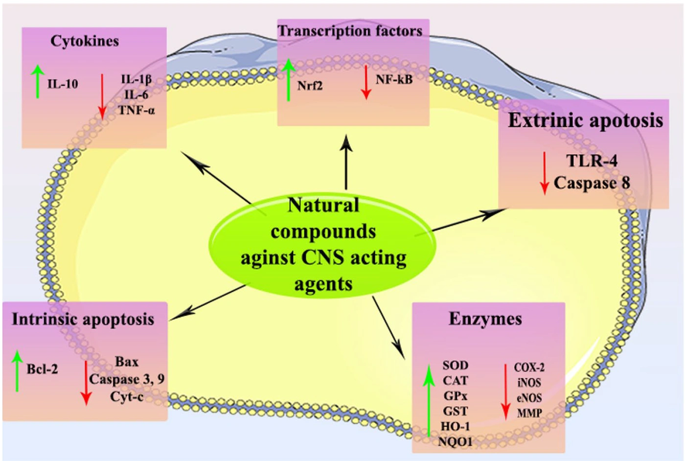
Figure 1.
Graphical abstract
2. Evidence Acquisition
This narrative review is based on a comprehensive but nonsystematic literature search; a formal systematic-review protocol, dual independent screening, and risk-of-bias/certainty-of-evidence assessment (e.g., PRISMA, GRADE) were not applied, consistent with the narrative review design. Relevant studies published from January 2000 to July 2025 were identified through searches of PubMed, Scopus, and Web of Science. A combination of controlled vocabulary and free-text keywords was used, grouped into four categories.
Search terms were grouped into four categories. Terms within each category were combined with OR, and the categories were combined with AND.
Intervention terms (OR): "natural products," "phytochemicals," "plant extracts," "medicinal plants," "polyphenols," "flavonoids," "alkaloids," curcumin, resveratrol, EGCG, quercetin. These four named compounds were included as representative examples of the broader intervention categories rather than as an exhaustive list; other natural compounds discussed in this review (e.g., sulforaphane, icariside II, sauchinone, taurine, oleuropein) were retrieved under the broader category terms (e.g., "flavonoid," "phytochemical," "plant extract") rather than through individual name-based searching.
Outcome terms (OR): "neuroprotection," "neurotoxicity," "neuronal damage," "neurodegeneration," "CNS toxicity."
Mechanistic terms (OR): "antioxidant," "anti-inflammatory," "apoptosis," "oxidative stress," "mitochondrial dysfunction," "neuroinflammation."
Agent terms (OR): "CNS drugs," "opioid agents," "antipsychotics," "stimulants," "drugs of abuse," methamphetamine, cocaine, MDMA, phencyclidine, morphine, tramadol.
Studies were included if they: 1) investigated at least one natural product with potential neuroprotective activity; 2) used preclinical (in vitro or in vivo) models; 3) employed a defined neurotoxic agent or pathological CNS model; and 4) reported measurable neuroprotective outcomes, such as oxidative stress markers, inflammatory markers, apoptotic markers, or behavioral and histopathological findings. Clinical studies were also considered during the search, but none met the inclusion criteria. Exclusion criteria included review articles, conference abstracts, studies without full-text availability, insufficient methodological detail, lack of an appropriate control group, or absence of measurable outcomes. Compounds discussed in this review were classified as natural products (plant-derived bioactive compounds and traditional herbal formulations), endogenous/dietary compounds (naturally occurring in the human body or diet, e.g., vitamins, taurine, coenzyme Q10), or synthetic comparator agents (deferoxamine). Deferoxamine was retained only because it was evaluated within the same experimental study as alpha-tocopherol under an identical methamphetamine-toxicity model, allowing direct mechanistic comparison between a natural antioxidant and a synthetic iron-chelating comparator; it is not included in any summary statement or conclusion regarding natural products.
In a few cases in which direct preclinical evidence for a specific CNS-toxic agent was limited (e.g., cocaine, PCP, MDMA, and opioids), studies using closely related experimental models with shared molecular pathways or receptor systems were included. These studies are clearly identified as indirect evidence within the relevant sections and are not presented as direct evidence for the specific agent.
# Records from the three databases were imported into EndNote (Clarivate), and duplicate records were identified and removed using its automated duplicate-detection function (based on title, author, and DOI matching), followed by manual verification of flagged records. The initial database search identified 883 records (PubMed, n = 500; Scopus, n = 227; Web of Science, n = 156). After removing 247 duplicate records, 636 unique articles remained for title and abstract screening. Following title and abstract screening, 575 records were excluded because they did not meet the predefined eligibility criteria. The full texts of 61 articles were assessed for eligibility. Of these, 32 articles were excluded due to the absence of a defined neurotoxic agent or CNS model (n = 13), lack of quantifiable neuroprotective outcomes (n = 8), insufficient methodological detail (n = 6), or unavailable full text (n = 5). Ultimately, 29 studies met the inclusion criteria and were included in this narrative review (Figure 2).
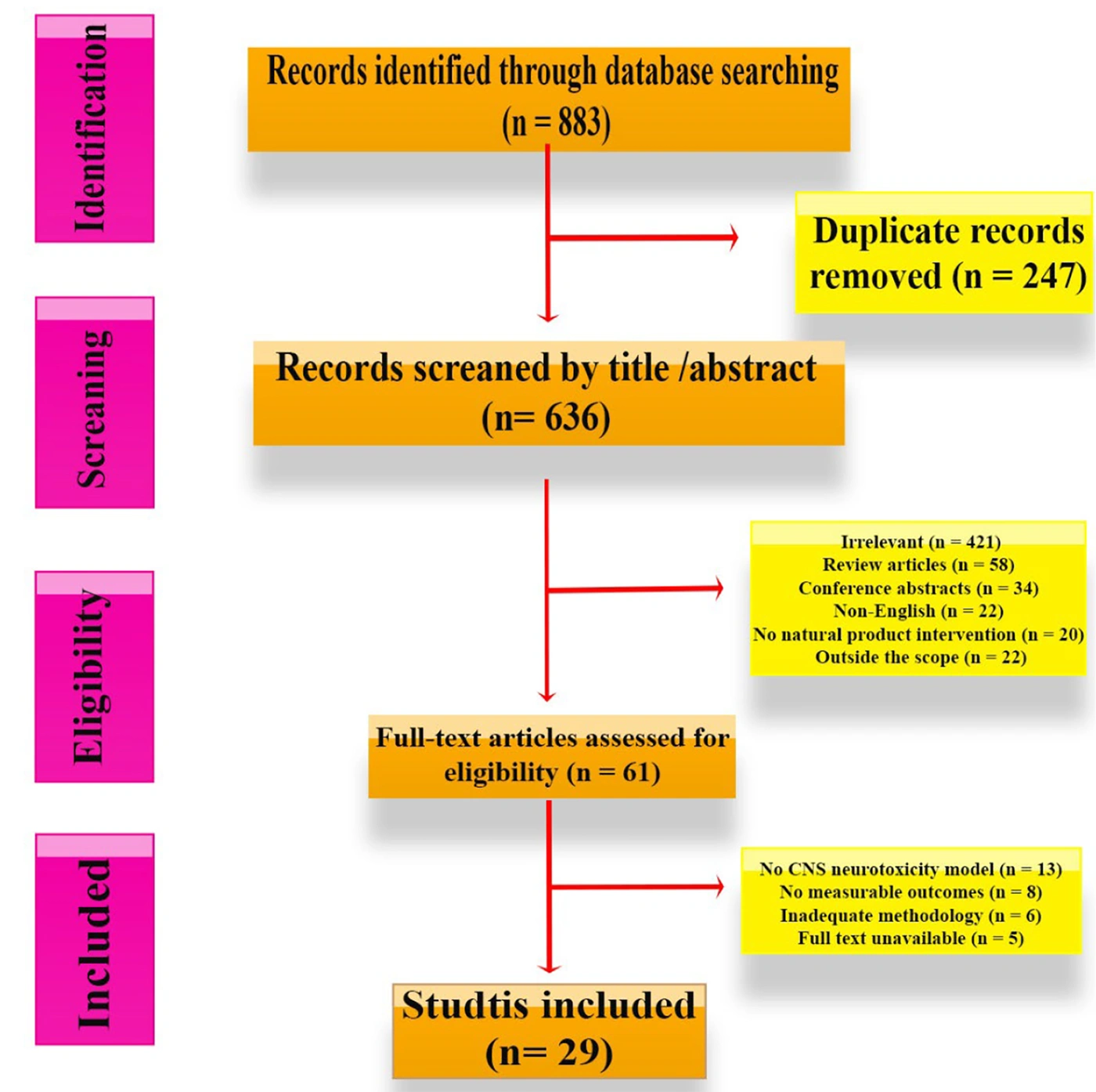
Figure 2.
Search strategy
3. Results
3.1. Antioxidant Activity
Antioxidant activity is a major mechanism by which natural products protect against neurotoxicity induced by CNS-active agents such as stimulants, hallucinogens, and opioids (31). Oxidative stress induced by these agents leads to the production of ROS, which can damage cellular components, including lipids, proteins, and DNA, ultimately contributing to neuronal cell death and cognitive deficits (32). The antioxidant effects of natural compounds are mainly attributed to their ability to neutralize ROS and enhance the activity of endogenous antioxidant enzymes, including superoxide dismutase (SOD), catalase (CAT), and glutathione peroxidase (GPx). By reducing ROS levels and strengthening cellular defense systems, these mechanisms help prevent oxidative damage to lipids, proteins, and DNA, protect neurons from apoptosis, and mitigate cognitive deficits induced by CNS-toxic agents, thereby supporting neuronal integrity and overall brain health (33).
Natural products are rich sources of antioxidants, including flavonoids, polyphenols, and vitamins C and E, which enhance the body’s defenses against oxidative damage. These antioxidants neutralize harmful free radicals and promote cellular repair mechanisms, thereby mitigating the harmful effects of nerve agent exposure (34, 35). For instance, studies have demonstrated that flavonoids can significantly reduce lipid peroxidation and improve mitochondrial function under oxidative stress induced by neurotoxic agents (36).
Chinonin, a natural flavonoid, exhibits neuroprotective effects primarily through antioxidant activity. It enhances endogenous defenses, including SOD and GSH, reduces ROS and LPO, and helps preserve neuronal integrity. These properties make chinonin a promising candidate for preventing or mitigating oxidative stress-related neurodegeneration (37). Lonicerae Japonicae Flos is rich in phenolic compounds and flavonoids with strong ROS-scavenging potential. Its antioxidant properties suggest neuroprotective effects, potentially reducing oxidative stress and inflammation in the CNS and offering therapeutic benefits against neurodegenerative conditions such as Alzheimer’s disease (38). Moreover, synergistic effects among multiple antioxidants in natural products may enhance their protective capacity. Research has indicated that combinations of different antioxidants may yield greater neuroprotective effects than individual compounds, potentially leading to improved recovery of neuronal function (39). Accordingly, incorporating antioxidant-rich natural products into therapeutic strategies may help counteract the effects of CNS neurotoxic agents by preserving neuronal integrity and supporting overall brain health (40).
In summary, the antioxidant effects of natural products against neurotoxic agents involve scavenging reactive species, reducing oxidative stress, and supporting neuronal survival. Further studies of these mechanisms may help identify more effective strategies to protect the nervous system from neurotoxic damage.
3.2. Inflammatory Mechanisms
Natural products have attracted considerable interest because of their potential to protect against neurotoxicity, particularly that induced by stimulant drugs, hallucinogens, and opioids. A key aspect of this protection is the modulation of inflammatory mechanisms, which play a significant role in the neurotoxic processes triggered by these substances (41, 42). Stimulants such as cocaine and methamphetamine, as well as hallucinogens and opioids, can induce inflammatory responses in the CNS (1, 43). These responses are not limited to the CNS, where activated microglia are major mediators, but may also involve systemic inflammatory processes that exacerbate neuroinflammation through peripheral immune signaling (44). This process involves microglial activation and the release of pro-inflammatory cytokines, which can lead to neuronal damage and apoptosis (45). Chronic activation of these inflammatory pathways has been implicated in various neurodegenerative diseases and cognitive impairments (46). Many natural products, including flavonoids, polyphenols, and certain alkaloids, exhibit robust anti-inflammatory properties. For example, curcumin (derived from turmeric) and resveratrol (found in grapes and berries) have been shown to inhibit NF-κB activation and reduce the production of pro-inflammatory cytokines (47). In addition to NF-κB, other signaling pathways, such as mitogen-activated protein kinases (MAPKs), are also targeted by natural products, leading to suppression of pro-inflammatory gene expression and attenuation of neuroinflammatory cascades (48). Natural products can also modulate specific cytokines involved in neuroinflammation by promoting the secretion of anti-inflammatory cytokines such as interleukin-10 (IL-10) while reducing levels of pro-inflammatory cytokines such as tumor necrosis factor-alpha (TNF-α) (49). For instance, berberine has been reported to inhibit inflammation through modulation of NF-κB and MAPK pathways, as demonstrated in both molecular and network pharmacology studies (50). Some natural products may also promote neurogenesis and repair of damaged neural circuits. Brain-derived neurotrophic factor (BDNF) can be upregulated by certain natural substances, enhancing neuronal growth and resilience; this neuroprotective effect can counteract the neurotoxic impacts of stimulants and opioids (51). Furthermore, natural products often exert protective effects through synergistic interactions with other biological pathways, including antioxidant defenses and apoptotic signaling (52). By modulating inflammation in concert with these pathways, natural products can provide a multifaceted approach to neuroprotection against the diverse neurotoxic effects of drugs (47).
In summary, the neuroprotective effects of natural products against stimulant-, hallucinogen-, and opioid-induced neurotoxicity are largely attributed to their ability to regulate inflammatory responses. These effects involve both local and systemic anti-inflammatory actions through pathways such as NF-κB and MAPK. By limiting neuroinflammation, supporting neuronal recovery, and strengthening antioxidant defenses, natural compounds may help preserve neuronal function during drug-induced neurotoxicity. Further research may provide new opportunities for developing neuroprotective therapies based on these natural products.
3.3. Apoptotic Mechanisms
Apoptosis, or programmed cell death, is essential for maintaining cellular homeostasis and is closely involved in the development of many neurological disorders; it is particularly relevant in the context of neurotoxicity induced by substances such as stimulants, hallucinogens, and opioids. In this context, the term natural products refers primarily to plant-derived bioactive compounds, including isolated phytochemicals such as flavonoids, alkaloids, polyphenols, and terpenoids, as well as certain traditional herbal formulations (53). The protective effects of natural products against this neurotoxicity are strongly associated with their ability to modulate apoptotic mechanisms (36). Exposure to neurotoxic agents in the nervous system frequently activates apoptotic pathways, which can result in neuronal cell death (54). Stimulants such as cocaine and methamphetamine, hallucinogens such as lysergic acid diethylamide (LSD) and psilocybin, and opioids including morphine and fentanyl have been shown to induce neuronal apoptosis through multiple pathways, including extrinsic and intrinsic apoptotic cascades (55, 56). These pathways involve caspase activation, which is essential for apoptosis execution, and the release of pro-apoptotic factors from mitochondria (55). Natural products, which are often rich in bioactive compounds, have been reported to exert neuroprotective effects by intervening in these apoptotic processes. For instance, curcumin inhibits apoptosis primarily by blocking caspase-3 activation and preventing mitochondrial release of cytochrome c, whereas resveratrol acts through activation of sirtuin-1 (SIRT1) signaling and upregulation of anti-apoptotic proteins such as Bcl-2 (57-59). Recent studies support these neuroprotective effects of natural products. For example, Hu et al. used a network pharmacology approach to show that Astragaloside IV can reduce neuronal apoptosis and inflammation in intracerebral hemorrhage models, highlighting molecular mechanisms relevant to neurotoxicity (60). Similarly, Zeng et al. reviewed how natural products modulate methamphetamine-induced neuronal apoptosis through oxidative stress, mitochondrial-dependent, and endoplasmic reticulum stress-mediated pathways, reinforcing the therapeutic relevance of these mechanisms within CNS-drug neurotoxicity (55). Furthermore, these natural products can induce the expression of anti-apoptotic proteins such as B-cell lymphoma 2 (Bcl-2), which counteract pro-apoptotic signals and promote cell survival (61). Apoptosis is regulated through extrinsic and intrinsic pathways, both of which can be targeted by natural products to enhance protective effects. The extrinsic pathway is initiated by extracellular death ligands (e.g., TRAIL, FasL) binding to cell surface death receptors (e.g., DR4/5, Fas), leading to caspase-8 activation via the death-inducing signaling complex (62). Natural compounds may inhibit extrinsic apoptosis by modulating death-receptor signaling and promoting antiapoptotic proteins (e.g., c-FLIP). In contrast, the intrinsic (mitochondrial) pathway is activated by intracellular stressors, including DNA damage and oxidative stress. This leads to mitochondrial outer membrane permeabilization (MOMP), release of cytochrome c, and subsequent activation of caspase-9. Bioactive agents may modulate Bcl-2 family proteins (e.g., decrease Bax/Bak and increase Bcl-2), restore p53 function, and inhibit ROS-mediated DNA damage, thereby reducing intrinsic apoptosis in damaged cells (36).
Apoptosis is also closely linked to the antioxidant and inflammatory mechanisms described above: oxidative stress (via Nrf2-related pathways, see the Antioxidant section) and chronic neuroinflammation (via NF-κB and pro-inflammatory cytokines, see the Inflammatory section) both increase susceptibility to apoptotic cell death (46). By limiting these upstream triggers, natural products may indirectly reduce apoptotic signaling in addition to their direct effects on caspases and Bcl-2 family proteins. Furthermore, natural products may also support neurogenesis, complementing the neurotrophic effects (e.g., BDNF upregulation) discussed in the Inflammatory Mechanisms section above, and further counterbalancing neuronal loss caused by drug-induced toxicity (63).
In summary, the role of apoptotic mechanisms in the protective effects of natural products against neurotoxicity induced by stimulants, hallucinogens, and opioids can be highlighted through three distinct biological contributions: 1) inhibition of apoptotic pathways via caspase regulation and Bcl-2 upregulation, 2) reduction of oxidative stress through direct radical scavenging and Nrf2 activation, and 3) promotion of neurogenesis and modulation of neuroinflammatory responses. Further studies are needed to determine whether these natural compounds could serve as potential therapeutic options for the prevention and treatment of neurotoxicity in vulnerable populations.
3.4. Amphetamine
Methamphetamine is a potent psychostimulant belonging to the amphetamine class that exerts its primary effects through excessive release of monoamines, particularly dopamine, in the CNS. Chronic or high-dose exposure leads to profound neurotoxicity, especially in dopaminergic and serotonergic pathways, through multiple mechanisms, including oxidative stress, mitochondrial dysfunction, excitotoxicity, and neuroinflammation. These processes ultimately result in neuronal apoptosis, synaptic dysfunction, and long-term cognitive and behavioral impairments (64, 65). The complexity of these overlapping pathological pathways has prompted extensive research into multi-target neuroprotective strategies, particularly using natural compounds with antioxidant and anti-inflammatory properties.
To counteract the harmful effects of methamphetamine on the brain, a diverse array of natural compounds has emerged as promising neuroprotective agents, each using distinct mechanisms to preserve neuronal health. Sulforaphane, a potent antioxidant found in cruciferous vegetables such as broccoli, protects against methamphetamine-induced neurotoxicity by enhancing the brain’s detoxification processes and minimizing oxidative stress. It helps prevent behavioral changes and neuronal damage by neutralizing harmful free radicals and strengthening cellular defenses (66). In addition to its antioxidant capacity, sulforaphane is well known for activating the Nrf2 signaling pathway, which regulates the expression of multiple cytoprotective genes. This mechanism provides a broader and more sustained neuroprotective response than direct free radical scavengers alone, making it particularly effective under chronic oxidative stress.
Curcumin, the active compound in turmeric, offers a multifaceted neuroprotective approach against methamphetamine-induced neurotoxicity through its antioxidant, anti-apoptotic, and anti-inflammatory properties. Curcumin demonstrates significant neuroprotective potential by influencing several pathways involved in neuronal injury, including oxidative stress, inflammation, and apoptosis. It restores antioxidant systems (SOD, GSH-Px), reduces LPO and pro-inflammatory cytokines (TNF-α), and attenuates neuronal cell death and glial activation in the hippocampus (67). Icariside II, derived from the Epimedium plant, plays a crucial role in mitigating methamphetamine-induced neurotoxicity through activation of the Keap1-Nrf2 pathway. This pathway enhances the brain’s antioxidant defenses by restoring dopaminergic function and suppressing oxidative stress and neuroinflammation. It increases TH, DAT, and dopamine levels while reducing GFAP, Iba-1, and α-synuclein expression, and improves neuronal structural integrity (68). Sauchinone, a compound extracted from Schisandra chinensis, provides robust protection against methamphetamine toxicity by reducing oxidative damage and maintaining neuronal health. Its mechanisms involve modulation of key molecular pathways such as NF-κB and regulation of apoptotic proteins, supporting its potential as a therapeutic candidate for preventing stimulant-induced neuronal damage (69). Notably, many of these natural compounds converge on common pathological pathways despite their structural diversity. This convergence, particularly on oxidative stress, inflammation, and apoptotic signaling, suggests that combination or multi-target approaches may offer superior neuroprotection compared with single-mechanism interventions.
Another investigation reported that alpha-lipoic acid, a well-known antioxidant, significantly improved outcomes related to anxiety-like behaviors and memory impairments associated with methamphetamine use. It prevented oxidative stress in the hippocampus, which is crucial for memory and emotional regulation, thereby supporting cognitive function and emotional stability (65). Linalool, an aromatic compound found in essential oils such as lavender, counteracts hypothalamic proteome disturbances caused by methamphetamine. Its antioxidant properties may reduce neurotoxicity by supporting cellular energy balance, reducing oxidative stress, preserving neuronal function, and limiting hypothalamic cellular damage under toxic conditions. These multi-target effects make linalool an attractive natural candidate for mitigating stimulant-induced neurotoxicity (70). Aromadendrin, a flavonoid with potent neuroprotective properties, protects neuronal cells from methamphetamine-induced damage by regulating endoplasmic reticulum stress and modulating the PI3K/Akt/mTOR signaling pathway, which is crucial for maintaining cellular health and function (71). Asiatic acid (AA), one of the major bioactive compounds of Centella asiatica, offers protection by blocking neuroinflammation and mitochondrial apoptosis pathways. AA counteracts methamphetamine-induced neuroinflammation and neurotoxicity by dampening key inflammatory pathways (NF-κB, STAT3, ERK) and blocking mitochondria-mediated apoptotic signals. It decreases harmful cytokine release, stabilizes mitochondrial function, and shifts apoptotic signaling toward cell survival, making it a promising natural agent for protecting neurons against stimulant-related damage (2).
Alpha-tocopherol (Vitamin E) and deferoxamine act together to combat methamphetamine-induced neurotoxicity. Alpha-tocopherol is a potent antioxidant, while deferoxamine diminishes oxidative stress by scavenging free radicals and limiting iron-dependent radical formation. Together, they preserve dopamine and serotonin levels, suppress LPO and GSH depletion, blunt hyperthermia, and normalize behavioral changes. This highlights that targeting oxidative stress pathways can effectively lessen stimulant-induced neuronal injury and support overall brain health and function (72). Luteolin, a flavonoid present in various plants, has been reported to alleviate neurotoxicity by regulating the PI3K/Akt signaling pathway and reducing both apoptosis and autophagy. Its ability to reduce cell death and support neuronal survival makes it a valuable compound for protecting against methamphetamine-induced damage (73). Another investigation indicated that L-carnitine, an amino acid derivative, helps address mitochondrial dysfunction associated with methamphetamine by improving mitochondrial energy metabolism and reducing oxidative and nitrosative stress. It lowers peroxynitrite formation and preserves neuronal integrity in mice, underscoring the key role of mitochondrial dysfunction in stimulant-induced neurotoxicity and suggesting that enhancing mitochondrial function may be a viable neuroprotective strategy (74). Melatonin, known for its role in regulating sleep, also provides neuroprotection against methamphetamine-induced damage. It reduces oxidative stress and supports neuronal health, thereby preserving cognitive function and mitigating neurotoxicity (75). Jitai tablet, a traditional Chinese medicine, provides protection and promotes recovery from methamphetamine-induced dopaminergic neurotoxicity, highlighting the potential of traditional approaches for mitigating drug-induced brain damage (30).
Lee et al. reported in 2009 that isoliquiritigenin protected dopaminergic neurons from methamphetamine-induced striatal damage in mice by blocking NF-κB activation and reducing glial inflammation. It also preserved dopamine transporter and tyrosine hydroxylase contents, highlighting its potential as a natural neuroprotective agent against stimulant-induced neurotoxicity (76).
Another investigation indicated that caffeic acid phenethyl ester (CAPE), a bioactive compound isolated from propolis, provides substantial neuroprotection by combating oxidative stress and supporting neuronal health, which is crucial for counteracting methamphetamine-induced neurotoxicity (64). Most available evidence is based on experimental and in vitro studies. Although these findings improve understanding of the underlying mechanisms, their clinical relevance has not yet been established. Further well-designed clinical studies are needed to evaluate the neuroprotective effects of these compounds in humans.
3.5. Cocaine
Cocaine, a potent CNS stimulant, exerts its neurotoxic effects primarily by blocking dopamine reuptake, leading to excessive synaptic dopamine accumulation and subsequent oxidative stress, mitochondrial dysfunction, and neuroinflammation (77). Natural products such as coenzyme Q10 (CoQ10) and isoliquiritigenin are emerging as promising neuroprotective agents against neurotoxicity. CoQ10, a key component of the mitochondrial electron transport chain, enhances the brain’s energy production by improving mitochondrial function and boosting ATP synthesis. This compound not only mitigated oxidative stress but also preserved striatal DA and 5-HT levels relative to untreated METH controls. Parallel in vitro studies using cultured neuronal cells showed that CoQ10 reduced METH-induced cytotoxicity and maintained mitochondrial membrane integrity (77). By restoring cellular energy homeostasis and protecting against oxidative damage, CoQ10 provides critical support against neurotoxic effects induced by stimulant drugs such as cocaine and methamphetamine (77). In addition, isoliquiritigenin, a flavonoid with antioxidant properties, has shown neuroprotective effects by reducing oxidative stress and neuronal damage. The original evidence was obtained from a methamphetamine model. It is discussed here because cocaine and methamphetamine share key neurotoxic mechanisms, including dopaminergic dysfunction, oxidative stress, and neuroinflammation, and no cocaine-specific study on natural products met our inclusion criteria. Therefore, this evidence is presented as indirect, mechanism-based support rather than direct evidence from a cocaine model (76). Overall, these findings suggest that targeting interconnected pathways, such as oxidative stress, mitochondrial impairment, and inflammation, may be relevant to cocaine-induced neurotoxicity. Natural compounds such as CoQ10 and isoliquiritigenin, through their multitarget actions, are candidate agents for mitigating stimulant-related neuronal damage and preserving brain function.
3.6. MDMA
MDMA (3,4-methylenedioxymethamphetamine), also known as ecstasy, is a psychoactive drug of abuse that produces neurotoxic effects by promoting excessive monoamine release, especially serotonin. This disruption initiates a series of pathological events, including oxidative stress, mitochondrial dysfunction, excitotoxicity, hyperthermia, and neuroinflammation, ultimately leading to long-term serotonergic and dopaminergic neuronal damage (78, 79).
Alpha-lipoic acid has shown neuroprotective effects against MDMA-induced neurotoxicity, mainly because of its antioxidant activity. It scavenges free radicals, reduces oxidative stress, and helps preserve neuronal integrity while protecting serotonergic neurons from damage. Ultimately, it may help maintain cognitive function and reduce behavioral deficits associated with MDMA exposure. Notably, although alpha-lipoic acid does not prevent the acute hyperthermic response induced by MDMA, it effectively counteracts the oxidative mechanisms responsible for long-term serotonergic deficits (80). Rhynchophylline, an alkaloid isolated from Uncaria rhynchophylla, has shown neuroprotective effects against neuroinflammation induced by DOI (2,5-dimethoxy-4-iodoamphetamine), a serotonergic hallucinogen commonly used as an experimental model. Although DOI is pharmacologically different from MDMA, both involve 5-HT2A receptor signaling. Because only a limited number of MDMA-specific studies on natural products met our inclusion criteria, this study is included as indirect, mechanism-based evidence. Rhynchophylline significantly improved behavioral abnormalities and reduced the levels of IL-6, IL-1β, and TNF-α in both serum and the striatum. It also suppressed key neuroinflammatory signaling pathways such as TLR/NLRP3/NF-κB. Supporting in vitro studies using BV2 microglial cells further confirmed its ability to inhibit inflammation-mediated neuronal damage, highlighting its role as a modulator of neuroinflammatory responses (81). This shift toward targeting neuroinflammation underscores the growing recognition that immune-related mechanisms play an essential role in the progression of stimulant-induced neuronal damage.
N-acetylcysteine (NAC) provides robust neuroprotection against MDMA-induced neurotoxicity through its antioxidant activity. It replenishes GSH levels, a crucial intracellular antioxidant, thereby reducing oxidative stress and preventing neuronal cell death. NAC also modulates inflammation and supports overall neuronal health, making it a valuable compound for protecting against MDMA-induced neurotoxicity (82, 83).
These findings indicate that a range of natural and synthetic compounds may help protect against MDMA-induced neurotoxicity. Their effects involve different mechanisms, including antioxidant activity, reduced inflammation, and direct neuronal protection, supporting their potential as therapeutic agents against MDMA-induced neurotoxicity.
3.7. Phencyclidine
Phencyclidine (PCP), a dissociative anesthetic and NMDA receptor antagonist, induces neurotoxicity primarily through disruption of glutamatergic signaling, leading to excitotoxicity, oxidative stress, and mitochondrial dysfunction. Ginsenoside Re, a major saponin component of ginseng, offers protection against PCP-induced neurotoxicity, which is often associated with behavioral disturbances and impaired mitochondrial function. It exerts its effects through interactive modulation of key oxidative stress-related enzymes, including GPx-1 and NADPH oxidase (84). By enhancing GPx-1 activity while inhibiting NADPH oxidase, ginsenoside Re effectively decreases oxidative stress and preserves mitochondrial integrity in the dorsolateral cortex of mice. This coordinated regulation supports neuronal survival and functional stability under toxic conditions (84). Because only a few PCP-specific studies of natural products met our inclusion criteria, we also included evidence from glutamate- and kainate-induced excitotoxicity models, which share key excitotoxic and NMDA receptor-related mechanisms with PCP. Gangliosides, a class of complex glycosphingolipids, have shown marked neuroprotective effects against glutamate- and kainate-induced excitotoxic neuronal injury. These findings are presented as indirect, mechanism-based evidence rather than direct evidence from a PCP model. In primary neuronal cultures derived from neonatal rat cerebellum and cortex, pretreatment with physiological concentrations of gangliosides significantly improved neuronal survival and preserved cellular morphology. These protective effects may be related to membrane stabilization, regulation of receptor activity, and suppression of apoptosis-related pathways. By limiting receptor-mediated excitotoxic stress, gangliosides play an important role in protecting neurons against both acute and chronic neurodegenerative processes (85). Overall, these findings highlight that targeting oxidative stress, mitochondrial dysfunction, and excitotoxicity represents an effective strategy for mitigating PCP-induced neurotoxicity. Compounds such as ginsenoside Re and gangliosides, through their multi-target actions, offer promising neuroprotective potential against glutamate-mediated neuronal damage.
3.8. Opioids
Opioids, including morphine and tramadol, are widely used for pain management but are also associated with significant neurotoxic effects, particularly following chronic exposure. These effects are largely mediated through oxidative stress, mitochondrial dysfunction, neuroinflammation, and apoptosis, ultimately leading to neuronal damage, especially in brain regions involved in cognition, such as the hippocampus and cortex (86, 87). Ginsenoside Re is included in this section because its mechanism of action involves the endogenous opioid system, although the original study was performed in a methamphetamine model rather than an exogenous opioid model (88). It showed neuroprotective effects by enhancing dynorphin-mediated κ-opioid receptor signaling while reducing substance P-mediated neurokinin-1 receptor signaling. This coordinated modulation helps attenuate neuroinflammatory responses and preserve dopaminergic neuronal integrity, ultimately improving behavioral outcomes in experimental models (88). Vitamin C, a well-established antioxidant, also provides significant protection against tramadol-induced neurotoxicity. It enhances cerebrocortical antioxidant defenses and modulates apoptotic pathways by reducing oxidative stress and regulating key proteins such as p53 and Bcl-2. These effects translate into improved histopathological outcomes and preservation of cortical neuronal function under toxic conditions (87). Oleuropein, a bioactive polyphenol found in olive derivatives, exhibits pronounced neuroprotective effects against morphine-induced hippocampal damage and cognitive deficits. It attenuates oxidative stress by reducing LPO and enhancing the activity of antioxidant enzymes. In addition, it regulates apoptotic signaling by decreasing Bax and caspase-3 expression while increasing Bcl-2 levels. These effects contribute to improved memory performance and preservation of hippocampal structure (86). Taurine, an amino sulfonic acid, reduces morphine-induced neurotoxicity in C6 glioma cells mainly through its antioxidant activity. By decreasing oxidative damage, it helps maintain cellular integrity and supports normal neuronal function. This makes taurine a potential therapeutic option for counteracting opioid-induced neuronal damage (89). 20-Deoxyingenol is a potent natural compound that exhibits significant neuroprotective effects against morphine-induced neurotoxicity in the hippocampus. In a recent study, 20-deoxyingenol was shown to attenuate hippocampal damage and improve memory impairments in rats exposed to morphine. This compound operates through multiple mechanisms, including reducing oxidative stress, mitigating inflammatory responses, and enhancing neuronal survival. By protecting hippocampal neurons from the harmful effects of morphine, 20-deoxyingenol helps preserve cognitive function and memory performance (90).
Overall, accumulating evidence indicates that targeting oxidative stress, mitochondrial dysfunction, inflammation, and apoptosis represents a comprehensive strategy for mitigating opioid- and stimulant-induced neurotoxicity. Natural compounds such as oleuropein, CoQ10, taurine, and emerging agents such as 20-deoxyingenol highlight the therapeutic potential of multi-target interventions for preserving neuronal integrity and cognitive function.
3.9. Discussion
This narrative review summarizes current evidence on the neuroprotective effects of natural products against CNS agent-induced neurotoxicity, including stimulants (methamphetamine, cocaine, and MDMA), the hallucinogen phencyclidine, and opioids (morphine and tramadol) (Figure 3). A total of 29 studies met the inclusion criteria and were included in the qualitative synthesis. Among these, 24 used in vivo animal models, mainly rats and mice, while 5 were conducted in vitro using primary neuronal cultures or cell lines such as C6 glioma and SH-SY5Y neuroblastoma cells. No eligible clinical studies were identified, indicating that the current evidence is limited to preclinical research and highlighting the need for well-designed human studies to assess efficacy, safety, and optimal dosing.
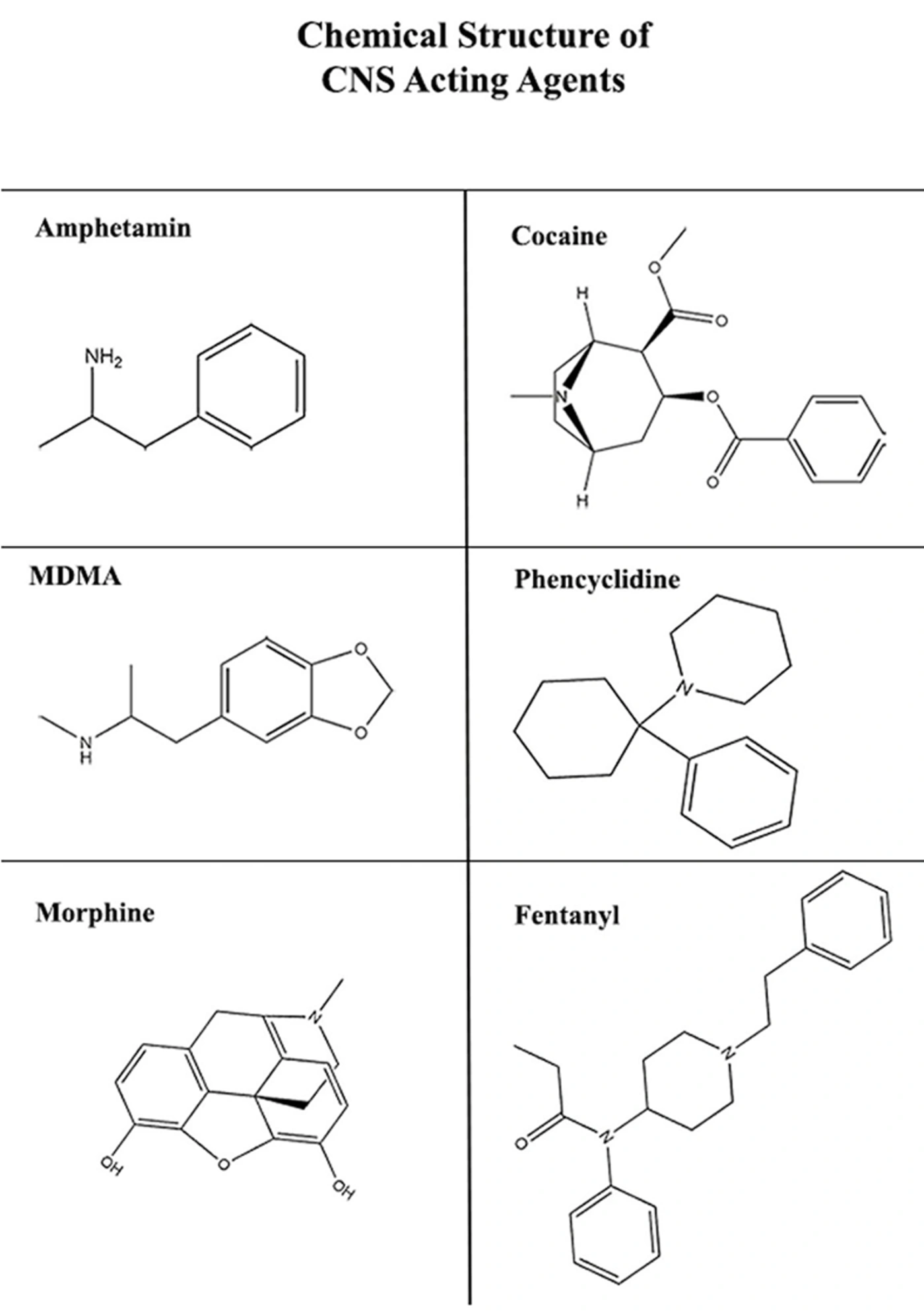
Figure 3.
Chemical structure of CNS-acting agents
The strength of evidence differed considerably among the reviewed compounds. Most compounds, including sulforaphane, icariside II, sauchinone, alpha-tocopherol, deferoxamine, luteolin, L-carnitine, caffeic acid phenethyl ester, asiatic acid, ginsenoside Re, oleuropein, vitamin C, taurine, and 20-deoxyingenol, have been investigated in only one preclinical study, usually using a single experimental model. Therefore, the evidence supporting these compounds should be considered preliminary. Likewise, coenzyme Q10 (77) has shown beneficial effects in more than one neurotoxicity model, providing relatively stronger support for its proposed mechanism of action. However, because only a limited number of studies are available for most compounds, we did not apply a formal three-level evidence classification (e.g., strong, moderate, or limited). Such a classification would add little beyond distinguishing compounds supported by a single study from those with replicated evidence and could suggest a level of precision not supported by the available data.
The most extensive evidence was available for methamphetamine-induced neurotoxicity. Natural products such as sulforaphane (66), curcumin (67), icariside II (68), asiatic acid (2), luteolin (73), and caffeic acid phenethyl ester (64) showed neuroprotective effects through related mechanisms, mainly involving Nrf2 activation, suppression of NF-κB-mediated inflammation, reduction of oxidative stress, and regulation of mitochondrial apoptotic pathways. Sulforaphane improved behavioral deficits and reduced striatal dopamine loss by activating Nrf2. Curcumin alleviated cognitive impairment by reducing oxidative stress and neuroinflammation, which contributed to its neuroprotective effects, whereas icariside II and asiatic acid restored dopaminergic function and reduced apoptosis through the Keap1-Nrf2 and NF-κB/STAT3/ERK pathways, respectively.
In opioid-induced neurotoxicity, oleuropein (86), vitamin C (87), and 20-deoxyingenol (90) reduced neuronal damage mainly by decreasing oxidative stress and regulating apoptotic signaling. In MDMA-induced neurotoxicity, alpha-lipoic acid (80) and N-acetylcysteine (82) exerted protective effects by reducing oxidative stress, inhibiting microglial activation, and restoring GSH levels. For PCP-induced neurotoxicity, ginsenoside Re (84) and gangliosides (85) improved neuronal survival by preserving mitochondrial function and limiting excitotoxic damage. In contrast, only coenzyme Q10 (77) was identified for cocaine-induced neurotoxicity, highlighting the limited evidence currently available for this agent.
Mechanistically, the neuroprotective effects of these natural products converge on three closely related pathways: antioxidant, anti-inflammatory, and anti-apoptotic mechanisms. Antioxidant activity, the most consistently reported effect, includes both direct scavenging of ROS and enhancement of endogenous antioxidant defenses through Nrf2 activation. This is supported by increased SOD, CAT, and GPx activities with compounds such as curcumin, sulforaphane, and icariside II, as well as restoration of GSH levels by alpha-lipoic acid and N-acetylcysteine across different experimental models. Anti-inflammatory effects are mainly associated with regulation of microglial activation and suppression of NF-κB-mediated cytokine production, including TNF-α, IL-1β, and IL-6.
These mechanisms have been reported for asiatic acid and isoliquiritigenin in methamphetamine models, and for ginsenoside Re through modulation of opioid- and substance P-related signaling. Anti-apoptotic effects primarily involve the mitochondrial pathway by regulating the Bcl-2/Bax ratio, preventing cytochrome c release, and inhibiting caspase-3 activation, as demonstrated for curcumin, asiatic acid, luteolin, oleuropein, vitamin C, and 20-deoxyingenol.
Although these pathways are closely interconnected, most included studies examined them individually rather than directly evaluating their interactions. Nevertheless, this interconnection is illustrated within individual studies. For example, the asiatic acid study demonstrated that reduced NF-κB/STAT3/ERK-mediated inflammation was accompanied by decreased caspase-3 fragmentation and preserved mitochondrial integrity (2). Similarly, the curcumin study showed that restoration of antioxidant enzymes (SOD, GSH-Px) occurred alongside reduced TNF-α and glial activation (67). These findings are consistent with the concept that oxidative stress contributes to downstream inflammatory and apoptotic signaling rather than acting as an isolated mechanism.
Compounds such as curcumin, asiatic acid, and 20-deoxyingenol have been shown to affect all three pathways within their respective experimental models, supporting the potential value of multi-target natural products. However, none of the included studies directly compared multi-target compounds with agents acting through a single mechanism, and any conclusion regarding their superiority would require dedicated head-to-head studies.
In addition to the mechanistic findings discussed above, several methodological limitations should be considered when interpreting the available evidence. Most studies evaluated only one or a limited range of doses, making it difficult to determine the optimal therapeutic dose. In addition, most in vivo studies were conducted only in male rodents; therefore, potential sex-related differences in the neuroprotective effects of natural products remain unclear. Beyond sex-related differences, extrapolating findings from rodents to humans has several additional limitations. Rodents often metabolize xenobiotics and natural compounds more rapidly than humans, meaning that the same dose on a milligram-per-kilogram basis may result in different systemic or CNS exposure between species. In addition, rodent models of CNS drug-induced neurotoxicity generally involve short and controlled exposure under standardized laboratory conditions, which may not reflect the variable exposure patterns, polydrug use, or comorbidities commonly observed in human substance users. Differences in the blood–brain barrier and drug-metabolizing enzyme systems between rodents and humans may also affect CNS penetration and the protective effects of natural compounds. These species-specific differences should therefore be considered when interpreting the translational relevance of the preclinical findings summarized in this review.
In many studies, protective effects were assessed mainly using biochemical markers, such as MDA, SOD, or inflammatory cytokines, without consistent confirmation by behavioral or histopathological findings. This may limit interpretation of functional benefits. Furthermore, none of the included studies directly compared different natural compounds under the same experimental conditions, making it difficult to determine whether one compound is more effective than another. Because direct head-to-head comparisons are lacking and dosing, treatment timing, and outcome measures differ substantially across studies, it is not possible to reliably compare the relative efficacy of compounds acting through similar pathways, such as Nrf2 activation or NF-κB inhibition. Standardized head-to-head studies are therefore needed to compare neuroprotective effects under comparable experimental conditions.
As with narrative reviews of published preclinical literature generally, publication bias cannot be excluded, as studies reporting positive neuroprotective effects are more likely to be published than those with null or negative findings; this may lead to an overestimation of the apparent efficacy of the compounds discussed here. These limitations should be considered when interpreting the overall findings and their potential clinical relevance.
Given these methodological limitations and the complete absence of eligible clinical studies, the findings summarized here should be regarded strictly as preclinical, hypothesis-generating evidence of biological plausibility, rather than an indication of clinical efficacy or readiness for therapeutic use.
Beyond their promising mechanisms of action, clinical translation of these compounds remains limited by pharmacokinetic and pharmaceutical challenges that are rarely addressed in preclinical studies. Many widely investigated compounds, including curcumin, resveratrol, luteolin, and several flavonoids, have poor oral bioavailability because of low aqueous solubility, extensive first-pass metabolism, and rapid systemic elimination. Curcumin, for example, is rapidly metabolized by glucuronidation and sulfation, resulting in very low plasma levels of the unmetabolized compound after oral administration in humans, despite the effective doses (100 - 200 mg/kg) reported in the rodent models reviewed here (67). This highlights the gap between effective experimental doses and clinically achievable human exposure. Another important limitation is blood–brain barrier (BBB) penetration.
Many polyphenols and flavonoids have limited CNS availability because of their physicochemical properties, such as molecular size, hydrophilicity, and active efflux by P-glycoprotein. As a result, beneficial effects observed in cell-based or animal studies, particularly those using local or parenteral administration, may not be fully reproduced after oral administration in humans, where CNS exposure is expected to be much lower. In addition, dosing protocols across the included studies were highly heterogeneous, differing in route of administration (oral, intraperitoneal, or subcutaneous), timing of treatment relative to toxicant exposure (pre-, co-, or post-treatment), and treatment duration (from a single dose to several weeks), as well as in laboratory methodology and outcome-assessment techniques. It should also be noted that some marine-derived natural products may contain environmental contaminants, including mercury and other pollutants. Therefore, careful quality control and safety assessment are essential before therapeutic application. Because this is a narrative rather than a systematic review, a formal risk-of-bias or certainty-of-evidence assessment was not performed; thus, the methodological quality of individual studies was not formally scored, which should be considered when interpreting the strength of evidence, particularly for compounds supported by only a single study. Predefined eligibility criteria were nonetheless applied consistently to all screened records, and every excluded study is reported with its reason for exclusion (Figure 2); however, the possibility of selective inclusion influencing the overall conclusions cannot be fully excluded. Furthermore, none of the included studies evaluated long-term safety, drug-drug interactions, or dose-related toxicity in humans. These issues are particularly important for patients who may be receiving multiple medications, such as those with substance use disorders. Therefore, the safety, pharmacokinetics, optimal dosing, and clinical efficacy of most compounds remain unclear, and the neuroprotective effects described in this review should be considered preclinical evidence rather than confirmed clinical benefits.
Although the safety of these compounds has not been specifically evaluated in the context of CNS drug-induced neurotoxicity, most have an established history of human use as dietary supplements. Vitamin C and vitamin E are generally well tolerated at standard doses, although high doses may cause gastrointestinal discomfort and increase the risk of oxalate kidney stones with vitamin C or bleeding with vitamin E (91, 92). N-acetylcysteine and coenzyme Q10 are also generally well tolerated, with mild gastrointestinal effects reported at higher doses (93). Curcumin is generally well tolerated when taken orally, although some individuals may experience gastrointestinal discomfort (94). Melatonin may cause headache or next-day drowsiness, while long-term safety data beyond several years remain limited (95). However, these safety profiles have not been adequately studied in individuals with substance use disorders, who may have altered drug metabolism or use other medications concurrently. Population-specific safety studies are therefore needed before these compounds can be considered for clinical use in this setting.
Together with the lack of a standardized approach for converting animal doses to human-equivalent doses, this variability makes it difficult to determine an optimal therapeutic range from the available evidence (Table 1). Using FDA body-surface-area conversion (human-equivalent dose ≈ rat dose × 0.16), the effective doses reported for several promising compounds appear to be within clinically achievable ranges. For example, the effective coenzyme Q10 dose of 10 mg/kg in rats corresponds to approximately 1.6 mg/kg in humans, or about 96 mg/day for a 60-kg adult, which is close to commonly used supplemental doses of 100 - 200 mg/day. Similarly, the effective doses reported for vitamin C and sulforaphane fall within or below typical human supplementation ranges. For curcumin, the effective doses of 100 - 200 mg/kg in rats correspond to approximately 16 - 32 mg/kg in humans, or about 960 - 1920 mg/day. Thus, the main challenge for translating curcumin to clinical use may be its limited oral bioavailability rather than the dose itself. Furthermore, none of the included studies assessed long-term safety, drug-drug interactions, or dose-related toxicity in humans, all of which are particularly important considerations for patients with substance use disorders who may be receiving multiple medications simultaneously. Accordingly, the neuroprotective effects described in this review should be considered as preclinical evidence rather than confirmed clinical benefits.
Table 1.
Summary of the Protective Mechanisms of Natural Products Against Neurotoxicity Induced by CNS Agents
| Toxin | Models | Dose & Exposure | Active Compound / Classification | Treatment Dose & Exposure | Key Outcomes | Evidence Type | Ref |
|---|---|---|---|---|---|---|---|
| Methamphetamine (chronic) | in vivo | Chronic exposure (mouse model) | Icariside II (ICS) / Natural product (plant flavonoid glycoside) | 10 & 30 mg/kg (in vivo) | ↑ TH, DAT, DA; ↓ GFAP, Iba-1, α-syn; improved motor function & dendritic structure; ↓ oxidative stress via Nrf2 | Direct | (68) |
| Methamphetamine | in vivo | 40 mg/kg (4 × 10 mg/kg, IP, acute) rat | Curcumin / Natural product (plant polyphenol) | 100 & 200 mg/kg, IP for 7 days post-exposure | ↓ MDA, TNF-α, caspase-3, GFAP, Iba-1; ↑ SOD, GSH-Px; improved memory | Direct | (67) |
| Methamphetamine | in vivo | Acute/repeated administration (mouse model) | Sauchinone / Natural product (plant lignan) | Systemic administration (pre-/co-treatment) | ↓ ROS, ↓ inflammation, ↓ apoptosis, improved neuronal survival | Direct | (69) |
| Methamphetamine | in vivo | 2 × 20 mg/kg i.p. (acute exposure), 2 h interval; evaluation ~48 h after | Alpha-Lipoic Acid (ALA) / Endogenous/dietary compound | 10 mg/kg & 40 mg/kg i.p. at 30 min, 24 h, 48 h post METH | ↓ hippocampal MDA, ↑ SOD & CAT, ↓ memory impairment & anxiety-like behavior | Direct | (65) |
| Methamphetamine | in vivo | Repeated exposure (rat model; hypothalamic toxicity) | Linalool / Natural product (plant terpenoid) | Pre-/co-treatment administration | Restored proteome balance, ↓ oxidative stress, improved neuronal function | Direct | (70) |
| Methamphetamine | in vitro | Repeated exposure (neuronal cells; duration: 24 - 72 h) | Aromadendrin / Natural product (plant flavonoid) | Co-treatment / pre-treatment (24 - 72 h) | ↓ ER stress, ↑ PI3K/Akt/mTOR signaling, ↓ apoptosis, improved cell survival | Direct | (71) |
| Methamphetamine | in vitro | 1 mM METH, ~24 h (neuroinflammation); ~12 h (neurotoxicity in cells) | Asiatic acid / Natural product (plant triterpene) | Pretreatment (e.g., 20 µM, 1 h) then co-exposure ~24 h | ↓ TNF-α/IL-6, ↓ NF-κB/STAT3/ERK activation, ↓ caspase-3 fragmentation, ↑ Bcl-xL, preserved mitochondria; reduced inflammation & cell death | Direct | (2) |
| Methamphetamine | in vivo | 10 mg/kg × 4 injections every 2 h (acute exposure in rats) | α-Tocopherol / Dietary compound (vitamin) | 20 mg/kg i.p. daily × 3 days + 30 min before METH | ↓ LPO, ↑ GSH, preserved DA/5-HT, ↓ hyperthermia & locomotor changes | Direct | (72) |
| Methamphetamine | in vivo | 10 mg/kg × 4 injections every 2 h (acute exposure in rats) | Deferoxamine / Synthetic comparator (microbial siderophore, pharmaceutical-grade) | 50 mg/kg s.c. 30 min before METH | ↓ LPO, ↑ GSH, preserved DA/5-HT, ↓ hyperthermia & locomotor changes | Direct | (72) |
| Methamphetamine | in vivo | 15 mg/kg i.p., 8 injections at 12-h intervals (~4 days) in rats | Luteolin / Natural product (plant flavonoid) | 100 mg/kg gavage daily × 3 days before METH | ↓ PI3K/Akt activation, ↓ p-Akt, ↓ p53/Bax/caspase-3, ↓ autophagy markers (Beclin-1, Atg5, LC3-II), reduced neurotoxicity | Direct | (73) |
| Methamphetamine | in vivo | 4 × 10 mg/kg i.p. each 2 h (acute, 1 day) with continued assessment 1 week after | L-Carnitine / Endogenous/dietary compound | 100 mg/kg orally, 1-week pre- + 1 week post- METH | ↓ 3-nitrotyrosine in striatum, reduced peroxynitrite formation; preserved mitochondrial energy metabolism | Direct | (74) |
| Methamphetamine | in vitro | Experimental exposure in rats/cell models (varies across studies and protocols) | Melatonin / Endogenous/dietary compound | Pre-/co-treatment (nanomolar range in vitro; mg/kg range in vivo; acute and chronic models) | ↓ oxidative stress & ROS, ↓ neuroinflammation (↓ TNF-α/NF-κB), preserved neurogenesis & cell survival, improved mitochondrial homeostasis | Direct | (75) |
| Methamphetamine | in vivo | 1 mg/kg/day i.p., 14 repeated days | Jitai Tablet (multi-herb formula) / Natural product (multi-herb formulation) | 0.84, 1.68, 3.36 g/kg orally, daily 7 days pre-treatment + throughout 14-day METH exposure | ↑ striatal DA, ↑ DAT & TH expression, ↓ LPO, improved dopaminergic function & behavior | Direct | (30) |
| Methamphetamine | in vivo | 10 mg/kg i.p., once daily for 5 consecutive days | Isoliquiritigenin / Natural product (plant flavonoid) | 10 mg/kg i.p., 30 min before each METH injection (5 days) | ↑ DAT & TH preservation, ↓ glial activation, ↓ NOS expression, ↓ NF-κB phosphorylation; neuroprotection | Direct | (76) |
| Methamphetamine | in vivo | 15 mg/kg i.p., twice daily × 4 days (8 injections total) | caffeic acid phenethyl ester (CAPE) / Natural product (propolis-derived) | 10 mg/kg i.p., once daily × 10 days (starting 2 days before and throughout METH exposure) | ↓ memory deficits & anxiety-like behavior, ↓ neurotoxicity markers (iNOS, PARP, caspase-3), ↑ neuronal preservation; modulation of hippocampal gene expression (ISG15) | Direct | (64) |
| Methamphetamine | in vivo | 3 mg/kg i.p., once daily for 5 days | Sulforaphane (SFN) / Natural product — plant-derived isothiocyanate | 1, 3, or 10 mg/kg i.p., 30 min before METH, once daily for 5 days | ↓ acute hyperlocomotion, ↓ behavioral sensitization, ↑ striatal DA & DOPAC, ↑ DAT immunoreactivity, ↓ microglial activation; neuroprotection | Direct | (66) |
| Methamphetamine | in vivo | 10 mg/kg i.p., once daily × 4 days | Coenzyme Q10 / Endogenous/dietary compound | 10 mg/kg orally once daily for 14 days (7 days before + 4 days during + 3 days after) | ↓ oxidative stress & LPO, ↑ GSH, preserved DA & 5-HT, improved mitochondrial integrity | Direct | (77) |
| MDMA | in vivo | 20 mg/kg i.p., single injection; effects assessed 7 days later | Alpha-lipoic acid / Endogenous/dietary compound | 100 mg/kg i.p., b.i.d. for 2 days (final dose 30 min pre-MDMA) | Complete prevention of ↓ 5-HT, ↓ 5-HT transporter; suppression of glial activation; antioxidant neuroprotection | Direct | (80) |
| MDMA | in vivo | 10 mg/kg i.p., 3 injections in 1 day at 2-h intervals | N-Acetylcysteine (NAC) / Semi-synthetic pharmaceutical derivative of an amino acid | 150 mg/kg i.p., once daily: 30 min before first MDMA + daily for 3 days post-MDMA | ↑ 5-HT & DA preservation, ↑ GSH levels, ↓ LPO, ↑ antioxidant enzymes; improved behavioral outcomes | Direct | (82) |
| MDMA | in vivo | 10 mg/kg i.p., 3 injections in 1 day at 2-h intervals | N-Acetylcysteine (NAC) / Semi-synthetic pharmaceutical derivative of an amino acid | 150 mg/kg i.p., once daily: 30 min before first MDMA + daily for 3 days post-MDMA | ↑ 5-HT & 5-HIAA preservation, ↑ GSH (GSH), ↓ LPO, improved behavior | Direct | (83) |
| Methamphetamine | in vivo | 35 mg/kg i.p., single dose | Ginsenoside Re / Natural product (plant saponin) | 20 mg/kg i.p. twice daily for 6 days (5 days pre-METH + 1 day post-METH); plus, administrations 2 h before & 10 h after METH | ↓ oxidative stress & ROS, ↓ microglial activation & apoptosis, ↑ dynorphin/κ-opioid receptor signaling, ↓ substance P/NK1 signaling; preserved dopaminergic function | Direct | (88) |
| DOI-induced TS model | in vivo | DOI to induce TS behavior (model specifics per protocol; behaviors assessed 24 h after last dose) | Rhynchophylline / Natural product (plant alkaloid) | 20 mg/kg & 40 mg/kg (daily during DOI-induction period) | ↓ stereotypic behaviors, ↓ IL-6/IL-1β/TNF-α in serum & striatum, ↓ TLR/NLRP3/NF-κB signaling | Indirect (mechanistic analog for MDMA) | (81) |
| Morphine-induced oxidative stress | in vitro | Morphine 0.2 mM & 0.5 mM, 24 h in rat C6 cells | Taurine / Endogenous/dietary compound (amino sulfonic acid) | 1, 5, 10 mM (30 min pre-treatment + co-incubation during 24 h morphine exposure) | ↑ cell viability, ↓ ROS & LPO, ↑ GSH & antioxidant enzyme activity, stabilized mitochondria | Direct | (89) |
| Morphine | in vivo | 45 mg/kg s.c., once daily for 4 weeks (chronic) | Oleuropein / Natural product (plant polyphenol) | 5, 15, 30 mg/kg i.p., daily co-administered throughout 4-week morphine exposure | ↑ spatial learning & memory, ↓ hippocampal oxidative stress (↓ MDA, ↑ SOD & GPx), ↓ Bax & cleaved caspase-3, ↑ Bcl-2; neuroprotection | Direct | (86) |
| Tramadol | in vivo | 50, 100, 150 mg/kg per oral, once daily for 30 days | Vitamin C / Dietary compound (vitamin) | 200 mg/kg per oral, once daily for 30 days (co-administered with tramadol) | ↓ MDA & NO (oxidative stress), ↑ CAT/SOD/GSH (antioxidant), ↓ p53 (pro-apoptotic), ↑ Bcl-2 (anti-apoptotic), improved cortical histology | Direct | (87) |
| Morphine | in vivo | 45 mg/kg s.c., once daily for 4 weeks | 20-Deoxyingenol (20-DOI) / Natural product (plant diterpene ester) | 10 or 20 mg/kg i.p., daily co-administered for 4 weeks | ↑ spatial learning & memory, ↓ apoptosis (cleaved caspase-3, Bax/Bcl-2), ↑ antioxidant enzymes (SOD2, HO-1, GPx), ↑ autophagy (TFEB-mediated restoration) | Direct | (90) |
| Glutamate | in vitro | 1 - 5 mM exposure in primary rat neuron culture (acute) | Gangliosides / Endogenous compound (glycosphingolipid) | 1.0 µg/mL pretreatment (before and during excitotoxin exposure) | ↑ neuronal survival, ↓ dendritic degeneration and cell death; membrane stabilization & reduced excitotoxic Ca2+ influx | Indirect (mechanistic analog for PCP) | (85) |
| Kainate | in vitro | 50 - 200 µM exposure in primary rat culture (acute) | Gangliosides / Endogenous compound (glycosphingolipid) | 1.0 µg/mL pretreatment | ↑ neuronal survival, ↓ excitotoxic injury | Indirect (mechanistic analog for PCP) | (85) |
| Phencyclidine (PCP) | in vivo | 10 mg/kg i.p., once daily × 7 days | Ginsenoside Re / Natural product (plant saponin) | 20 mg/kg orally once daily × 14 days (7 days pre-PCP + 7 days concurrent) | ↓ behavioral deficits, ↑ GPx-1, ↓ NADPH oxidase, improved mitochondrial function & redox balance | Indirect (mechanistic analog for Opioids) | (84) |
3.10. Future Research Directions
Future research should proceed in a stepwise manner. First, preclinical models should be better standardized, particularly with respect to dosing, route of administration, treatment timing, and outcome measures, to improve comparability between studies. Second, pharmacokinetic and bioavailability studies should be conducted for the most promising compounds identified above to determine whether the exposures associated with neuroprotection in experimental models can be achieved in humans before proceeding to efficacy studies. Third, multicenter preclinical studies should independently replicate the most promising findings across laboratories and, where appropriate, across different rodent strains, to provide stronger evidence of reproducible neuroprotective effects. Following these steps, well-designed, dose-ranging early-phase clinical trials should be considered, beginning with pharmacokinetic and safety evaluations before progressing to efficacy studies. Based on the current evidence, compounds such as curcumin, asiatic acid, sulforaphane, and coenzyme Q10 could be considered candidates for this staged evaluation. Among the compounds reviewed, coenzyme Q10, N-acetylcysteine, and vitamins C and E may have greater potential for near-term clinical translation because of their established safety profiles and known dosing in humans. Coenzyme Q10 is further supported by evidence from more than one neurotoxicity model. In contrast, compounds such as curcumin, despite showing promising effects across several mechanisms, have limited oral bioavailability and may require advanced delivery approaches before their clinical potential can be properly evaluated. Given the limited oral bioavailability and poor BBB penetration of many natural compounds discussed in this review, several delivery approaches may help improve their pharmacokinetic and CNS availability. Nanoparticle-based systems, including polymeric and lipid nanoparticles, can improve the solubility and stability of compounds such as curcumin and resveratrol and may reduce their rapid first-pass metabolism. Human pharmacokinetic studies have also shown improved plasma bioavailability with nanoformulated curcumin compared with conventional curcumin. Liposomal formulations may provide similar benefits by incorporating poorly soluble compounds into a lipid bilayer, potentially increasing circulation time and facilitating BBB delivery. Another approach involves structural modification of the parent compound or development of prodrugs designed to improve membrane permeability and subsequently release the active compound after absorption. This strategy has been explored for flavonoids with limited CNS penetration. Intranasal administration may also be considered for selected compounds because it can provide access to the brain through olfactory and trigeminal pathways. Future preclinical studies should directly compare these delivery approaches with conventional oral administration to determine which strategies can most effectively reduce the gap between the experimental doses used in animal studies and clinically achievable exposure in humans.
Because the available evidence suggests considerable overlap among antioxidant, anti-inflammatory, and anti-apoptotic mechanisms, future studies should directly compare combination therapies or standardized multi-component formulations with single-agent treatments in well-controlled head-to-head experiments, rather than inferring synergistic effects from separate studies. In addition, greater standardization of dosing regimens, treatment schedules, and outcome measures across preclinical models would improve study comparability and facilitate stronger evidence synthesis. Finally, several CNS-toxic agents remain poorly investigated, particularly cocaine, for which only one natural compound, coenzyme Q10, has been evaluated. Expanding research to include a wider range of neurotoxic agents and natural compounds will help strengthen the preclinical evidence base and support future translational studies (Table 2).
Table 2.
Comparative Effects of Standard and Natural Antioxidants on Neuroprotection
| Type | Classification | Examples | Primary Mechanism | Key Neuroprotective Effects | References |
|---|---|---|---|---|---|
| Standard Antioxidants | Dietary compound / Semi-synthetic derivative | Vitamin C, Vitamin E (α-tocopherol), N-Acetylcysteine (NAC) | Direct scavenging of ROS, regeneration of other antioxidants, replenishing GSH | Prevent LPO, protect DNA/protein from oxidative damage; demonstrated against tramadol-, methamphetamine-, and MDMA-induced neurotoxicity | (34, 72, 82, 87) |
| Natural Flavonoids | Natural product (plant flavonoid) | Chinonin | Scavenging ROS, enhancing SOD and GSH activity, reducing LPO | Preserving neuronal integrity, improving mitochondrial function | (37) |
| Polyphenols | Natural product (plant polyphenol) | Curcumin | Activation of Nrf2 pathway, modulation of antioxidant enzymes | Reduce oxidative stress, improve neuronal survival, anti-inflammatory synergy | (37) |
| Traditional Herbal Extracts | Natural product (traditional herbal formulation) | Lonicerae Japonicae Flos | Rich in phenolics/flavonoids; strong ROS-scavenging and enzyme modulation | Reduce oxidative stress and inflammation, potential benefit in Alzheimer's disease | (38) |
4. Conclusions
Natural products have demonstrated neuroprotective potential against neurotoxicity induced by CNS neurotoxic agents, including stimulants, hallucinogens, and opioids, via interconnected antioxidant, anti-inflammatory, and anti-apoptotic mechanisms. Compounds such as curcumin, asiatic acid, and 20-deoxyingenol illustrate how single agents may modulate multiple pathological pathways simultaneously, providing a mechanistic rationale for multi-target neuroprotective strategies.
However, the evidence base remains almost entirely preclinical, and most compounds discussed are supported by only a single study; no clinical trials met the inclusion criteria. Translational barriers, including poor oral bioavailability, limited BBB penetration, and highly heterogeneous dosing regimens, currently constrain the extrapolation of these findings to humans. Accordingly, the available preclinical evidence should be interpreted as indicating biological plausibility and mechanistic promise rather than established clinical efficacy. Well-designed, dose-ranging human trials, improved delivery systems to enhance bioavailability and CNS penetration, and head-to-head comparisons of combination versus single-agent approaches will be essential before the neuroprotective potential of these natural products can be responsibly translated into clinical practice (Figure 4).
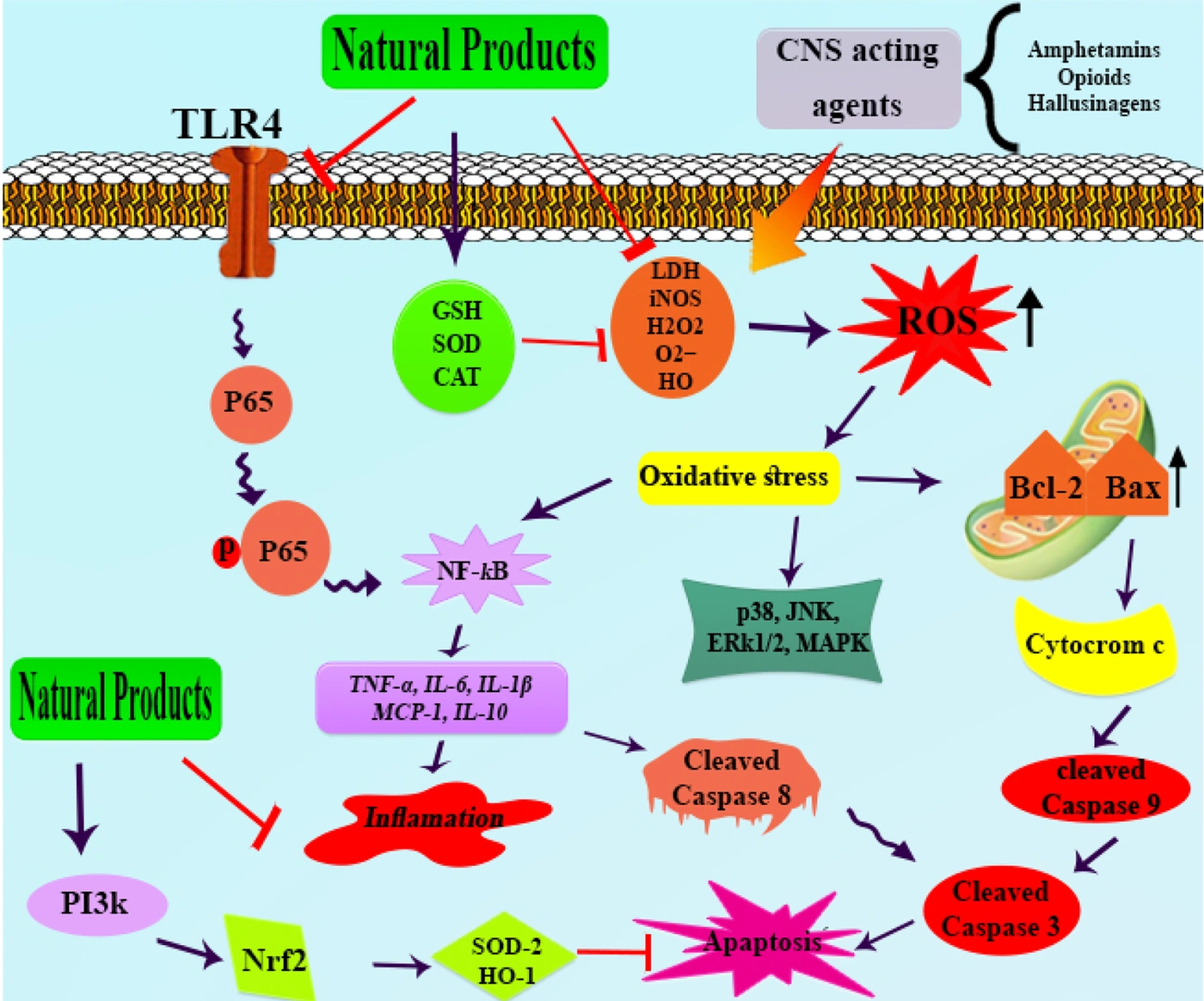
Figure 4.
Molecular Mechanisms of CNS Agent-Induced Neurotoxicity and the Neuroprotective Effects of Natural Products