Cover image of the article: Synergistic Effects of High-Intensity Interval Training and Omega-3 on Modulating Neuroinflammation and Improving Cognitive Function in an Alzheimer's Disease Rat Model

Image Credit:J Kermanshah Univ Med Sci

Synergistic Effects of High-Intensity Interval Training and Omega-3 on Modulating Neuroinflammation and Improving Cognitive Function in an Alzheimer's Disease Rat Model

Authors

Mohammad Sadegh EbrahimiMohammad Sadegh Ebrahimi ORCID1, Abdolhossein Taheri KalaniAbdolhossein Taheri Kalani ORCID1,*, Fardin FatahiFardin Fatahi ORCID1
1Department of Exercise Physiology, Il.C., Islamic Azad University, Ilam, Iran
*Corresponding Author: Department of Exercise Physiology, Il.C., Islamic Azad University, Ilam, Iran. Email: htaheriedu@gmail.com

Journal of Kermanshah University of Medical Sciences:Vol. 30, issue 3; e172491
Published online:Sep 30, 2026
Article type:Research Article
Received:Jun 13, 2026
Accepted:Sep 05, 2026
How to Cite:Ebrahimi MS, Taheri Kalani A, Fatahi F. Synergistic Effects of High-Intensity Interval Training and Omega-3 on Modulating Neuroinflammation and Improving Cognitive Function in an Alzheimer's Disease Rat Model. J Kermanshah Univ Med Sci. 2026;30(3):e172491. doi: https://doi.org/10.5812/jkums-172491

Abstract

Background:

Previous studies have demonstrated the role of inflammatory processes in the pathogenesis and progression of Alzheimer's disease (AD). Because currently available medications have insufficient clinical efficacy for the treatment of AD, lifestyle modifications, including exercise and dietary interventions, have become increasingly important for prevention and treatment.

Objectives:

This study aimed to determine the independent and combined effects of high-intensity interval training (HIIT) and omega-3 (OMG3) supplementation on amyloid beta precursor protein (App) mRNA expression, interleukin-1 beta (IL-1β) gene expression in brain tissue, and spatial memory in rats with AD.

Methods:

In this experimental study, 30 male Wistar rats aged 12 weeks and weighing 180 - 200 g were randomly assigned to five groups: Control, AD, AD + HIIT, AD + OMG3, and AD + HIIT + OMG3 (combined). To induce the AD model and memory impairment, scopolamine was administered intraperitoneally at 2 mg/kg/day for 9 days. The HIIT protocol consisted of 3 - 4 bouts of treadmill running at 30 m/min, 5 days per week for 8 weeks. The supplemented groups received Mega DHA, containing 10% eicosapentaenoic acid and 50% docosahexaenoic acid, via daily gavage for 8 weeks at a total product dose of 500 mg/kg/day. Forty-eight hours after the final interventions, the rats were sacrificed, and App mRNA and IL-1β gene expression in the brain were measured using real-time polymerase chain reaction. Spatial memory was assessed as the percentage of spontaneous alternation in the Y-maze test.

Results:

AD induction increased App mRNA and IL-1β gene expression in brain tissue. HIIT and OMG3 supplementation, alone or in combination, were associated with decreased IL-1β gene expression. App mRNA expression was reduced relative to that in the AD group only in the combined group. HIIT and OMG3 supplementation alone did not significantly alter App mRNA expression compared with that in the AD group.

Conclusions:

These findings suggest that combining HIIT with OMG3 supplementation improves cognitive function in the AD model by modulating neuroinflammation.

1. Background

Neurodegenerative diseases are age-related disorders characterized by progressive neuronal loss in the central or peripheral nervous system, accompanied by alterations in synaptic transmission and plasticity. These increasingly prevalent diseases represent a major public health concern because of their adverse effects on cognitive and physical health and quality of life (1, 2). Alzheimer's disease (AD) is a neurodegenerative disorder that impairs cognition, memory, thinking, judgment, and behavior. No treatment has yet been identified to delay AD progression, and treatment and care costs continue to increase as its prevalence rises with population aging (1, 3).
AD is characterized by synaptic loss, neuronal atrophy, and the accumulation of amyloid plaques and tau neurofibrillary tangles. Deposition of amyloid-beta (Aβ) and tau proteins in the brain leads to neuronal destruction (4). Aβ is a major protein implicated in AD, and its abnormal accumulation promotes amyloid plaque formation, neuronal destruction, and cognitive impairment. These plaques also induce neuroinflammation, damaging neurons and disrupting synaptic connections (5). In particular, Aβ can disrupt glial cell function and activate the brain's immune system, ultimately contributing to AD development. Reducing brain Aβ levels is therefore an effective strategy for reducing brain damage and slowing AD progression (6).
Studies have shown that Aβ peptides act as proinflammatory agents and increase AD risk by activating inflammatory mediators, including cytokines, in the brain. The presence of activated microglia and astroglia around senile plaques further supports the role of Aβ in inflammation. Microglia and astrocytes provide neuroprotection by removing Aβ deposits from the brain (7, 8). Senile plaques can cause neurotoxicity through the release of proinflammatory cytokines, chemokines, and reactive oxygen species (8). Similarly, interleukin-1 beta (IL-1β), an inflammatory cytokine, increases amyloid precursor expression, exacerbating amyloidosis and creating a vicious cycle (9). IL-1β is an important inflammatory cytokine released in response to brain Aβ accumulation and plays a key role in AD development. It can activate microglia and astrocytes, which subsequently release other inflammatory cytokines, leading to neuronal damage and synaptic destruction (10). Modulating Aβ and IL-1β levels in the central nervous system could therefore represent a therapeutic strategy for AD.
Because current AD medications have not achieved sufficient clinical efficacy, attention has shifted toward lifestyle modifications for prevention and treatment. Exercise, particularly aerobic exercise, is recognized as a nonpharmacological approach that can modulate brain inflammation in AD (11, 12). Exercise may improve behavioral impairments by reducing Aβ42 peptide levels through increased production of neurotrophic factors, including NGF, BDNF, and IGF-1, which support neuronal survival and proliferation, as well as neuronal and synaptic plasticity (13, 14). Exercise can also reduce microglial activity and thereby prevent inflammation-induced neuronal damage (2). In this regard, 4 weeks of aerobic training before and after AD induction have been associated with decreased IL-1 gene expression in the hippocampus of male Wistar rats (15).
Omega-3 fatty acids (OMG3), including eicosapentaenoic acid (EPA) and docosahexaenoic acid (DHA), are essential fatty acids and components of neuronal cell membranes in the brain (16). Several studies have highlighted their neuroprotective roles in Huntington's disease (17), Parkinson's disease (18), AD, and mild cognitive impairment (19, 20). Similarly, 8 weeks of aerobic training and OMG3 administration reduced hippocampal Aβ levels in male rats with AD (21).

2. Objectives

Despite the importance of inflammatory pathways in AD progression and evidence supporting the beneficial effects of exercise and OMG3 administration on inflammatory markers, few studies have evaluated their combined effects on neuroinflammation in this disease model. In addition, information remains insufficient regarding the optimal dose and duration of OMG3 administration, as well as the mode and intensity of aerobic training, in patients with AD. Therefore, this study aimed to determine the separate and combined effects of high-intensity interval training (HIIT) and OMG3 supplementation on amyloid beta precursor protein (App) mRNA expression, IL-1β gene expression in brain tissue, and spatial memory in an AD rat model.

3. Methods

3.1. Animals

In this experimental study, 30 male Wistar rats aged 12 weeks and weighing 180 - 200 g were obtained from the Pasteur Institute and transferred to the animal laboratory. After 2 weeks of acclimatization, the animals were randomly assigned to five groups: Control, AD, AD + HIIT, AD + OMG3, and AD + HIIT + OMG3 (combined). Animals were housed at 22 ± 2 °C and 50% - 55% humidity under a 12:12-hour light/dark cycle, with free access to water and standard food throughout the experiment.

3.2. Induction of the Alzheimer's Disease Model

Scopolamine was administered intraperitoneally at 2 mg/kg/day for 9 days to induce the AD model and memory impairment. Each day, 200 μL of scopolamine dissolved in 1% carboxymethyl cellulose was injected into the animals. The healthy control group received 200 μL of the vehicle daily. After 9 days, spatial memory was assessed using the Y-maze test, and the exercise and nutritional supplementation interventions were initiated (22).

3.3. High-Intensity Interval Training Protocol

Before the main training protocol, the rats underwent treadmill familiarization for 2 weeks, 5 days per week, running for 10 - 15 minutes per day at 5 - 15 m/min. The HIIT protocol lasted 8 weeks, with five sessions per week (Table 1). During Weeks 1 - 4, the main protocol consisted of three 4-minute running bouts at 30 m/min. During Weeks 5 - 8, four 4-minute bouts were performed at the same speed. Between bouts, the animals ran for 3 minutes at 20 m/min. Each session began and ended with 5 minutes of running at 10 - 15 m/min for warm-up and cool-down (23).
Table 1.
High-Intensity Interval Training Protocol a
Variable/Week12345678
Speed (m/min)3030303030303030
Time (min)44444444
Sets (number)33334444
a Source: Adapted from Costa et al. (23).

3.4. Omega-3 Administration

Mega DHA® (Vitafor, Brazil), containing 10% EPA and 50% DHA, was administered by daily gavage for 8 weeks at a total product dose of 500 mg/kg/day (24).

3.5. Spatial Memory Assessment

The Y-maze test was used to assess spatial memory. This test assesses short-term spatial memory and is based on rodents' innate curiosity to explore new environments. The maze apparatus consisted of a central platform with a diameter of 25 cm and eight equally spaced arms. In the standard test, the animals were first familiarized with the environment for 3 days. They were placed in the center of the apparatus and allowed to explore it for 15 minutes. On the first and second days, food was distributed in the arms. On the third day, the amount of food was reduced by half and placed in a container at the end of an arm. The session ended when the rats had explored all eight arms (25).

3.6. Brain Tissue Collection

Forty-eight hours after the final interventions and following a 12-hour fast, the rats were anesthetized by intraperitoneal injection of ketamine (100 mg/kg) and xylazine (10 mg/kg). After deep anesthesia and the absence of responses to external stimuli were confirmed, the animals were decapitated using special scissors. The skull was then opened with a surgical blade, and the brain was carefully removed. Brain tissue was frozen in liquid nitrogen and stored at -80 °C. After homogenization, the tissue was centrifuged for 10 minutes at 12,000 rpm.

3.7. Gene Expression Measurement

Real-time polymerase chain reaction (PCR) was used to measure App mRNA and IL-1β gene expression. The procedures are described below.

3.7.1. RNA Extraction

RNA was extracted using a total RNA extraction kit (Pars Tous, Iran) according to the manufacturer's instructions. First, samples were handled under sterile conditions using liquid nitrogen and a porcelain powder mortar and stored in a -70 °C freezer.

3.7.2. Assessment of Extracted RNA Quality

To measure RNA concentration, 1 μL of RNA solution was diluted with 49 μL of sterile Tris buffer, and its absorbance at 260 nm was measured using a microspectrophotometer (Eppendorf, Germany). The A260/A280 absorbance ratio was used to assess protein contamination. Ratios above 1.8 were considered indicative of desirable RNA quality. The A260/A230 absorbance ratio was used to assess phenol contamination, with values above 1.8 also considered desirable.

3.7.3. cDNA Synthesis

The Easy™ cDNA synthesis kit (Pars Tous, Iran) was used to synthesize cDNA from RNA.

3.7.4. Real-Time Polymerase Chain Reaction

Real-time PCR was used to evaluate App mRNA and IL-1β gene expression. The primers, their sequences and lengths, amplified product lengths, and GenBank accession numbers are listed in Table 2. Primers were designed using Primer3 software. Reactions were performed using the 2X SYBR Green Real Time PCR kit (Pars Tous, Iran) and the Lava96 Real-time PCR Detection System (DaanGene Co Ltd). Results were evaluated using the comparative ΔΔCt method and Light Cycler SW1.1 software. GAPDH was used as the calibrator. Results were reported according to the general formula 2-ΔΔCt.
Table 2.
Primer Sequences for the Studied Genes
Gene Name and Primer TypePrimer SequencePrimer LengthProduct LengthAccession Number
App117NM_019288.2
ForwardAGGTTATGACACACCTCCGT20
ReverseTCTGAAGCAGCTCGTCAACT20
IL-1β127NM_031512
ForwardTGTCTGAAGCAGCTATGGCA20
ReverseAGGGCTTGGAAGCAATCCTTA21
GAPDH119NM_017008.4
ForwardAGTTCAACGGCACAGTCAAG20
ReverseTACTCAGCACCAGCATCACC20

3.8. Statistical Analysis

Data normality was assessed using the Shapiro-Wilk test, and homogeneity of variances was assessed using Levene's test. One-way analysis of variance (ANOVA) and Tukey's post hoc tests were used to compare group means. All statistical analyses were performed using SPSS version 27, with significance set at P < 0.05.

3.9. Ethical Considerations

The study was conducted in accordance with the Declaration of Helsinki and approved by the Ethics Committee of Islamic Azad University of Ilam, Iran (IR.IAU.ILAM.REC.1404.049). The authors avoided data fabrication, falsification, plagiarism, and misconduct.

4. Results

The Y-maze results showed significantly lower spatial memory in all intervention groups than in the healthy control group (all P < 0.0001). Spatial memory was significantly higher in the AD + HIIT and AD + OMG3 groups than in the AD group, and in the combined group than in the AD, AD + HIIT, and AD + OMG3 groups (all P < 0.0001) (Figure 1).
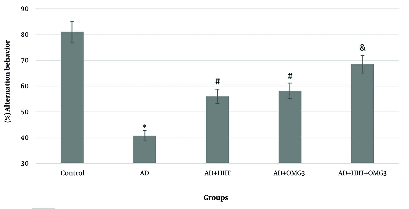
Spatial memory scores in the study groups (mean ± SEM). *Significant difference relative to all groups (P &lt; 0.0001). #Significant difference from the control and AD groups (P &lt; 0.0001). &amp;Significant difference from the control, AD, AD + HIIT, and AD + OMG3 groups (P &lt; 0.0001).
Figure 1.
Spatial memory scores in the study groups (mean ± SEM). *Significant difference relative to all groups (P < 0.0001). #Significant difference from the control and AD groups (P < 0.0001). &Significant difference from the control, AD, AD + HIIT, and AD + OMG3 groups (P < 0.0001).
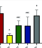
One-way ANOVA and Tukey's post hoc tests showed that brain App mRNA expression was significantly higher in all treatment groups than in the healthy control group (all P < 0.001). Only the combined treatment significantly reduced App mRNA expression compared with the AD group (P = 0.003). Although HIIT (P = 0.469) and OMG3 supplementation (P = 0.264) alone showed trends toward reduced App mRNA expression, these changes were not statistically significant (Figure 2).
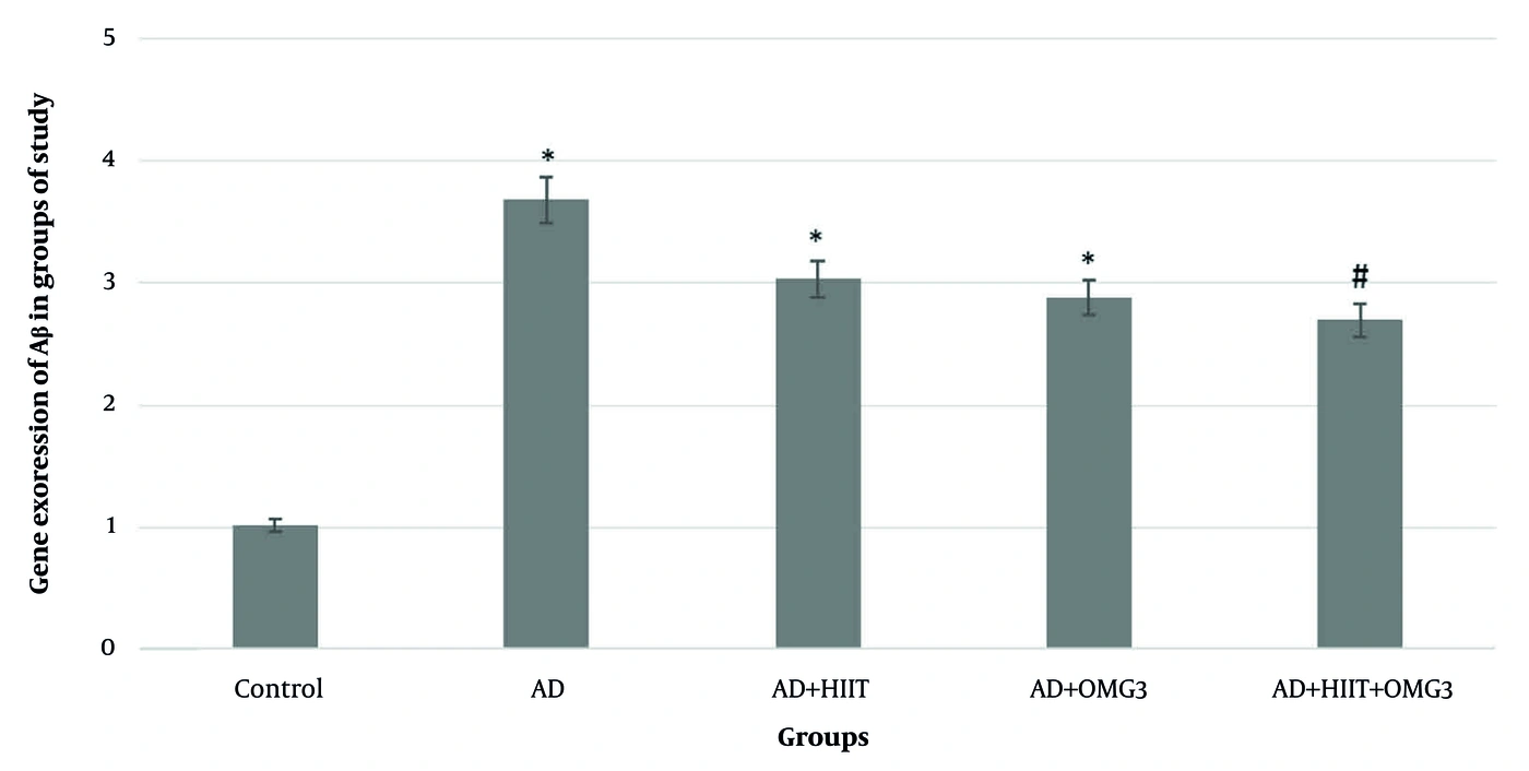
Brain App mRNA expression in the study groups (mean ± SEM). *Significant difference relative to the control group (P &lt; 0.001). #Significant difference from the control and AD groups (P = 0.003).
Figure 2.
Brain App mRNA expression in the study groups (mean ± SEM). *Significant difference relative to the control group (P < 0.001). #Significant difference from the control and AD groups (P = 0.003).
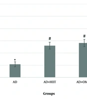
As shown in Figure 3, AD induction significantly increased IL-1β gene expression compared with that in the other study groups (all P < 0.001). IL-1β gene expression was also significantly higher in all treatment groups than in the healthy control group (P < 0.001). However, compared with the AD group, IL-1β gene expression was significantly lower in the AD + HIIT, AD + OMG3, and AD + HIIT + OMG3 groups (all P < 0.001). There was no significant difference in IL-1β gene expression among the AD + HIIT, AD + OMG3, and AD + HIIT + OMG3 groups (all P < 0.174).
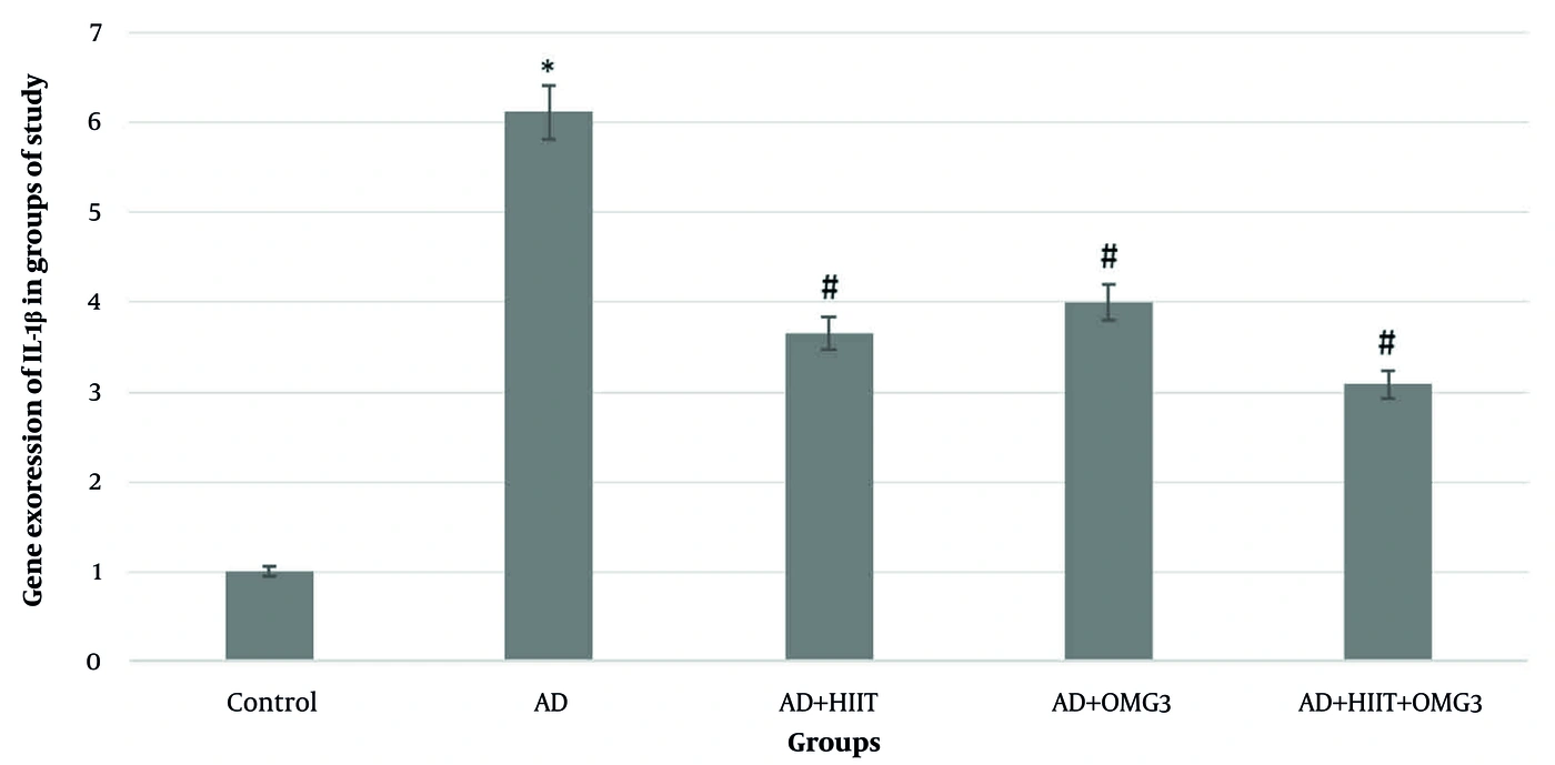
Brain IL-1β gene expression in the study groups (mean ± SEM). *Significant difference relative to the other groups (P &lt; 0.0001). #Significant difference from the control and AD groups (P &lt; 0.0001).
Figure 3.
Brain IL-1β gene expression in the study groups (mean ± SEM). *Significant difference relative to the other groups (P < 0.0001). #Significant difference from the control and AD groups (P < 0.0001).

5. Discussion

This study showed that AD induction increased brain App mRNA and IL-1β gene expression and impaired spatial memory in rats. HIIT and OMG3 administration, alone and in combination, reduced IL-1β gene expression and improved spatial memory. App mRNA expression was significantly reduced relative to the AD group only with the combined treatment. IL-1β gene expression did not differ significantly among the treatment groups.
Previous studies have reported that exercise can counteract AD progression by regulating neuronal apoptosis, intercellular communication, oxidative stress, mitochondrial autophagy, synaptic plasticity, and neurotoxicity (11, 26). OMG3 administration also provides neuroprotection and promotes cellular regeneration in neurological diseases such as AD by modulating inflammation and subsequently reducing oxidative stress (3, 26). Thus, the combined intervention may enhance the effects of these approaches on AD-related pathological changes and memory impairment, as observed in this study.
AD induction increased brain App mRNA and IL-1β gene expression in the present study. Previous studies have reported increased hippocampal Aβ (21) and IL-1β (27). Other studies have also reported increased serum (28) and hippocampal (29) levels of inflammatory factors, including Aβ and IL-1β, in AD. The amyloidogenic pathway is active in AD, producing Aβ through cleavage of amyloid precursor protein by β- and γ-secretases (30). Aβ also acts as an inflammatory factor by activating mediators, including IL-1β, IL-6, and TNF-α, in the brain, thereby increasing AD risk. The presence of activated microglia and astroglia around senile plaques supports the role of Aβ in neuroinflammation. Microglia and astrocytes may provide neuroprotection by clearing brain Aβ plaques (7, 8). These plaques can also mediate neurotoxicity through the release of proinflammatory cytokines, chemokines, reactive oxygen species, and protein complexes (8). Our findings are therefore consistent with previous studies implicating amyloid-related pathways in neuroinflammation and AD development.
Although HIIT and OMG3 supplementation alone did not significantly alter App mRNA expression, they reduced IL-1β gene expression. Changes in inflammatory markers following exercise training have been reported previously (27, 28). Treadmill running for 12 weeks increased the conversion of microglia from the proinflammatory M1 phenotype to the anti-inflammatory M2 phenotype, reducing hippocampal inflammation and oxidative damage and ultimately improving cognitive function in mice (31). Exercise inhibits neuroinflammation by upregulating Aβ transporter activity and promoting Aβ clearance (13, 14). In old age, chronic aerobic exercise supports the neurovascular unit, reduces the entry of inflammatory substances into the brain, and improves behavioral capacity in mice by enhancing synaptic plasticity (32). Although no study was identified that investigated the effects of OMG3 on neuroinflammation in AD, its effectiveness in reducing inflammatory biomarkers has been reported in individuals with diabetes (33), atherosclerosis (34), and old age (35). Under inflammatory conditions, OMG3 fatty acids are converted into anti-inflammatory metabolites and mediators, including resolvins, protectins, and maresins (36). Arachidonic acid, an omega-6 fatty acid, is released from membrane phospholipids by inflammatory stimuli. After release, it is converted into prostaglandins, leukotrienes, and lipoxins through the action of enzymes such as cyclooxygenases and lipoxygenases and exerts proinflammatory effects. EPA and DHA, two OMG3 fatty acids, reduce inflammation by competing with proinflammatory arachidonic acid substrates and receptors (37).
Finally, 8 weeks of combined HIIT and OMG3 administration significantly reduced brain App mRNA and IL-1β gene expression compared with the AD group. Few studies have examined the combined effects of aerobic training and OMG3 on inflammatory markers in AD. Previous research has reported that combining aerobic training with OMG3 administration reduces hippocampal Aβ in rats with AD (21). Similarly, IL-6 and TNF-α expression decreased in elderly rats (35) and obese mice (38) after 8 weeks of aerobic training and OMG3 intake (35). Fish oil, which contains high levels of OMG3, combined with aerobic training also reduced NF-κB, IL-6, and TNF-α expression more than either intervention alone in obese rats (39). This evidence suggests that aerobic training and OMG3 may enhance each other's anti-inflammatory effects in conditions such as AD, aging, and obesity. Further controlled studies are needed to confirm this hypothesis.

5.1. Conclusions

AD induction increased brain App mRNA and IL-1β gene expression. After 8 weeks, HIIT and OMG3 supplementation, alone and in combination, reduced IL-1β gene expression, whereas App mRNA expression was significantly reduced relative to the AD group only with the combined treatment. The combined treatment improved spatial memory more than either treatment alone, but IL-1β gene expression did not differ significantly among the treatment groups. HIIT and OMG3 supplementation may therefore help counteract the adverse effects of AD on the central nervous system by modulating inflammation. Further research is needed.

5.2. Limitations

This study used a scopolamine-induced animal model of AD. Although this model is considered one of the most valid approaches for simulating AD, it cannot reflect all aspects of the disease in humans. Human AD is often multifactorial and chronic and is associated with diverse genetic and environmental factors, whereas the scopolamine-induced model primarily reflects acute, inducible changes. Direct generalization to humans should therefore be approached cautiously.
Another limitation was the 8-week intervention period. Although this duration demonstrated beneficial effects of HIIT and OMG3, longer-term effects and the persistence of changes after the intervention were not determined. The study also included only male rats; females were not examined, although hormonal differences may influence responses to exercise and OMG3 administration.
Finally, the study was conducted under controlled laboratory conditions and did not account for real-world factors such as stress, unrestricted dietary intake, and daily human activity. Clinical application therefore requires larger human studies with longer follow-up. Despite these limitations, careful control of variables and standardized methods provided high internal validity and may support future research.
Footnotes

  • AI Use Disclosure:The authors declare that no generative AI tools were used in the creation of this article.

  • Authors' Contribution:Study Concept and Design: A. T. K.; Data Acquisition: M. S. E.; Data Analysis and Interpretation: A. T. K. and F. F.; Manuscript Drafting: A. T. K. and F. F.; Critical Revision for Important Intellectual Content: A. T. K.; Statistical Analysis: A. T. K. and F. F.; Administrative, Technical, and Material Support: M. S. E.; Study Supervision: A. T. K.

  • Conflict of Interests Statement:The authors declare that they have no competing interests.

  • Data Availability:The dataset presented in the study is available on request from the corresponding author during submission or after publication.

  • Ethical Approval:Ethical approval was granted by the Islamic Azad University Ethics Committee (IR.IAU.ILAM.REC.1404.049).

  • Funding/Support:No funding was received for this study.

References

  • 1.
    Ricci C. Neurodegenerative disease: from molecular basis to therapy. Int J Mol Sci. 2024;25(2):967. [PubMed ID: 38256040]. [PubMed Central ID: PMC10815646]. https://doi.org/10.3390/ijms25020967.
  • 2.
    Twarowski B, Herbet M. Inflammatory processes in Alzheimer's disease-pathomechanism, diagnosis and treatment: A review. Int J Mol Sci. 2023;24(7):6518. [PubMed ID: 37047492]. [PubMed Central ID: PMC10095343]. https://doi.org/10.3390/ijms24076518.
  • 3.
    Calderon Martinez E, Zachariah Saji S, Salazar Ore JV, Borges‐Sosa OA, Srinivas S, Mareddy NSR, et al. The effects of omega-3, DHA, EPA, Souvenaid® in Alzheimer's disease: A systematic review and meta-analysis. Neuropsychopharmacol Rep. 2024;44(3):545-556. [PubMed ID: 38924283]. [PubMed Central ID: PMC11544444]. https://doi.org/10.1002/npr2.12455.
  • 4.
    Scheltens P, De Strooper B, Kivipelto M, Holstege H, Chételat G, Teunissen CE, et al. Alzheimer's disease. Lancet. 2021;397(10284):1577-1590. [PubMed ID: 33667416]. [PubMed Central ID: PMC8354300]. https://doi.org/10.1016/S0140-6736(20)32205-4.
  • 5.
    Sebastián-Serrano Á, De Diego-García L, Díaz-Hernández M. The neurotoxic role of extracellular tau protein. Int J Mol Sci. 2018;19(4):998. [PubMed ID: 29584657]. [PubMed Central ID: PMC5979432]. https://doi.org/10.3390/ijms19040998.
  • 6.
    Guan PP, Cao LL, Wang P. Elevating the levels of calcium ions exacerbate Alzheimer's disease via inducing the production and aggregation of β-amyloid protein and phosphorylated tau. Int J Mol Sci. 2021;22(11):5900. [PubMed ID: 34072743]. [PubMed Central ID: PMC8198078]. https://doi.org/10.3390/ijms22115900.
  • 7.
    Agostinho P, A. Cunha R, Oliveira C. Neuroinflammation, oxidative stress and the pathogenesis of Alzheimer's disease. Curr Pharm Des. 2010;16(25):2766-78. [PubMed ID: 20698820]. https://doi.org/10.2174/138161210793176572.
  • 8.
    Lee CYD, Landreth GE. The role of microglia in amyloid clearance from the AD brain. J Neural Transm (Vienna). 2010;117(8):949-60. [PubMed ID: 20552234]. [PubMed Central ID: PMC3653296]. https://doi.org/10.1007/s00702-010-0433-4.
  • 9.
    Chen Y, Yu Y. Tau and neuroinflammation in Alzheimer's disease: Interplay mechanisms and clinical translation. J Neuroinflammation. 2023;20(1). 165. [PubMed ID: 37452321]. [PubMed Central ID: PMC10349496]. https://doi.org/10.1186/s12974-023-02853-3.
  • 10.
    Calsolaro V, Edison P. Neuroinflammation in Alzheimer's disease: Current evidence and future directions. Alzheimers Dement. 2016;12(6):719-32. [PubMed ID: 27179961]. https://doi.org/10.1016/j.jalz.2016.02.010.
  • 11.
    Bonanni R, Cariati I, Cifelli P, Frank C, Annino G, Tancredi V, et al. Exercise to counteract Alzheimer's disease: What do fluid biomarkers say? Int J Mol Sci. 2024;25(13):6951. [PubMed ID: 39000060]. [PubMed Central ID: PMC11241657]. https://doi.org/10.3390/ijms25136951.
  • 12.
    Köbe T, Witte AV, Schnelle A, Lesemann A, Fabian S, Tesky VA, et al. Combined omega-3 fatty acids, aerobic exercise and cognitive stimulation prevents decline in gray matter volume of the frontal, parietal and cingulate cortex in patients with mild cognitive impairment. Neuroimage. 2016;131:226-38. [PubMed ID: 26433119]. https://doi.org/10.1016/j.neuroimage.2015.09.050.
  • 13.
    Pahlavani HA. Exercise therapy to prevent and treat Alzheimer's disease. Front Aging Neurosci. 2023;15. 1243869. [PubMed ID: 37600508]. [PubMed Central ID: PMC10436316]. https://doi.org/10.3389/fnagi.2023.1243869.
  • 14.
    Cantón-Suárez A, Sánchez-Valdeón L, Bello-Corral L, Cuevas MJ, Estébanez B. Understanding the molecular impact of physical exercise on Alzheimer's disease. Int J Mol Sci. 2024;25(24):13576. [PubMed ID: 39769339]. [PubMed Central ID: PMC11677557]. https://doi.org/10.3390/ijms252413576.
  • 15.
    Saboury M, Kordi MR, Shabkhiz F. Effect of aerobic training before and after induction of Alzheimer on interleukin-1ß and CREB gene expression in the hippocampus of Wistar male rats. J Pract Studies Biosci Sport. 2021;9(20):68-83. https://doi.org/10.22077/jpsbs.2018.1008.1316.
  • 16.
    Layé S, Nadjar A, Joffre C, Bazinet RP. Anti-inflammatory effects of omega-3 fatty acids in the brain: Physiological mechanisms and relevance to pharmacology. Pharmacol Rev. 2018;70(1):12-38. [PubMed ID: 29217656]. https://doi.org/10.1124/pr.117.014092.
  • 17.
    Clifford JJ, Drago J, Natoli AL, Wong JYF, Kinsella A, Waddington JL, et al. Essential fatty acids given from conception prevent topographies of motor deficit in a transgenic model of Huntington's disease. Neuroscience. 2002;109(1):81-8. [PubMed ID: 11784701]. https://doi.org/10.1016/s0306-4522(01)00409-2.
  • 18.
    Barros AS, Crispim RYG, Cavalcanti JU, Souza RB, Lemos JC, Cristino Filho G, et al. Impact of the chronic omega-3 fatty acids supplementation in hemiparkinsonism model induced by 6-hydroxydopamine in rats. Basic Clin Pharmacol Toxicol. 2017;120(6):523-31. [PubMed ID: 27883274]. https://doi.org/10.1111/bcpt.12713.
  • 19.
    Grimm MOW, Michaelson DM, Hartmann T. Omega-3 fatty acids, lipids, and apoE lipidation in Alzheimer's disease: a rationale for multi-nutrient dementia prevention. J Lipid Res. 2017;58(11):2083-101. [PubMed ID: 28528321]. [PubMed Central ID: PMC5665674]. https://doi.org/10.1194/jlr.R076331.
  • 20.
    Olivera‐Perez HM, Lam L, Dang J, Jiang W, Rodriguez F, Rigali E, et al. Omega-3 fatty acids increase the unfolded protein response and improve amyloid-β phagocytosis by macrophages of patients with mild cognitive impairment. FASEB J. 2017;31(10):4359-69. [PubMed ID: 28634213]. [PubMed Central ID: PMC5602894]. https://doi.org/10.1096/fj.201700290R.
  • 21.
    Yaghoubi A. Changes in Aβ42, neprilysin, and γ-secretase in the hippocampus of male rats alzheimer's model: the effects of aerobic training and omega-3 intake. J Kerman Univ Med Sci. 2023;30(3):136-45. https://doi.org/10.34172/jkmu.2023.23.
  • 22.
    Nedaei SE, Pourmotabbed A, Aeenfar M, Seifi Z. The therapeutic effect of Crocin on Scopolamine induced retrograde amnesia in male rats. J Kermanshah Univ Med Sci. 2012;16(2). e78821.
  • 23.
    Costa LR, Castro CAD, Marine DA, Fabrizzi F, Furino VDO, Malavazi I, et al. High-Intensity interval training does not change vaspin and omentin and does not reduce visceral adipose tissue in obese rats. Front Physiol. 2021;12. 564862. [PubMed ID: 33716759]. [PubMed Central ID: PMC7952996]. https://doi.org/10.3389/fphys.2021.564862.
  • 24.
    Neto J, Jantsch J, de Oliveira S, Braga MF, Castro LFDS, Diniz BF, et al. DHA/EPA supplementation decreases anxiety-like behaviour, but it does not ameliorate metabolic profile in obese male rats. Br J Nutr. 2022;128(5):964-74. [PubMed ID: 34605386]. https://doi.org/10.1017/S0007114521003998.
  • 25.
    Kraeuter AK, Guest PC, Sarnyai Z. The Y-maze for assessment of spatial working and reference memory in mice. Methods Mol Biol. 2019;1916:105-11. [PubMed ID: 30535688]. https://doi.org/10.1007/978-1-4939-8994-2_10.
  • 26.
    Kousparou C, Fyrilla M, Stephanou A, Patrikios I. DHA/EPA (Omega-3) and LA/GLA (Omega-6) as bioactive molecules in neurodegenerative diseases. Int J Mol Sci. 2023;24(13):10717. [PubMed ID: 37445890]. [PubMed Central ID: PMC10341783]. https://doi.org/10.3390/ijms241310717.
  • 27.
    Rashet A, Abdi A, Barari A. The effect of aerobic training with resveratrol on the inflammatory and antioxidant indices of the hippocampus of rats with Alzheimer's disease. Metab Exer. 2024;14(2):105-126. https://doi.org/10.22124/jme.2024.26320.342.
  • 28.
    Medhat E, Rashed L, Abdelgwad M, Aboulhoda BE, Khalifa MM, El-Din SS. Exercise enhances the effectiveness of vitamin D therapy in rats with Alzheimer's disease: Emphasis on oxidative stress and inflammation. Metab Brain Dis. 2020;35(1):111-20. [PubMed ID: 31691146]. https://doi.org/10.1007/s11011-019-00504-2.
  • 29.
    Wu C, Yang L, Tucker D, Dong YAN, Zhu L, Duan RUI, et al. Beneficial effects of exercise pretreatment in a sporadic Alzheimer's rat model. Med Sci Sports Exerc. 2018;50(5):945-56. [PubMed ID: 29232315]. [PubMed Central ID: PMC5899011]. https://doi.org/10.1249/MSS.0000000000001519.
  • 30.
    Querfurth HW, LaFerla FM. Alzheimer's disease. N Engl J Med. 2010;362(4):329-44. [PubMed ID: 20107219]. https://doi.org/10.1056/NEJMra0909142.
  • 31.
    Zhang X, He Q, Huang T, Zhao N, Liang F, Xu B, et al. Treadmill exercise decreases Aβ deposition and counteracts cognitive decline in APP/PS1 mice, possibly via hippocampal microglia modifications. Front Aging Neurosci. 2019;11. 78. [PubMed ID: 31024293]. [PubMed Central ID: PMC6461026]. https://doi.org/10.3389/fnagi.2019.00078.
  • 32.
    Soto I, Graham LC, Richter HJ, Simeone SN, Radell JE, Grabowska W, et al. APOE stabilization by exercise prevents aging neurovascular dysfunction and complement induction. PLoS Biol. 2015;13(10). e1002279. [PubMed ID: 26512759]. [PubMed Central ID: PMC4626092]. https://doi.org/10.1371/journal.pbio.1002279.
  • 33.
    Malekshahi Moghadam A, Saedisomeolia A, Djalali M, Djazayery A, Pooya S, Sojoudi F. Efficacy of omega-3 fatty acid supplementation on serum levels of tumour necrosis factor-alpha, C-reactive protein and interleukin-2 in type 2 diabetes mellitus patients. Singapore Med J. 2012;53(9):615-9. [PubMed ID: 23023905].
  • 34.
    Aliasghari F, Eftekhari MH, Babaei Beigi MA, Hasanzadeh J, Mazooji N. The effect of conjugated linoleic acids and omega-3 fatty acids supplementation on some inflammatory and oxidative stress markers in atherosclerosis. Iranian Journal of Nutrition Sciences & Food Technology. 2013;7(4):35-44.
  • 35.
    Torabi Palat Kaleh G, Abdi A, Abbassi Daloii A. The effect of aerobic exercise and omega-3 on inflammatory and oxidative stress in the heart tissue of elderly HFD rats. J Isfahan Med Sch. 2023;41(716):277-86. https://doi.org/10.48305/jims.v41.i716.0277.
  • 36.
    Ishihara T, Yoshida M, Arita M. Omega-3 fatty acid-derived mediators that control inflammation and tissue homeostasis. Int Immunol. 2019;31(9):559-567. [PubMed ID: 30772915]. https://doi.org/10.1093/intimm/dxz001.
  • 37.
    Jeong HY, Moon YS, Cho KK. ω-6 and ω-3 polyunsaturated fatty acids: Inflammation, obesity and foods of animal resources. Food Sci Anim Resour. 2024;44(5):988-1010. [PubMed ID: 39246544]. [PubMed Central ID: PMC11377208]. https://doi.org/10.5851/kosfa.2024.e65.
  • 38.
    Anbari K, Taheri Kalani A, Omidi M. The effect of intensive aerobic training with omega-3 supplementation on level of pro-inflammatory cytokines in liver tissue of obese mice. Sport Physiol. 2025;17(68):91-106. https://doi.org/10.22089/spj.2025.18512.2402.
  • 39.
    Veras ASC, Gomes RL, Almeida Tavares ME, Giometti IC, Cardoso APMM, da Costa Aguiar Alves B, et al. Supplementation of polyunsaturated fatty acids (PUFAs) and aerobic exercise improve functioning, morphology, and redox balance in prostate obese rats. Sci Rep. 2021;11(1). 6282. [PubMed ID: 33737530]. [PubMed Central ID: PMC7973565]. https://doi.org/10.1038/s41598-021-85337-9.

Copyright

Copyright © 2026, Journal of Kermanshah University of Medical Sciences. This open-access article is available under the Creative Commons Attribution 4.0 (CC BY 4.0) International License (https://creativecommons.org/licenses/by/4.0/), which allows for the copying and redistribution of the material only for noncommercial purposes, provided that the original work is properly cited.

Similar Articles

25
Dec
2018
The Effects of Six Weeks High Intensity Interval Training on Amyloid Beta1-42 Peptide in Hippocampus of Rat Model of Alzheimer's Disease Induced with STZ

The Effects of Six Weeks High Intensity Interval Training on Amyloid Beta1-42 Peptide in Hippocampus of Rat Model of Alzheimer's Disease Induced with STZ

Shokufeh Naderi,
Abdolhamid Habibi,
Mahnaz Kesmati,
Anahita Rezaie,
Mohsen Ghanbarzadeh

Naderi S, Habibi A, Kesmati M, Rezaie A, Ghanbarzadeh M. The Effects of Six Weeks High Intensity Interval Training on Amyloid Beta1-42 Peptide in Hippocampus of Rat Model of Alzheimer's Disease Induced with STZ. J Clin Res Paramed Sci. 2018;7(2):e86866. doi: https://doi.org/10.5812/jcrps.86866

28
Sep
2016
Effects of eight weeks aerobic training on levels of amyloid β42, neprilysin and γ- secretase in the hippocampus of male rat Alzheimer’s model by homocysteine injection

Effects of eight weeks aerobic training on levels of amyloid β42, neprilysin and γ- secretase in the hippocampus of male rat Alzheimer’s model by homocysteine injection

A. li yaghoubi,
Marziyeh Saghebjoo,
Z. ia Fallah-Mohammadi,
Mehdi Hedayati,
Akbar Hajizadeh Moghaddam

yaghoubi AL, Saghebjoo M, Fallah-Mohammadi ZI, Hedayati M, Hajizadeh Moghaddam A. Effects of eight weeks aerobic training on levels of amyloid β42, neprilysin and γ- secretase in the hippocampus of male rat Alzheimer’s model by homocysteine injection. koomesh. 2016;17(4):e151195. doi:

17
Jul
2022
Are There Any Differences Between the Effect of Resistance and Aerobic Training on Spatial Learning and Memory in the Rat Model of AD?

Are There Any Differences Between the Effect of Resistance and Aerobic Training on Spatial Learning and Memory in the Rat Model of AD?

Khadijeh Ebrahimi,
Behrouz Baghaiee

Ebrahimi K, Baghaiee B. Are There Any Differences Between the Effect of Resistance and Aerobic Training on Spatial Learning and Memory in the Rat Model of AD?. J Clin Res Paramed Sci. 2022;11(1):e123329. doi: https://doi.org/10.5812/jcrps-123329

13
Mar
2024
Effects of Aerobic Interval Training Along with Ginkgo biloba Supplementation on Passive Avoidance Memory and GDNF Gene Expression in Alzheimer Rats

Effects of Aerobic Interval Training Along with Ginkgo biloba Supplementation on Passive Avoidance Memory and GDNF Gene Expression in Alzheimer Rats

Sepideh Basirat Dehkordi,
Mehdi Mogharnasi,
Majid Vahidian-Rezazadeh,
Mohsen Saravani

Basirat Dehkordi S, Mogharnasi M, Vahidian-Rezazadeh M, Saravani M. Effects of Aerobic Interval Training Along with Ginkgo biloba Supplementation on Passive Avoidance Memory and GDNF Gene Expression in Alzheimer Rats. Jundishapur J Nat Pharm Prod. 2024;19(2):e144050. doi: https://doi.org/10.5812/jjnpp-144050

9
Sep
2024
Synergistic Role of Aerobic Training and Resveratrol on AMPK/PGC1-α/SIRT1 Pathway in the Hippocampus of Rats with Alzheimer's Disease

Synergistic Role of Aerobic Training and Resveratrol on AMPK/PGC1-α/SIRT1 Pathway in the Hippocampus of Rats with Alzheimer's Disease

Ammar Rashet,
Ahmad Abdi,
Alireza Barari

Rashet A, Abdi A, Barari A. Synergistic Role of Aerobic Training and Resveratrol on AMPK/PGC1-α/SIRT1 Pathway in the Hippocampus of Rats with Alzheimer's Disease. J Arch Mil Med. 2024;12(1):e144281. doi: https://doi.org/10.5812/jamm-144281

More by these authors

Mohammad Sadegh EbrahimiPubMedScholar
Abdolhossein Taheri KalaniPubMedScholar
Fardin FatahiPubMedScholar
Share
Cited by
Metrics