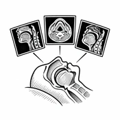
Cover image of the article: Obstructive Sleep Apnea and Radiologic Assessment: The Good, the Bad, and the Ugly (AI-generated illustration)

Image Credit:AI-generated illustration

Obstructive Sleep Apnea and Radiologic Assessment: The Good, the Bad, and the Ugly

Authors

Maziar Motiee-LangroudiMaziar Motiee-Langroudi ORCID1, Ebrahim KarimiEbrahim Karimi ORCID1, Mohammad Rasoul GhadamiMohammad Rasoul Ghadami ORCID1,*
1Otorhinolaryngology Research Center, Tehran University of Medical Sciences, Tehran, Iran
*Corresponding Author: Otorhinolaryngology Research Center, Tehran University of Medical Sciences, Tehran, Iran. Email: mr_ghadami@yahoo.com

Journal of Kermanshah University of Medical Sciences:Vol. 30, issue 3; e175696
Published online:Sep 30, 2026
Article type:Letter
Received:Aug 23, 2026
Accepted:Sep 06, 2026
How to Cite:Motiee-Langroudi M, Karimi E, Ghadami MR. Obstructive Sleep Apnea and Radiologic Assessment: The Good, the Bad, and the Ugly. J Kermanshah Univ Med Sci. 2026;30(3):e175696. doi: https://doi.org/10.5812/jkums-175696

Dear Editor,
Obstructive sleep apnea (OSA) is a prevalent disorder with significant systemic consequences. Diagnosis primarily relies on overnight polysomnography (PSG), which provides objective measures of airway obstruction and sleep fragmentation. In selected patients, drug-induced sleep endoscopy (DISE) can further refine the evaluation by visualizing dynamic airway collapse. Although most cases of OSA result from multifactorial airway collapsibility, a subset arises from discrete structural abnormalities of the head and neck. In this context, radiologic assessment plays a nuanced yet critical role that may be framed as “the good, the bad, and the ugly.”
The “good” lies in the ability of imaging modalities, such as computed tomography and magnetic resonance imaging, to identify clinically significant but often overlooked causes of OSA. Conditions such as aberrant carotid arteries, vallecular cysts, Thornwaldt cysts, parapharyngeal space tumors, and other mass lesions may directly contribute to airway obstruction. Recognition of these conditions is essential because their management differs substantially from standard therapies, such as continuous positive airway pressure. Radiologic evaluation enables precise anatomic delineation, facilitates surgical planning, and may ultimately lead to curative treatment in selected patients, particularly thin patients with long necks and no clear risk factors for OSA (1).
However, the “bad” emerges when imaging is overused without adequate clinical justification. Not all patients with suspected or confirmed OSA require radiologic evaluation. Indiscriminate referral for imaging, particularly in the absence of a detailed history and focused physical examination, can impose an unnecessary financial burden on healthcare systems, increase patient exposure to radiation, and lead to incidental findings that complicate clinical decision-making. Therefore, a more judicious approach is warranted, with imaging reserved for atypical presentations or cases with suspected structural abnormalities (2, 3).
The “ugly” represents the most concerning scenario: failure to identify or correctly interpret clinically relevant pathology. Missed diagnoses, such as parapharyngeal or nasopharyngeal tumors, may result in incomplete or ineffective treatment, leaving patients with unresolved symptoms despite standard therapy. Furthermore, unrecognized anatomic variations, such as aberrant carotid artery courses, may pose significant risks during surgical intervention, leading to delayed diagnosis, increased morbidity, or serious complications (1).
In conclusion, radiologic assessment in OSA should neither be neglected nor indiscriminately applied. There is an urgent need to develop clear guidelines for appropriate imaging use in OSA evaluation. Integrating radiologic assessment with clinical history, PSG, and DISE, in collaboration with sleep specialists, otolaryngologists, and radiologists, can optimize diagnosis and improve patient outcomes.
Footnotes

  • AI Use Disclosure:The authors declare that no generative AI tools were used in the creation of this article.

  • Authors' Contribution:All authors contributed equally to this article.

  • Conflict of Interests Statement:The authors have no conflict of interest.

  • Funding/Support:No funding was received for this study.

References

Copyright

Copyright © 2026, Journal of Kermanshah University of Medical Sciences. This open-access article is available under the Creative Commons Attribution 4.0 (CC BY 4.0) International License (https://creativecommons.org/licenses/by/4.0/), which allows for the copying and redistribution of the material only for noncommercial purposes, provided that the original work is properly cited.

Similar Articles

16
Jul
2019
Comments on “Cardiovascular Complications in Obstructive Sleep Apnea Syndrome”

Comments on “Cardiovascular Complications in Obstructive Sleep Apnea Syndrome”

Mahsa Mirdamadi,
Ali Safaei

Mirdamadi M, Safaei A. Comments on “Cardiovascular Complications in Obstructive Sleep Apnea Syndrome”. Multidiscip Cardio Annal. 2019;10(2):e94876. doi: https://doi.org/10.5812/mca.94876

19
Dec
2022
Clinical Audit of Obstructive Sleep Apnea: A Case Study in Sleep Disorders

Clinical Audit of Obstructive Sleep Apnea: A Case Study in Sleep Disorders

Mehdi Mirzaei Alavijeh,
Habibolah Khazaie,
Ali Alipourian,
Sepideh Khazaie,
Negar Karimi,
Nafiseh Mirzaei Alavijeh
,et al.

Mirzaei Alavijeh M, Khazaie H, Alipourian A, Khazaie S, Karimi N, et al. Clinical Audit of Obstructive Sleep Apnea: A Case Study in Sleep Disorders. J Health Rep Technol. 2022;8(4):e131339. doi: https://doi.org/10.5812/jhrt-131339

18
Aug
2019
Letter to Editors About “Cardiovascular Complications in Obstructive Sleep Apnea Syndrome and It’s Comments”

Letter to Editors About “Cardiovascular Complications in Obstructive Sleep Apnea Syndrome and It’s Comments”

Ali Seyed Resuli

Seyed Resuli A. Letter to Editors About “Cardiovascular Complications in Obstructive Sleep Apnea Syndrome and It’s Comments”. Multidiscip Cardio Annal. 2019;10(2):e96701. doi: https://doi.org/10.5812/mca.96701

16
Feb
2018
Anesthesia Preoperative Clinic Evaluation of Obstructive Sleep Apnea Using Nasal Fiberoptic Videoendoscopy: A Pilot Study Comparison with Polysomnography

Anesthesia Preoperative Clinic Evaluation of Obstructive Sleep Apnea Using Nasal Fiberoptic Videoendoscopy: A Pilot Study Comparison with Polysomnography

Sandeep Jain,
Peter J Kallio,
Kenneth Less,
Jutta Novalija,
Paul S Pagel,
Thomas J Ebert

Jain S, Kallio PJ, Less K, Novalija J, Pagel PS, et al. Anesthesia Preoperative Clinic Evaluation of Obstructive Sleep Apnea Using Nasal Fiberoptic Videoendoscopy: A Pilot Study Comparison with Polysomnography. Anesth Pain Med. 2018;8(1):e63546. doi: https://doi.org/10.5812/aapm.63546

24
Mar
2024
Validity of Airway Ultrasound in Correlation with Cormack-Lehane Grading in Obese Patients: A Cross-Sectional Study

Validity of Airway Ultrasound in Correlation with Cormack-Lehane Grading in Obese Patients: A Cross-Sectional Study

Ahmed El-Tawansy,
Ahmed Mohamed Salama Elnajar,
Hossam Abdel Baky Mahmoud,
Mohamed Ibrahim Amin,
Ahmed Abd Elmohsen Bedewy

El-Tawansy A, Mohamed Salama Elnajar A, Abdel Baky Mahmoud H, Ibrahim Amin M, Abd Elmohsen Bedewy A. Validity of Airway Ultrasound in Correlation with Cormack-Lehane Grading in Obese Patients: A Cross-Sectional Study. Anesth Pain Med. 2024;14(2):e142701. doi: https://doi.org/10.5812/aapm-142701

More by these authors

Maziar Motiee-LangroudiPubMedScholar
Ebrahim KarimiPubMedScholar
Mohammad Rasoul GhadamiPubMedScholar
Share
Cited by
Metrics