
1. Background
Cancer is a leading cause of mortality in developing countries, second only to cardiovascular diseases. The prevalence of colon cancer is increasing worldwide (1-3). Nearly 2 million new cases of colon cancer are diagnosed each year, and 950 thousand deaths are attributed to colon cancer annually (4-6). Despite advances in cancer diagnosis and treatment, chemotherapy, the most common cancer treatment, remains associated with numerous debilitating adverse effects and limitations (7-11).
Protein-based drug delivery systems, particularly albumin nanoparticles, have emerged as promising tools for cancer drug delivery because of their biocompatibility, biodegradability, and tumor-targeting capability through the enhanced permeability and retention (EPR) effect (12-17). Albumin naturally binds hydrophobic drugs, making it an ideal carrier for compounds such as curcumin (CUR) and paclitaxel (PTX). Curcumin, a polyphenolic compound derived from Curcuma longa, exhibits potent anti-inflammatory, antioxidant, and anticancer properties; however, its clinical application is hindered by poor bioavailability and rapid metabolism (18-21). Similarly, paclitaxel, a widely used chemotherapeutic agent that inhibits cell division by stabilizing microtubules, presents challenges related to low solubility and systemic toxicity (22). Incorporating both agents into albumin nanoparticles improves their solubility, stability, and tumor accumulation while minimizing adverse effects (23). In colon cancer, the co-delivery of curcumin and paclitaxel via albumin-based nanocarriers has shown combined anticancer effects, including reduced tumor proliferation and metastasis in experimental models (24), suggesting a promising strategy for improving therapeutic outcomes.
2. Objectives
This study aimed to develop bovine serum albumin (BSA) nanoparticles incorporating a cobalt ferrite magnetic core, encapsulated PTX, and a CUR coating. The synthesized nanoparticles were characterized by scanning electron microscopy (SEM) and Fourier-transform infrared spectroscopy (FTIR). Ls174T cells were cultured and treated with different concentrations of free drugs and nanoparticle formulations for 24, 48, and 72 h. Their effects were evaluated using methylthiazolyl tetrazolium (MTT) assays and by calculating half-maximal inhibitory concentrations (IC50) and P values. The aim was to design an albumin nanoparticle drug-delivery system to improve the effectiveness of PTX and CUR against cancer cells. Nontoxicity is an essential requirement for nanocarriers used in drug delivery. Various studies have shown that albumin nanoparticles alone are not toxic. For example, Tirkey et al. (8) demonstrated the nontoxicity of albumin nanoparticles with an average diameter of 212.6 nm in L132 cells.
3. Methods
Nanoparticle size and mean diameter are critical parameters in drug delivery systems because they directly influence cellular uptake and tumor penetration. Nanoparticles smaller than 200 nm generally accumulate more efficiently within the tumor microenvironment. In this study, appropriate concentrations of raw materials were selected to control nanoparticle size and achieve the desired physicochemical properties. The following sections describe solution preparation and the synthesis of albumin nanoparticles, magnetic albumin nanoparticles, and single- and dual-drug-loaded nanoparticle formulations.
3.1. Synthesis of Albumin Nanoparticles
Albumin nanoparticles were prepared using the desolvation method. Briefly, 80 mg of BSA was dissolved in 1 mL of double-distilled water under magnetic stirring until a clear solution was obtained. Ethanol was then added dropwise to induce nanoparticle formation, as indicated by the appearance of turbidity.
Chemical crosslinking was performed by adding 4% glutaraldehyde to stabilize the nanoparticles. The suspension was stirred for 24 h to ensure complete solvent removal. The nanoparticles were collected by sequential centrifugation at 2000 rpm for 10 min and then at 10,000 rpm for 20 min. The resulting pellet was used for further analyses.
3.2. Synthesis of BSA-CoFe2O4 Nanoparticles
BSA-CoFe2O4 nanoparticles were synthesized using the desolvation method. Briefly, 80 mg of BSA was dissolved in 1 mL of double-distilled water under magnetic stirring until a clear solution was obtained. A sonicated cobalt ferrite suspension containing 30 mg of CoFe2O4 in 1 mL of water was then added. Ethanol was subsequently introduced dropwise until turbidity appeared, indicating nanoparticle formation.
Chemical crosslinking was performed by adding 4% glutaraldehyde, and the mixture was stirred for 24 h at room temperature. The nanoparticles were then collected by centrifugation at 2000 rpm for 10 min and then at 10,000 rpm for 20 min. The resulting pellet was used for further analyses and experiments.
3.3. Synthesis of Drug-Loaded BSA-CoFe2O4 Nanoparticles
BSA-CoFe2O4 nanoparticles were prepared using the desolvation method. Briefly, 80 mg of BSA was dissolved in 1 mL of double-distilled water under magnetic stirring until a clear solution was obtained. Sonicated cobalt ferrite nanoparticles were then added to the protein solution. A paclitaxel solution (20 mg/mL in ethanol; 1 mL, corresponding to 20 mg of PTX) was added dropwise, followed by gradual addition of ethanol until turbidity appeared, indicating nanoparticle formation with encapsulated PTX. Crosslinking was performed by adding 4% glutaraldehyde, and the reaction mixture was stirred for 24 h at room temperature.
The nanoparticles were collected by centrifugation at 2000 rpm for 10 min and then at 10,000 rpm for 20 min, washed three times with deionized water to remove unbound drug, and resuspended. For CUR surface loading, 50 mg of CUR was dissolved in 20 mL of ethanol, and 10 mL of this solution, corresponding to 25 mg of CUR, was added to the nanoparticle suspension. The mixture was stirred for 2 - 3 h at room temperature at 300 rpm, followed by centrifugation at 10,000 rpm to remove unbound CUR. The final nanoparticles were washed three times with distilled water and freeze-dried after storage at −20 °C for 24 h.
Single-drug formulations (PTX-loaded or CUR-loaded nanoparticles) were prepared using the same procedure, with omission of the second drug-loading step.
3.4. Drug Loading and Encapsulation Efficiency
The drug loading (DL) of PTX- and CUR-loaded BSA-CoFe2O4 nanoparticles was evaluated using a gravimetric procedure. Briefly, 1 g of freeze-dried drug-loaded nanoparticles was dispersed in 10 mL of ethanol and stirred at room temperature for 45 min. The suspension was then centrifuged at 5000 rpm for 15 min, and the recovered nanoparticle pellet was collected, dried, and weighed. Based on this procedure, the estimated DL values were 17 ± 2% for PTX-loaded nanoparticles and 10 ± 2% for CUR-loaded nanoparticles. All measurements were performed in triplicate (n = 3), and the results were expressed as mean ± standard deviation.
Because the gravimetric procedure does not allow drug-specific quantification of PTX and CUR, these values are approximate gravimetric estimates rather than direct measurements of actual drug content. The recovered pellet may contain nanoparticle components in addition to the drug; therefore, its mass cannot be used to accurately determine the actual amount of PTX or CUR in the nanoparticles.
Encapsulation efficiency (EE%) was not quantitatively determined in this study. During nanoparticle preparation, 1 mL of PTX solution (20 mg/mL in ethanol), corresponding to 20 mg of PTX, and 10 mL of CUR solution prepared by dissolving 50 mg of CUR in 20 mL of ethanol, corresponding to 25 mg of CUR, were used. However, the encapsulated and unencapsulated fractions of each drug were not independently quantified using a validated, drug-specific analytical method. Therefore, no experimentally verified EE% values are reported. Accurate determination of drug-specific DL and EE would require a validated analytical method, such as high-performance liquid chromatography, which was not performed in this study. This limitation is acknowledged.
The experimental procedure and data analysis were performed by Dr. Mehdi Haghi, Associate Professor at the University of Tabriz, Tabriz, Iran.
3.5. Cell Viability Assay
A cell viability assay was performed to evaluate the potential cytotoxic effects of the nanoparticles on Ls174T cells. The cells were purchased from the Pasteur Institute, Tehran, Iran. RPMI 1640 medium used for cell culture was supplemented with 10% fetal bovine serum (Gibco) and 1% antibiotic solution containing 10,000 U/mL penicillin and 10,000 µg/mL streptomycin (Gibco). Cell morphology, adherence patterns, and growth characteristics were routinely monitored using an inverted light microscope and were consistent with those expected for Ls174T cells. Low-passage cells (passages 3 - 8) were used, and periodic testing for mycoplasma infection yielded negative results. Cells were cultured under humidified conditions with 5% CO2 at 37 °C. They were harvested using 0.25% trypsin/1 mM EDTA and seeded into 96-well plates at a density of 1.0 × 10⁴ cells per well. Cells were incubated to allow attachment and to reach approximately 80% confluence before treatment.
An MTT-based assay was used to assess cell viability and cytotoxicity. Cells were exposed to nanoparticles prepared from stock solutions made by dissolving the compounds in a 1:1 mixture of dimethyl sulfoxide (0.1%) and RPMI medium. No solvent control was used; however, the results obtained in this study indicate that the solvent did not significantly affect the therapeutic effects. These stock solutions were serially diluted in the same medium to obtain final concentrations of 0.00, 0.01, 0.02, 0.04, and 0.08 μg/mL. Three replicates were assigned to each dilution of each agent and to the untreated cell-containing controls. These replicates were used to calculate IC50 values. Treatments were not randomly assigned, and no blinding or masking was performed.
After the cells were allowed to adhere for 24 h, the prepared nanoparticle solutions and other agents were added to achieve a final volume of 100 μL per well. After incubation for 24, 48, or 72 h, 10 μL of MTT solution (5 mg/mL in phosphate-buffered saline) was added to each well, followed by incubation for an additional 3 h. The medium was then removed, and 100 μL of dimethyl sulfoxide was added to each well in the dark at room temperature to solubilize the purple formazan crystals. Absorbance, indicative of cell viability, was recorded at 570 nm using a microplate reader.
3.6. Statistical Analysis
Statistical analyses were performed using GraphPad Prism software. One-way analysis of variance followed by Tukey's post hoc multiple-comparison test was used for dose-response analyses. Time points were compared using unpaired t tests. IC50 values were calculated using nonlinear regression with a four-parameter logistic dose-response model. Statistical significance was defined as P < 0.05, and P values were indicated as *P ≤ 0.05, **P ≤ 0.01, and ***P ≤ 0.001.
All experimental procedures described in this study were performed at the Faculty of Natural Sciences, University of Tabriz, Tabriz, Iran.
3.7. Scanning Electron Microscopy and Fourier-Transform Infrared Spectroscopy
The morphology and particle size of the prepared nanoparticles were evaluated using a TESCAN MIRA3 field-emission scanning electron microscope (FE-SEM; TESCAN, Brno, Czech Republic) at the SEM Laboratory, Faculty of Natural Sciences, University of Tabriz, Tabriz, Iran. The SEM measurements and image analysis were performed by Dr. Mehdi Haghi, Associate Professor at the University of Tabriz. FTIR spectra were recorded using a Bruker Tensor 27 FTIR spectrometer (Bruker, Germany) at the FTIR Laboratory, Faculty of Natural Sciences, University of Tabriz, Tabriz, Iran. The FTIR measurements and spectral analysis were also performed by Dr. Mehdi Haghi.
4. Results
4.1. Scanning Electron Microscopy
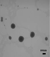
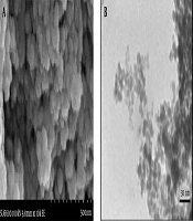
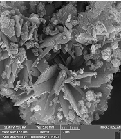
As shown in Figure 1, the size and morphology of BSA nanoparticles (BSA NPs), magnetic cobalt ferrite nanoparticles (CoFe2O4 NPs), magnetic albumin nanoparticles (BSA-CoFe2O4 NPs), and drug-loaded formulations, including CUR-loaded nanoparticles (BSA-CoFe2O4 NPs + CUR), PTX-loaded nanoparticles (BSA-CoFe2O4 NPs + PTX), and dual-drug-loaded nanoparticles (BSA-CoFe2O4 NPs + PTX and CUR), were investigated by FE-SEM.

Figure 1.
Scanning electron microscopy images showing the morphology of different nanoparticle formulations. (A) BSA NPs, (B) CoFe2O4 NPs, (C) BSA-CoFe2O4 NPs, (D) BSA-CoFe2O4 NPs + PTX, and (E) BSA-CoFe2O4 NPs + PTX and CUR. Abbreviations: BSA, bovine serum albumin; NPs, nanoparticles; PTX, paclitaxel; CUR, curcumin.
SEM images were analyzed using ImageJ software by randomly selecting approximately 50 nanoparticles from different regions of each formulation. Particle size ranged from approximately 120 to 220 nm, with a mean diameter of 170 ± 30 nm. The nanoparticles exhibited generally uniform morphology with minimal aggregation.
The observed morphology and size distribution were consistent with those reported in previous studies of similar albumin-based magnetic nanoparticle systems.
Dynamic light scattering, zeta potential, and magnetic responsiveness analyses were not performed in this study.
4.2. Fourier-Transform Infrared Spectroscopy
The FTIR spectra of BSA powder and different nanoparticle formulations are shown in Figure 2.
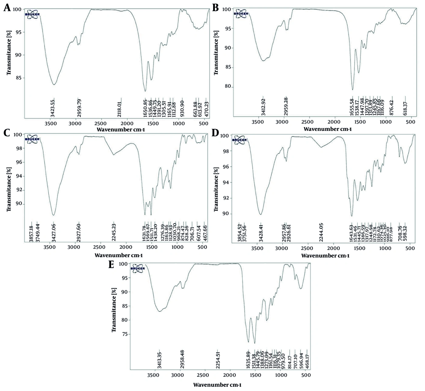
Figure 2.
Fourier-transform infrared spectra of different samples showing characteristic functional groups. (A) BSA powder, (B) BSA-CoFe2O4 NPs, (C) BSA-CoFe2O4 NPs + CUR, (D) BSA-CoFe2O4 NPs + PTX, and (E) BSA-CoFe2O4 NPs + PTX and CUR. Abbreviations: BSA, bovine serum albumin; NPs, nanoparticles; CUR, curcumin; PTX, paclitaxel.
The FTIR spectra of BSA, BSA-CoFe2O4 nanoparticles, and drug-loaded formulations were used to confirm nanoparticle formation and drug incorporation. In the spectrum of native BSA, characteristic amide I and amide II bands were observed at approximately 1650 cm-1 and 1536 cm-1, corresponding to C=O stretching and N–H bending vibrations, respectively.
For BSA-CoFe2O4 nanoparticles, these peaks shifted slightly to 1655 cm-1 and 1538 cm-1, indicating interactions between BSA and the magnetic nanoparticles. In addition, metal–oxygen (Fe–O and Co–O) stretching vibrations appeared in the low-frequency region of 580 - 460 cm-1, confirming the incorporation of cobalt ferrite.
In PTX-loaded nanoparticles (BSA-CoFe2O4 NPs + PTX), characteristic PTX peaks were observed at 1716 cm-1, 1248 cm-1, and 1074 cm-1, corresponding to C=O, C–N, and C–O functional-group vibrations, respectively, along with preserved BSA amide bands and metal–oxygen vibrations.
Similarly, CUR-loaded nanoparticles (BSA-CoFe2O4 NPs + CUR) exhibited characteristic CUR peaks at approximately 2927 cm-1, 1722 cm-1, 1620 - 1580 cm-1, and 1074 cm-1, corresponding to C–H, C=O, aromatic C=C, and C–O stretching vibrations, respectively.
In dual-drug-loaded nanoparticles (BSA-CoFe2O4 NPs + PTX + CUR), the FTIR spectrum displayed characteristic peaks of both drugs, along with BSA and CoFe2O4 signatures, confirming co-loading without substantial structural disruption of the nanoparticle matrix.
4.3. Cell Viability Assay
The primary MTT assay comparison included dual-drug-loaded nanoparticles (BSA-CoFe2O4 NPs + PTX + CUR), free PTX, free CUR, single-drug-loaded nanoparticles (BSA-CoFe2O4 NPs + PTX and BSA-CoFe2O4 NPs + CUR), and unloaded nanoparticles (BSA NPs, CoFe2O4 NPs, and BSA-CoFe2O4 NPs) to evaluate their relative cytotoxic effects.
Cells were treated with increasing concentrations (0.01, 0.02, 0.04, and 0.08 µg/mL) of each formulation for 24, 48, and 72 h. Treatment concentrations for nanoparticle groups were calculated based on total nanoparticle mass rather than drug-equivalent concentrations. The results, including cell viability, IC50 values, and statistical analysis, are presented in Figure 3 and Table 1.
Table 1.
Half-Maximal Inhibitory Concentrations (IC50) in Ls174T Cells Following Exposure to Free Drugs, Single-Drug-Loaded Nanoparticles, Dual-Drug-Loaded Nanoparticles, and Unloaded Nanocarriers for 24, 48, and 72 H a
| Agents | 24 h (µg/mL) | 48 h (µg/mL) | 72 h (µg/mL) |
|---|---|---|---|
| BSA | 0.32 (NS) | 0.155 (NS) | 0.112 (NS) |
| PTX | 0.074 b | 0.036 b | 0.019 c |
| CUR | 0.213 (NS) | 0.091 (NS) | 0.073 (NS) |
| BSA NPs | 0.257 (NS) | 0.102 (NS) | 0.099 (NS) |
| CoFe2O4 NPs | 0.105 (NS) | 0.087 (NS) | 0.032 b |
| BSA-CoFe2O4 NPs | 0.098 (NS) | 0.106 (NS) | 0.079 (NS) |
| BSA-CoFe2O4 NPs + PTX | 0.071 b | 0.025 c | 0.016 d |
| BSA-CoFe2O4 NPs + CUR | 0.191 (NS) | 0.061 b | 0.072 (NS) |
| BSA-CoFe2O4 NPs + PTX and CUR | 0.069 b | 0.02 c | 0.013 d |
a Abbreviations: BSA, bovine serum albumin; PTX, paclitaxel; CUR, curcumin; NPs, nanoparticles; NS, not significant.
b A P value ≤ 0.05 was considered statistically significant.
c A P value ≤ 0.01 was considered statistically significant.
d A P value ≤ 0.001 was considered statistically significant.
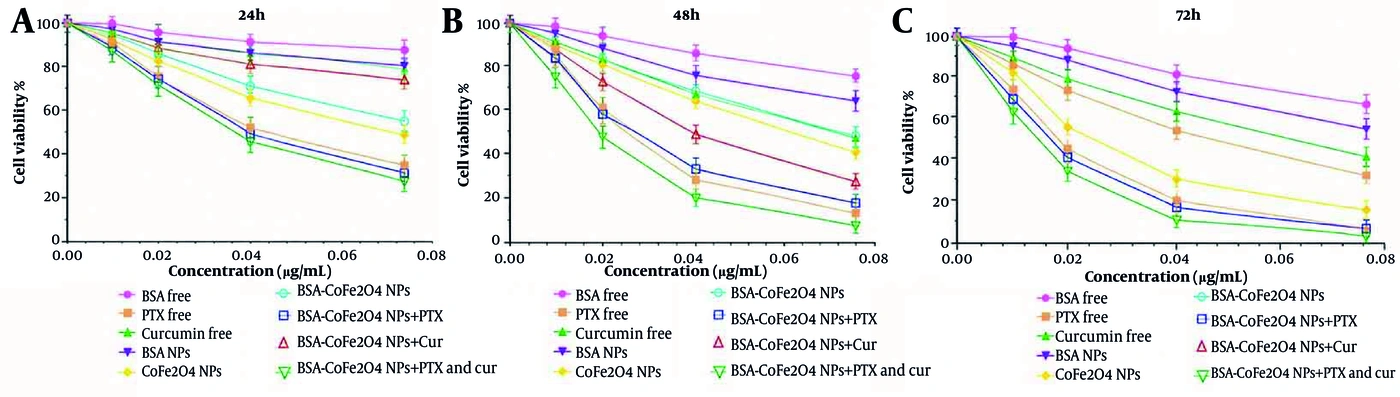
Figure 3.
Methylthiazolyl tetrazolium (MTT) assay results showing the viability of Ls174T cells after treatment with various concentrations of the indicated formulations. (A) Treatment for 24 h, (B) treatment for 48 h, and (C) treatment for 72 h. Cell viability was assessed after exposure to free drugs, single-drug-loaded nanoparticles, dual-drug-loaded nanoparticles, and unloaded carriers.
Untreated cell-containing control wells exhibited approximately 100% cell viability, confirming normal cell growth conditions. Free PTX and PTX-loaded formulations demonstrated concentration- and time-dependent cytotoxic effects against Ls174T cells. Curcumin and CUR-loaded nanoparticles showed minimal cytotoxicity at the tested concentrations; however, co-administration with PTX in dual-loaded nanoparticles resulted in enhanced cytotoxicity compared with the single-drug formulations.
Approximate gravimetric estimates of DL were 17 ± 2% for PTX-loaded nanoparticles and 10 ± 2% for CUR-loaded nanoparticles. Because this gravimetric procedure does not provide drug-specific quantification, these values should be interpreted as approximate estimates rather than experimentally verified drug-loading values.
These findings indicate that the dual-loaded nanoparticle formulation exhibited enhanced cytotoxic activity under the tested conditions. The dual-drug-loaded nanoparticles showed the greatest overall inhibitory effect. Although prolonged exposure for 48 and 72 h generally increased cytotoxicity, the time-dependent pattern was not uniform across all formulations.
The unloaded BSA and BSA-CoFe2O4 carriers exhibited negligible cytotoxicity within the tested concentration range. In contrast, CoFe2O4 NPs had a reported IC50 of 0.032 µg/mL at 72 h, within the tested range, indicating cytotoxic activity under these conditions.
5. Discussion
Protein-based drug delivery systems, particularly albumin-based carriers, have been widely investigated as promising platforms for anticancer drug delivery because of their biocompatibility, biodegradability, and capacity for drug encapsulation. However, whether these properties translate into reduced systemic toxicity depends on the formulation and requires experimental validation. Paclitaxel is a well-established chemotherapeutic agent with broad clinical application; however, its use remains associated with systemic adverse effects and drug resistance. In this study, CUR, a natural compound with reported anticancer activity, was incorporated into the formulation. Despite its relatively mild cytotoxicity and rapid metabolic degradation, CUR has demonstrated combined effects with PTX. Considering the reported biological activities of CUR and the cytotoxic effects observed in this study, co-delivery of PTX and CUR using albumin-based nanocarriers may provide a potential approach for combined anticancer drug delivery. However, the present in vitro findings do not establish reduced systemic toxicity or improved therapeutic efficacy in vivo. These outcomes require further investigation in appropriate preclinical models (25-29). A previous study of a doxorubicin-loaded lipid-based formulation (DepoDOX) demonstrated that appropriate particle size, high drug encapsulation, physical stability, and prolonged drug release can enhance the potential of nanoformulations for localized cancer drug delivery. Although doxorubicin differs from PTX, these findings highlight the importance of physicochemical properties and drug-loading capacity in developing effective nanocarriers. In this study, PTX was incorporated into BSA-CoFe2O4 nanoparticles, providing a protein-based nanocarrier for PTX delivery, while co-loading with CUR further enhanced the cytotoxic effect against Ls174T cells (30).
Curcumin exhibits multiple anticancer mechanisms, including inhibition of NF-κB signaling, induction of apoptosis, and suppression of angiogenesis (28, 31, 32). A 2025 study found that nano-curcumin increased p53 gene expression in A549 lung cancer cells compared with control groups, demonstrating anticancer and antiproliferative effects in these cells (33). Reduced cyclooxygenase-2 (COX-2) expression was also reported when Shirmohammadzadeh et al. treated colon cancer in BALB/c mice using chitosan–curcumin nanoparticles (34). Vahedian Sadeghi et al. showed that nano-curcumin can modulate key cancer-related pathways by upregulating the tumor suppressors P53 and Rb and downregulating the oncogenes E6 and E7 more effectively than free CUR, while exerting limited effects on normal fibroblast cells. These findings suggest that nanoformulation may enhance the anticancer activity of CUR and support its potential as a therapeutic strategy (35). Hadadian et al. also reported that combining CUR with photometric laser treatment can further reduce cell viability, suggesting a potential enhancing effect of CUR as a therapeutic agent (36). However, poor solubility and low systemic bioavailability have limited the standalone clinical efficacy of CUR (37, 38). Loading CUR onto albumin nanoparticles has been reported to improve its stability, solubility, and cellular uptake (39). Previous research has also demonstrated that protein-associated CUR can exhibit greater in vitro anticancer activity than free CUR, supporting the potential role of protein-based carriers in enhancing its biological activity (40).
Similarly, PTX, a microtubule-stabilizing chemotherapeutic agent, is associated with systemic toxicity and multidrug resistance in solid tumors, including colorectal cancer (41). Previous studies have suggested that albumin-based delivery systems may interact with tumor-associated transport mechanisms, including the EPR effect and albumin-associated pathways involving proteins such as gp60 and SPARC (42). However, the involvement of these mechanisms in the cellular uptake, tumor accumulation, or biodistribution of the BSA-CoFe2O4 formulation developed in this study was not investigated. Their contribution to the observed cytotoxic effects therefore remains to be established.
The greater cytotoxic effect of BSA-CoFe2O4 NPs co-loaded with PTX and CUR observed in this study, as shown in Figure 4, is consistent with earlier findings suggesting that co-delivery of PTX and CUR may enhance cancer cell sensitivity to apoptosis, reduce drug resistance, and modulate key survival pathways such as PI3K/Akt and MAPK (43). Amphiphilic diblock copolymer poly(ethylene glycol)-block-poly(lactide-co-glycolide) (PEG-PLGA) nanoparticles encapsulating PTX and CUR have also been reported to exert cytotoxic effects on 4T1 and MDA-MB-231 cells through suppression of the NF-κB signaling pathway (44). In addition, significant cytotoxicity has been observed in MCF-7 cells after treatment with F127 nanogels loaded with PTX and CUR (45). Although combination therapy and nanoparticle-based drug delivery have been investigated as approaches to improve anticancer treatment, this study did not evaluate whether the developed formulation permits dose reduction, reduces systemic toxicity, or improves drug accumulation in tumor tissues. Furthermore, although approximate gravimetric estimates of DL were obtained, drug-specific content and EE were not quantitatively determined. Consequently, no conclusions can be drawn regarding dose-sparing effects or reductions in systemic adverse effects. These questions warrant further investigation using validated drug quantification methods and appropriate in vivo models (46-48). Such investigations are particularly relevant to colorectal cancer, in which chemoresistance and treatment-associated toxicity remain important clinical challenges.
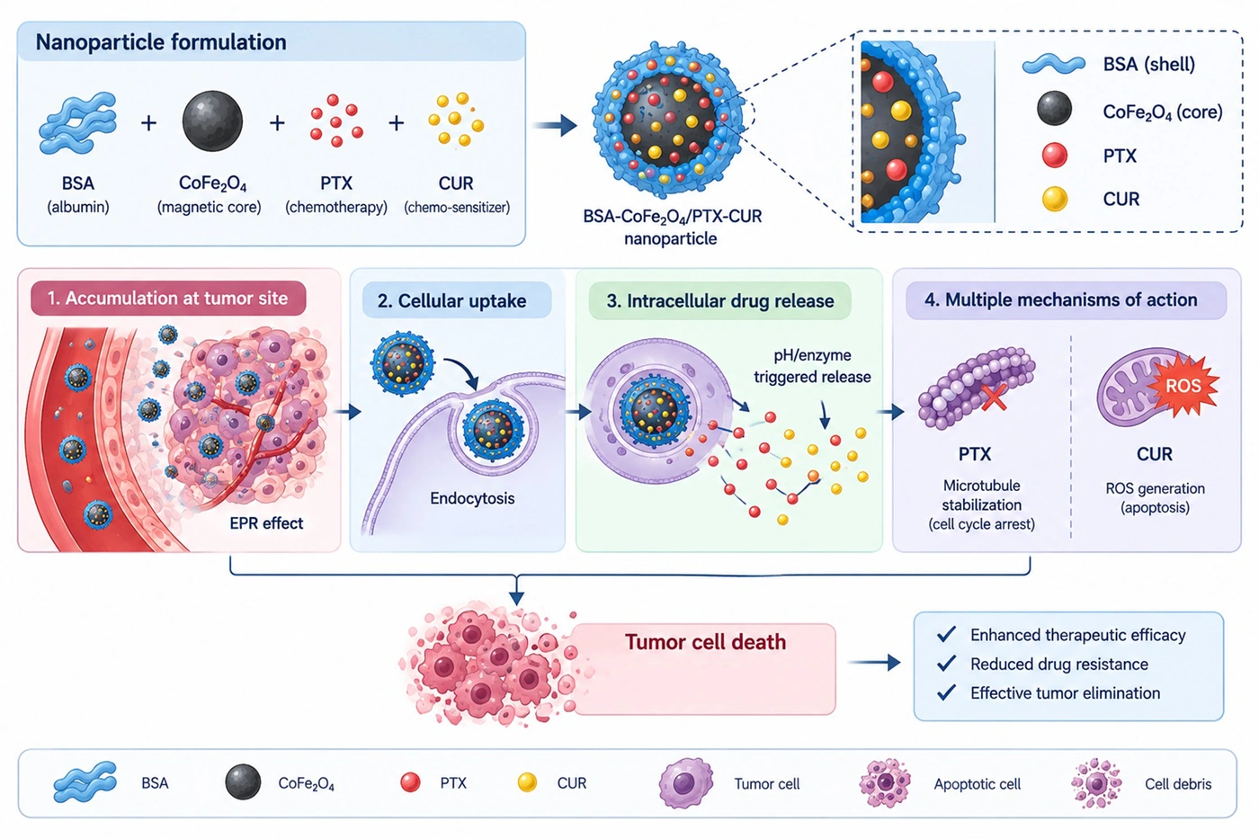
Figure 4.
Schematic illustration of the proposed mechanism of action of bovine serum albumin (BSA)-CoFe2O4 nanoparticles co-loaded with paclitaxel (PTX) and curcumin (CUR) in Ls174T colorectal cancer cells. The proposed mechanism involves cellular uptake of the nanoparticle formulation and intracellular delivery of PTX and CUR. Paclitaxel may contribute to microtubule stabilization and cell-cycle arrest, whereas CUR may contribute to modulation of oxidative stress and apoptotic pathways. Their combined effects may contribute to the reduced cell viability observed in this study. These proposed intracellular mechanisms were not directly investigated and require further experimental validation.
A limitation of this study is the use of a single cancer cell line, which restricts the generalizability of the findings. In addition, in vivo experiments were not performed; therefore, the systemic behavior, biodistribution, and therapeutic efficacy of the developed nanoparticles remain unvalidated. Furthermore, in vitro drug release profiles and cytotoxic selectivity against normal, noncancerous cells were not evaluated. These aspects should be addressed in future studies to comprehensively assess the clinical potential of the proposed nanocarrier system.
Overall, this study demonstrates the in vitro cytotoxic activity of BSA-CoFe2O4 nanoparticles co-loaded with PTX and CUR against Ls174T colorectal cancer cells. The findings support further investigation of this formulation as a potential platform for combined anticancer drug delivery. However, these results do not establish tumor-specific targeting, enhanced tumor accumulation, reduced systemic toxicity, or improved therapeutic efficacy in vivo. The use of a single cancer cell line, the absence of normal cell controls, and the lack of in vivo validation limit the interpretation of the findings. Future studies should investigate drug release kinetics, cytotoxic selectivity, biodistribution, tumor accumulation, and systemic toxicity using validated analytical methods and appropriate preclinical models before conclusions regarding targeted delivery or therapeutic safety can be drawn.