
1. Background
Cancer is one of the leading causes of morbidity and mortality worldwide and represents a major public health challenge in both developed and developing countries. The global number of new cancer cases was estimated at 19.3 million in 2020 and is projected to reach nearly 30 million by 2040, primarily because of population growth, aging, and continued exposure to modifiable risk factors (1, 2). Despite advances in early detection and conventional treatments, including surgery, chemotherapy, and radiotherapy, many malignancies remain resistant or relapse because of drug resistance, systemic toxicity, and the inability of treatments to selectively target tumor cells (3).
In recent years, gene therapy has emerged as a promising alternative for treating cancer and other gene-related diseases because it targets the molecular basis of disease rather than its symptoms (4). This approach delivers therapeutic nucleic acids, such as plasmid DNA (pDNA), small interfering RNA, or gene-editing constructs, into target cells to regulate, repair, replace, or silence disease-associated genes (5). In cancer, one strategy is to induce apoptosis by introducing suicide or proapoptotic genes that activate programmed cell death pathways. These genes can sensitize tumor cells to chemotherapy or act as independent antitumor agents (6). Natural death-inducing genes that activate intrinsic and extrinsic apoptotic cascades are particularly promising for promoting selective tumor cell death while sparing normal tissue. The expression of these genes, typically delivered via pDNA, leads to the synthesis of proapoptotic proteins that can either sensitize tumor cells to chemotherapeutic drugs or directly induce apoptosis, making them highly attractive for cancer gene therapy (7, 8). The success of gene therapy, however, depends critically on the development of safe and efficient gene delivery systems capable of transporting nucleic acids into target cells with minimal toxicity and high transfection efficiency (9). Although viral vectors exhibit high gene transfer efficiency, their potential immunogenicity and mutagenic risks have limited their clinical use (10). Consequently, nonviral delivery systems, particularly cationic polymers such as polyethylenimine (PEI), have received increasing attention because of their strong DNA-binding capacity, efficient endosomal escape, and low cost (11). Nevertheless, the high cytotoxicity of PEI, especially in its high-molecular-weight forms, restricts its biomedical applications (12).
To address these limitations, researchers have developed various nanocomposite-based delivery systems that combine cationic polymers such as PEI with functional nanomaterials, including gold nanoparticles (NPs), silica NPs, carbon nanotubes, and graphene derivatives, to enhance nucleic acid binding, cellular uptake, and biocompatibility (13, 14). Among these materials, graphene oxide quantum dots (GOQDs) are notable for their large specific surface area, high dispersibility in aqueous media, good biocompatibility, and capacity for functional modification. GOQDs can serve as versatile platforms for gene or drug delivery, enhancing PEI-mediated transfection while reducing cytotoxicity (15).
Several previous studies have used PEI and graphene oxide (GO) for gene or drug delivery. In 2011, Zhang et al. reported enhanced chemotherapy efficacy through the delivery of Bcl-2-targeted short interfering RNA (siRNA) and the anticancer drug doxorubicin using PEI-grafted GO (16). In another study, a hybrid gene carrier fabricated by conjugating low-molecular-weight branched PEI (BPEI) to GO increased the effective molecular weight of BPEI; consequently, it improved DNA binding and condensation and enhanced transfection efficiency (17). Previous work by this research group has investigated PEI-decorated GOQD NPs as carriers for suicide gene delivery to a three-dimensional breast cancer model (18) and aptamer-guided GOQDs for targeted suicide gene therapy in an organoid model of luminal breast cancer (19).
However, the fabrication and characterization of GOQD/PEI nanocarriers functionalized with hyaluronic acid (HA), a naturally occurring polysaccharide with a strong binding affinity for CD44 receptors that are overexpressed on the surface of many tumor cells, have not yet been reported. Incorporating HA as a surface modification strategy is particularly attractive in targeted cancer therapy because HA-CD44 interactions facilitate receptor-mediated endocytosis, enabling selective cellular uptake by malignant cells while minimizing nonspecific internalization and off-target cytotoxicity (20, 21).
2. Objectives
Accordingly, this study developed a hyaluronic acid-modified GOQD/PEI nanocarrier as a biocompatible and efficient nonviral platform for gene delivery (Figure 1). The carrier effectively condensed pDNA and exhibited significantly improved cell viability following HA modification, confirming enhanced cytocompatibility. These results indicate that it has strong potential as a safe and targetable vehicle for future gene therapy applications, particularly in advanced 3D or organoid models.
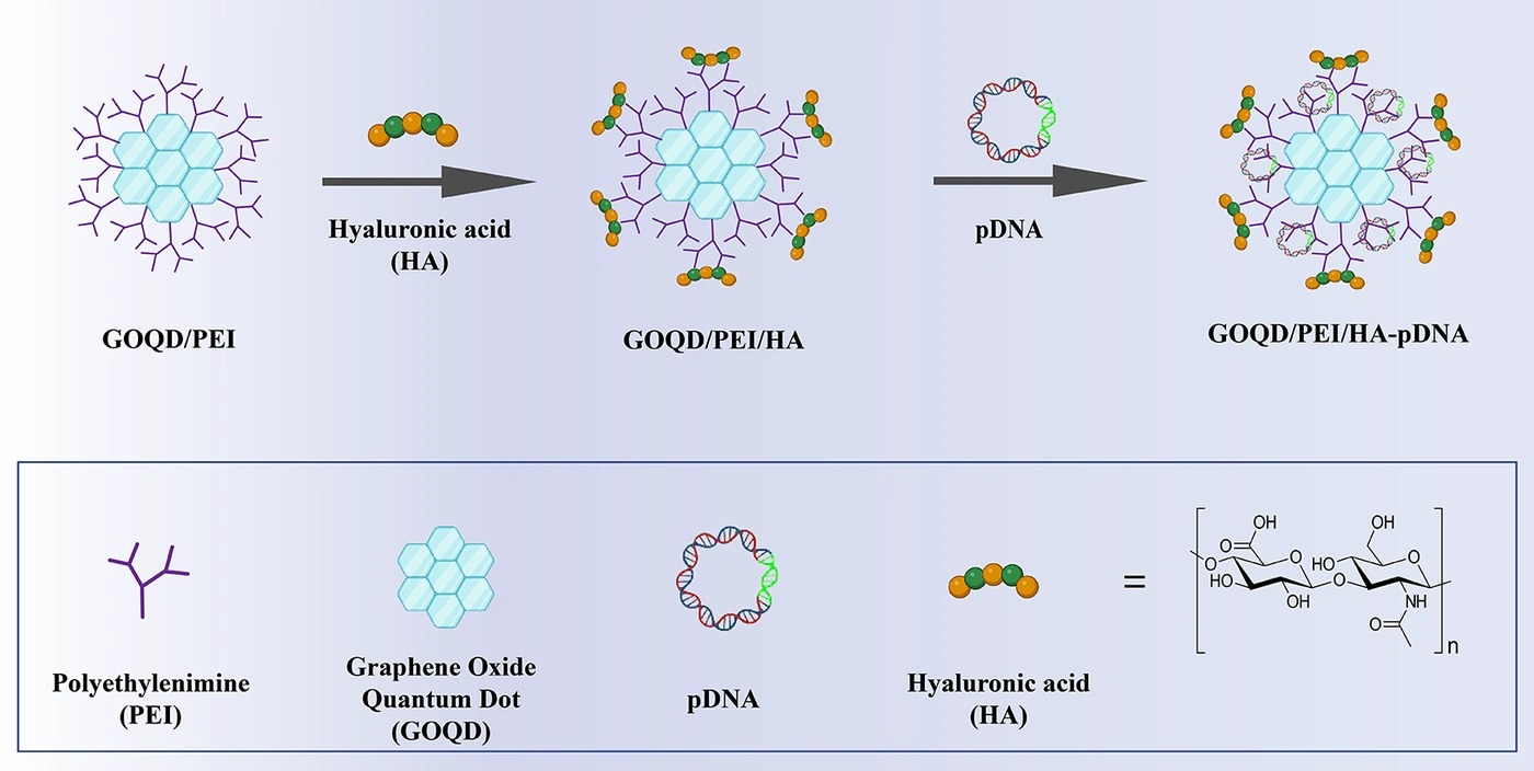
Figure 1.
Schematic illustration of the preparation of GOQD/PEI/HA-pDNA nanocomplexes. The diagram depicts the sequential assembly of GOQD/PEI nanoparticles, followed by surface modification with hyaluronic acid (HA) and complexation with plasmid DNA (pDNA).
3. Methods
3.1. Materials
The Escherichia coli (E. coli) strain and pIRES2-EGFP vector were obtained from Invitrogen, USA, and the Research Institute of Biotechnology of Mashhad, Iran, respectively. The plasmid extraction kit, safe stain, and DNA ladder were purchased from Sinaclon, Iran. Dulbecco's Modified Eagle's Medium F-12 (DMEM-F12) and fetal bovine serum (FBS) were purchased from Bioidea, Iran, and GIBCO, respectively. The cervical cancer HeLa cell line was obtained from the Pasteur Institute of Iran (catalog number: IBRC C11311). Cells were cultured in DMEM-F12 supplemented with 10% FBS and 1% penicillin/streptomycin. 3-(4,5-Dimethylthiazol2-yl)-2,5-diphenyltetrazolium bromide (MTT), 1-ethyl-3-(3-dimethyleaminopropyl) carbodiimide (EDC), N-hydroxysuccinimide (NHS), branched polyethyleneimine (PEI), and a 3.5-kDa dialysis bag were purchased from Sigma-Aldrich. Hydrochloric acid (HCl), HA (MW = 100 - 120 kDa), agarose, kanamycin, and dimethyl sulfoxide (DMSO) were obtained from Merck (Darmstadt, Germany).
3.2. Synthesis of GOQD/PEI/HA NPs
To synthesize GOQD/PEI/HA, 12.3 mg of HA was dispersed in 6 mL of distilled water, and 6 mL of distilled water was added to 12 mg of the previously synthesized GOQD/PEI (18). The solutions were mixed, and the pH was adjusted to 6.5 by adding 1 M HCl.
Next, to perform the amidation reaction between PEI amino groups and HA carboxylic groups, 1 mL of a solution prepared by dispersing 24 mg of EDC and 14.41 mg of NHS in 500 μL of water and 500 μL of DMSO was added to the prepared solution. The resulting solution was stirred for 24 hours at room temperature. The reaction mixture was then dialyzed against water using a 3.5-kDa dialysis bag for 3 days and finally lyophilized to remove excess residues.
3.3. Characterization of GOQD/PEI/HA Structural Features
3.3.1. Fourier Transform Infrared Spectroscopy
After lyophilization, the dried GOQD/PEI/HA sample was characterized using FT-IR spectroscopy (Bruker Vector 22 spectrophotometer). KBr pellets were prepared, and spectra were recorded from 400 to 4000 cm-1. The results were compared with those of the GOQD and GOQD/PEI samples.
3.3.2. Thermogravimetric Analysis
The thermal stability of the GOQD/PEI/HA sample was assessed by thermogravimetric analysis (NETZSCH STA 409 PC/PG) under a nitrogen flow of 20 mL/min at a heating rate of 10 °C/min over a range of 25 - 600 °C. The results were compared with those of the GOQD and GOQD/PEI samples.
3.4. Bacterial Culture and pDNA Extraction
Bacteria were cultured on Luria-Bertani (LB) agar medium containing kanamycin at 37 °C overnight. A single colony of E. coli transformed with the pIRES2-EGFP plasmid was cultured in LB broth medium with shaking at 250 rpm in a shaker incubator (Binder; CB-150) overnight. pDNA was extracted from the cultured bacteria using a plasmid extraction kit (Sinaclon). The concentration and purity of the extracted plasmid were assessed and verified using agarose gel electrophoresis (Paya Pagoohesh).
3.5. Plasmid Loading on GOQD/PEI/HA and Determination of Absorbed pDNA
Aqueous suspensions of GOQD/PEI/HA at concentrations of 9, 37.5, 150, 300, 450, 600, and 750 ng/μL were prepared. To construct different ratios of GOQD/PEI/HA-pDNA complexes, various amounts of GOQD/PEI/HA NPs were added to a constant amount of pDNA (150 ng/μL) at ratios of i.e., 1 to 0.6, not 0.6 in another format. The mixtures were gently pipetted twice and then incubated at room temperature for 40 minutes. Absorbed and free plasmids were analyzed using a gel retardation assay. Agarose gel electrophoresis was used to evaluate the mobility of each complex, followed by visualization under UV illumination (UV tech, Germany).
3.6. Complex Stability Over Time and at Different Temperatures
To determine the stability of GOQD/PEI/HA-pDNA complexes over time and at different temperatures, a gel retardation assay was performed for each sample at various ratios (i.e., 1 to 0.6, not 0.6 in another format) after incubation for 40 minutes and 24 hours at 25 and 37 °C. The gel retardation assay indicated the stability of the complexes.
3.7. Charge and Size Distribution Analysis of the GOQD/PEI/HA-pDNA Complex
3.7.1. Zeta Potential and Dynamic Light Scattering Measurement
The surface charge and particle size distribution of the GOQD/PEI/HA-pDNA complex at a 1:4 ratio were measured by zeta potential analysis and DLS using a Malvern Nano ZS90 instrument and DTS software (Malvern Instruments, UK). This complex ratio was prepared using the flash mixing procedure described above, and surface charge and hydrodynamic particle size were measured at room temperature.
3.7.2. Field-Emission Scanning Electron Microscopy
The morphology and average diameter of the GOQD/PEI/HA-pDNA complexes (prepared at a mass ratio of 1:4) were assessed using field-emission scanning electron microscopy (FE-SEM). Samples were deposited onto a clean conductive substrate and subsequently vacuum-coated with a thin gold layer. FE-SEM imaging was performed using a TESCAN MIRA3-LMU instrument.
3.8. Hemolysis Assay
Fresh blood samples were obtained from the Kermanshah Blood Bank Center and collected in EDTA-containing tubes to prevent coagulation. Samples were centrifuged at 2000 rpm for 10 minutes to separate red blood cells (RBCs) from plasma. The RBCs were then diluted with PBS at a 1:9 ratio. Next, 1 mL of diluted blood was mixed with different concentrations of GOQD/PEI/HA in microtubes and incubated for 1 hour at 37 °C. After incubation, the microtubes were centrifuged, and the supernatant was transferred to a 96-well plate. Hemolytic activity was evaluated by comparison with negative and positive controls. PBS and distilled water, in conjunction with blood, served as the negative (0% lysis) and positive controls (100% lysis), respectively. Hemoglobin release, indicating RBC damage, was measured at 545 nm using an enzyme-linked immunosorbent assay (ELISA) reader and evaluated according to the ASTM F756 - 00 standard (22).
3.9. GOQD/PEI/HA NPs and Complex Cytotoxicity Test
HeLa cells were seeded in 96-well plates at a density of 7 x 10³ cells/well and incubated overnight in DMEM-F12 supplemented with 10% FBS and 1% penicillin-streptomycin under standard mammalian cell culture conditions. Cells were then treated for 24 hours with different concentrations (150, 300, 450, and 600 ng) of GOQD/PEI, GOQD/PEI/HA, and their corresponding complexes at ratios of 1:1, 1:2, 1:3, and 1:4. The medium was then replaced with 100 μL of culture medium containing 0.5 mg/mL MTT and incubated for 4 hours. The MTT solution was removed, and 100 μL of DMSO was added and gently pipetted to dissolve the formazan crystals. After 10 minutes of incubation, absorbance at 570 nm was measured using an ELISA reader (CytationTM 5, BioTek).
3.10. Statistical Analysis
All experiments were performed in triplicate (n = 3), and data are presented as mean ± SD. Cell viability (MTT assay) was analyzed by one-way ANOVA followed by Tukey's post hoc test. Differences were considered significant at P < 0.05. All analyses and graphs were generated using GraphPad Prism 9.0.
4. Results
4.1. GOQD/PEI/HA NPs Synthesis
GOQDs were synthesized via incomplete carbonization of citric acid, an efficient, rapid, and straightforward method that produces sheet-like nanostructures enriched with oxygen-containing functional groups, which confer strong hydrophilicity and chemical reactivity to the GOQDs (23). PEI, a widely used cationic polymer with excellent DNA-binding capacity and high transfection efficiency, was subsequently grafted onto GOQDs to form GOQD/PEI nanostructures previously applied in gene delivery studies (18, 19). To further improve biocompatibility, HA was conjugated to GOQD/PEI using EDC/NHS chemistry. Briefly, an aqueous dispersion of HA was mixed with an equal volume of the previously synthesized GOQD/PEI suspension under mild aqueous conditions (pH 6.5) to activate the HA carboxyl groups, followed by stirring at room temperature for 24 hours to enable stable covalent attachment of HA chains to the GOQD/PEI surface. The resulting GOQD/PEI/HA NPs were thoroughly purified by dialysis and lyophilization and subsequently characterized using a series of analytical techniques.
GOQDs were synthesized via incomplete carbonization of citric acid, yielding highly hydrophilic nanosheets rich in oxygen-containing functional groups (23). PEI was then covalently grafted onto GOQDs via amide bond formation between carboxyl groups on GOQDs and primary amines of PEI, forming GOQD/PEI nanostructures previously applied in gene delivery studies (18, 19). To further improve biocompatibility and targeting, HA was conjugated to GOQD/PEI using EDC/NHS chemistry under mild aqueous conditions (pH 6.5) to activate the HA carboxyl groups, followed by overnight stirring at room temperature. The resulting reaction mixture was purified by dialysis (3.5-kDa cutoff, 3 days) and lyophilized to obtain a dry GOQD/PEI/HA nanocarrier. The successful formation of this hybrid structure was subsequently confirmed by FT-IR and TGA analyses.
4.2. GOQD/PEI/HA NPs Structural Properties
4.2.1. Fourier Transform Infrared Spectroscopy
FT-IR spectra of GOQD and GOQD-PEI were analyzed to confirm the conjugation of PEI chains to the GOQD surface. Briefly, the intensity of peaks at 3500, 1577, and 1390 cm-1 (attributed to the C = O bonds of carboxylic acid and the asymmetric and symmetric vibrations of C = O bonds of carboxylate groups, respectively) decreased due to the reaction of PEI amine groups with carboxylate/carboxylic acid groups of GOQDs, forming linking amide groups (17, 24). Furthermore, upon addition of HA to GOQD/PEI, new vibrational peaks appeared, in addition to the characteristic peaks of PEI and GOQD, indicating the successful attachment of HA chains to the PEI chains in the GOQD/PEI structure (25). These new peaks correspond to the N-H stretching vibration of the amide bond at 3115 cm-1, the C = O stretching vibration of HA carboxylic acid groups at 1730 cm-1, and the C = O stretching vibration of the newly formed acetamide and amide bonds at 1641 cm-1, which overlap with the GOQD/PEI amide bond (Figure 2A) (24, 25).
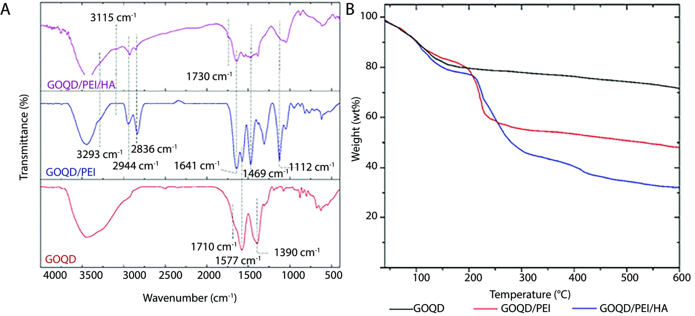
Figure 2.
Characterization of GOQD/PEI/HA structural features; A, FT-IR spectra of GOQD, GOQD/PEI, and GOQD/PEI/HA showing characteristic shifts associated with PEI conjugation with GOQD and the introduction of HA; B, TGA curves of GOQD, GOQD/PEI, and GOQD/PEI/HA demonstrating multistep weight-loss patterns corresponding to the sequential degradation of GOQD surface groups, PEI chains, and HA components.
4.2.2. Thermogravimetric Analysis
The thermal degradation results for GOQD/PEI/HA, shown in Figure 2B, illustrate the degradation temperature and total weight loss. The results indicate that GOQD exhibited a weight loss of approximately 18% up to around 180 °C, which appears to be related to adsorbed water and the removal of GOQD surface functional groups, such as hydroxyl and carboxyl groups. For the GOQD/PEI structure, two weight reductions were observed: one up to about 180 °C, corresponding to adsorbed surface water and GOQD surface functional groups, and another at about 200 - 230 °C, attributed to PEI chain degradation. In the GOQD/PEI/HA structure, three weight-loss events were identified: the first, up to around 180 °C, corresponding to the removal of GOQD surface functional groups; the second, between about 200 and 260 °C, resulting from overlapping degradation of PEI chains and the initial stage of HA chain decomposition; and the third, at about 400 °C, associated with the second stage of HA chain degradation. Measurements and calculations indicate that the weight contents of PEI and HA in the final GOQD/PEI/HA structure were 24.1 wt% and 18.7 wt%, respectively.
4.3. Determination of the Amount of Absorbed pDNA by GOQD/PEI/HA NPs
Agarose gel electrophoresis confirmed the accuracy and integrity of the extracted pDNA, with a concentration of approximately 150 ng/μL used for all subsequent experiments and ratio calculations. To evaluate the DNA-binding capacity of the synthesized GOQD/PEI/HA NPs, a gel retardation assay was performed at various NP-to-pDNA mass ratios. As shown in Figure 3A, increasing the ratio of GOQD/PEI/HA to pDNA progressively reduced plasmid migration, with complete retardation observed at ratios above 1:2, indicating full complexation and condensation of pDNA. In our previous study (18), the minimum ratio required for complete pDNA condensation using GOQD/PEI nanocarriers was reported to be approximately 1:0.06, suggesting that the inclusion of HA in our system slightly decreased condensation efficiency compared with unmodified GOQD/PEI. This difference is likely due to the negatively charged carboxyl groups of HA, which may partially compete with pDNA for interaction with PEI, thereby reducing electrostatic binding at lower ratios. At higher ratios (1:4 and 1:5), reduced staining intensity was observed, likely due to restricted accessibility of pDNA to the nucleic acid dye, further confirming stable complex formation (26).
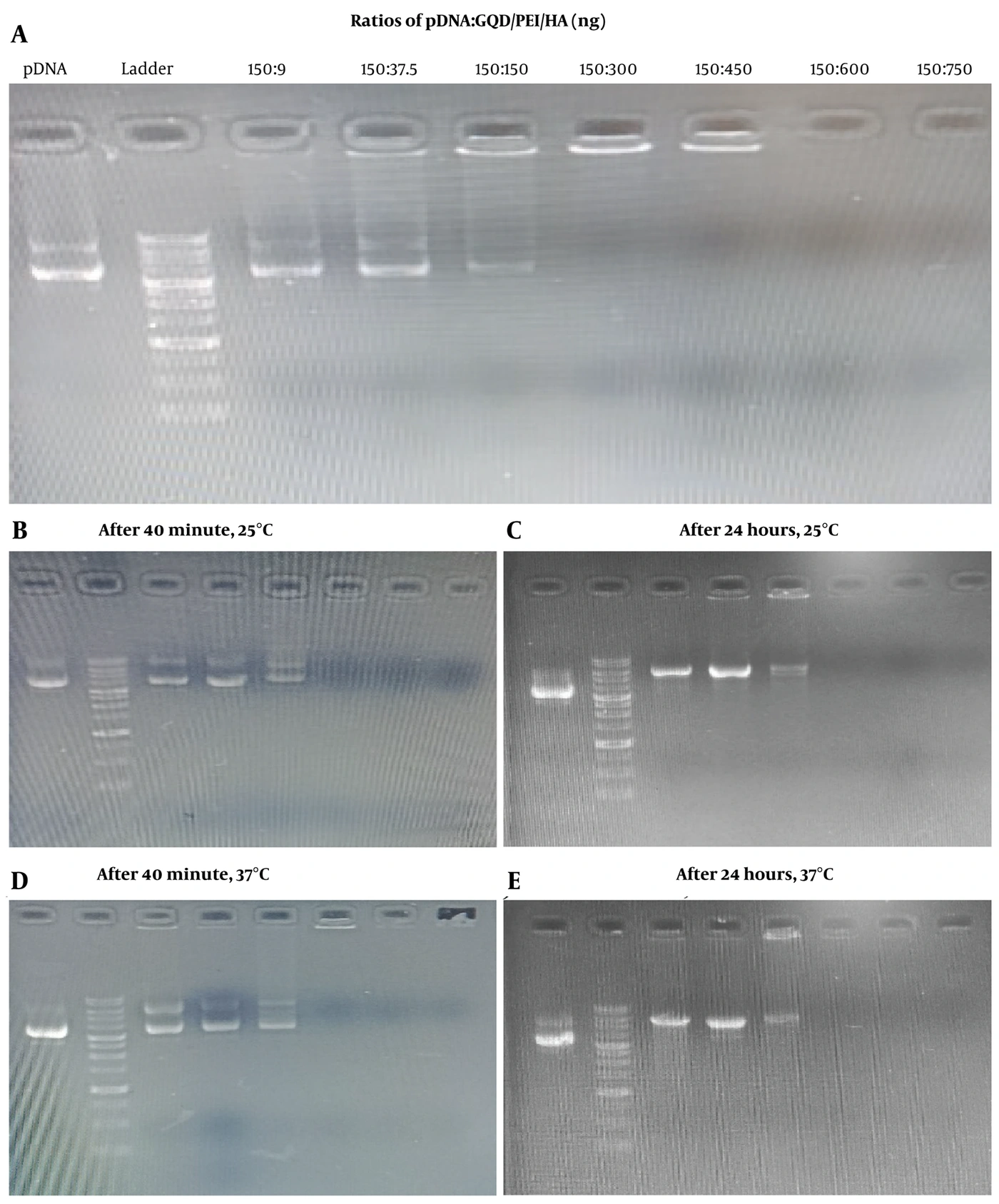
Figure 3.
Agarose gel retardation assay. A, Agarose gel electrophoresis of pDNA complexed with GOQD/PEI/HA at different ratios (1:0.06, 1:0.25, 1:1, 1:2, 1:3, 1:4, and 1:5), showing progressive plasmid retardation and complete complexation at higher nanoparticle-to-pDNA ratios. B-E, Agarose gel electrophoresis for evaluating the stability of GOQD/PEI/HA-pDNA complexes over time, at different temperatures, and across various ratios (1:0.06, 1:0.25, 1:1, 1:5, 1:7, and 1:10). The gels show the mobility pattern of the complexes after 40 minutes (initial complex formation) and 24 hours (prolonged incubation) at 25 °C (B, C) and 37 °C (D, E). Visualization for all assays was performed under UV illumination.
4.4. Complex Stability Over Time and at Different Temperatures
Complex integrity is a prerequisite for protecting DNA during the gene delivery pathway in vitro and in vivo. Therefore, the stability of GOQD/PEI/HA-pDNA complexes at different ratios was evaluated over 24 hours at 25 and 37 °C. None of the samples released pDNA after 24 hours of incubation at either temperature (Figure 3C and E), and at all studied ratios, the NPs retained pDNA similarly to the complexes at 40 minutes (Figure 3B and D). In addition, increasing the incubation time from 40 minutes to 24 hours did not affect complex formation, and complexes still did not form at low ratios (Figure 3C and E).
4.5. Surface Charge, Particle Size, and Morphology of the GOQD/PEI/HA-pDNA Complex
Surface charge is a critical physicochemical parameter influencing the interaction of gene delivery systems with biological membranes, thereby affecting cellular uptake and transfection efficiency. To assess these properties, the surface charge of GOQD/PEI/HA-pDNA complexes was evaluated by zeta potential measurement, yielding a zeta potential of +26.7 mV (Figure 4A). This positive charge suggests favorable electrostatic interactions with cell membranes, which can enhance cellular uptake and transfection.
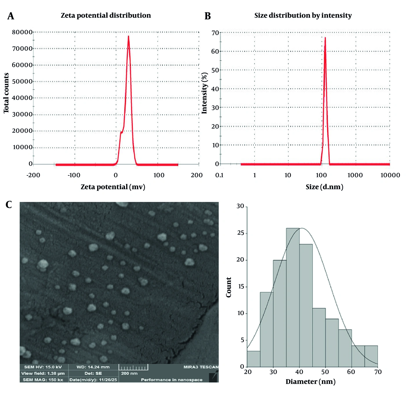
Figure 4.
Surface charge, particle size, and morphology of the GOQD/PEI/HA-pDNA complex at the mass ratio of 1:4; A, Zeta potential measurement of GOQD/PEI/HA-pDNA nanocomplexes obtained using a Malvern Zetasizer Nano ZS90, showing a surface charge of +26.7 mV; B, Dynamic light scattering (DLS) analysis of the GOQD/PEI/HA-pDNA nanocomplex, showing a narrow size distribution with an average hydrodynamic diameter of 125 nm; C, FE-SEM micrograph of the GOQD/PEI/HA-pDNA complexes illustrating their uniform spherical morphology (left). Particle size distribution obtained from FE-SEM images and quantitatively analyzed using ImageJ software (version 8), revealing a nearly normal distribution with an average diameter of 40.96 ± 10.52 nm (right).
The size and morphology of the GOQD/PEI/HA-pDNA complexes were characterized by DLS and FE-SEM. DLS showed an average hydrodynamic size of 125 nm for the 1:4 ratio of the GOQD/PEI/HA-pDNA complex (Figure 4B). The FE-SEM micrograph of the GOQD/PEI/HA-pDNA complexes illustrates a uniform spherical morphology. Particle size was quantitatively analyzed using ImageJ software version 8, and the resulting histogram showed the size distribution of the complexes. As shown in Figure 4C, the complexes exhibited a nearly normal size distribution with an average particle diameter of 40.96 ± 10.52 nm.
4.6. Hemolysis Assay
The hemolysis assay conducted on GOQD/PEI/HA at concentrations of 50 and 200 μg/mL demonstrated hemolysis rates of less than 2% in both cases, as shown in Figure 5A. These results indicate minimal RBC membrane disruption, suggesting that the tested nanocarrier exhibits excellent hemocompatibility and can be classified as nonhemolytic according to standard biomedical criteria (22).
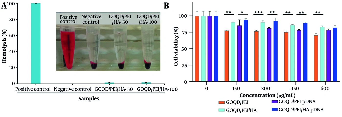
Figure 5.
Assessment of hemocompatibility and cellular biocompatibility; A, Hemolysis evaluation of GOQD/PEI/HA nanocarrier at concentrations of 50 and 200 μg/mL; B, Cytotoxicity analysis of GOQD/PEI, GOQD/PEI/HA, GOQD/PEI-pDNA, and GOQD/PEI/HA-pDNA at various ratios (1:1, 1:2, 1:3, and 1:4) after 24 hours of incubation with HeLa cells, assessed using the MTT assay. Data are presented as mean ± SD (n = 3), and statistical significance was determined by one-way ANOVA followed by Tukey's post hoc test. P < 0.05 was considered statistically significant.
4.7. Cytotoxicity of GOQD/PEI/HA NPs and pDNA-Loaded NPs
Nanocarrier biocompatibility is a critical parameter for assessing suitability for gene delivery applications, as excessive cytotoxicity can substantially compromise transfection efficiency and limit clinical translation (27). To evaluate the biocompatibility of the synthesized carriers, the viability of HeLa cells treated with different concentrations (150 - 600 ng/μL) of GOQD/PEI, GOQD/PEI/HA, and their pDNA-loaded complexes was assessed using the MTT assay after 24 hours of incubation (Figure 5B). The results showed that HA-modified nanocarriers consistently exhibited higher biocompatibility than their unmodified counterparts across all tested concentrations. For example, at 300 ng/μL, the concentration associated with optimal DNA condensation, cell viability values were 76.6% for GOQD/PEI, 90.09% for GOQD/PEI/HA, 81.08% for GOQD/PEI-pDNA, and 92.5% for GOQD/PEI/HA-pDNA. These findings indicate that HA functionalization substantially enhances the biocompatibility of the GOQD/PEI system while maintaining strong gene delivery capability.
5. Discussion
Despite remarkable advances in diagnostic and therapeutic strategies, cancer continues to pose a major global health challenge, with increasing incidence and mortality. Conventional therapies, such as chemotherapy and radiotherapy, are limited by poor selectivity, systemic toxicity, and drug resistance (28). In this context, gene therapy offers a promising alternative by delivering nucleic acids to regulate or silence disease-related genes (29, 30). Its success, however, depends on the development of safe and efficient carriers capable of protecting genetic material, promoting cellular uptake, and minimizing cytotoxicity, which remain key challenges for nonviral gene delivery systems (31).
Polyethyleneimine is a highly efficient and widely used nonviral cationic polymer for gene delivery in vitro and in vivo. However, its application is limited by cytotoxicity, which can cause membrane damage and apoptosis. Chemical modification, particularly with targeting ligands, can effectively reduce this toxicity (32). The incorporation of PEI into hybrid nanomaterials, such as gold, silica, magnetic NPs, and graphene derivatives, has been shown to enhance both safety and transfection efficiency (33-36). For example, Chen et al. developed a graphene oxide-PEI (PEI-GO) hybrid carrier with improved gene transfer performance and reduced cytotoxicity compared with unmodified PEI (37).
In this study, a HA-modified GOQD/PEI (GOQD/PEI/HA) nanocarrier was developed to enhance the transfection efficiency and biocompatibility of PEI-based systems. HA was covalently conjugated to GOQD/PEI via amide bond formation between HA carboxyl and PEI amino groups using EDC/NHS chemistry, and successful synthesis was verified by FT-IR and TGA analyses. Gel-retardation assays confirmed effective pDNA condensation, achieving complete complexation at a 1:2 pDNA-to-carrier ratio (150 ng pDNA: 300 ng carrier), indicating stable complex formation at low concentrations. MTT and hemolysis assays demonstrated that HA conjugation markedly enhanced biocompatibility, increasing HeLa cell viability from approximately 76% to approximately 90.09% and maintaining hemolysis below 2% across the tested concentrations, confirming reduced PEI-related cytotoxicity and excellent blood compatibility.
These findings align with and extend several recent reports. The aptamer-guided GOQD-PEI carrier developed by Taghizadeh-Tabarsi et al. showed effective DNA binding, robust transfection, and negligible cytotoxicity in 2D and organoid models, illustrating how surface ligands can enable target selectivity without added toxicity (19).
The HA-modified GOQD/PEI nanocarrier demonstrated excellent biocompatibility and hemocompatibility, indicating its potential as a safe and effective platform for future gene delivery applications. Given its strong DNA condensation ability and high cellular viability, further investigations could evaluate its performance in more physiologically relevant models, such as three-dimensional (3D) tumor spheroids and organoid systems, in which cell-cell and matrix interactions more closely mimic in vivo environments. Similar strategies have been applied in related studies, including the work of Soheila Mohammadi et al. (18), who examined GOQD/PEI nanocarriers in a 3D breast cancer model, and Reza Taghizadeh-Tabarsi et al. (19), who employed an aptamer-guided GOQD system in a luminal breast cancer organoid model. Both studies underscored the importance of evaluating nanocarrier cytocompatibility and penetration in 3D cultures, supporting the extension of such approaches to the HA-modified carrier developed here.
5.1. Study Limitations
However, this study has several limitations that should be acknowledged. First, although HA was incorporated to enable potential CD44-mediated targeting, this mechanism was not experimentally verified, and dedicated assays will be needed to confirm receptor-specific uptake. Second, all biological evaluations were performed in 2D in vitro systems, which do not replicate the complexity of physiological environments or account for protein corona formation that may influence nanocarrier behavior in vivo. These limitations highlight the need for future studies in 3D models and in vivo settings to evaluate targeting specificity, biodistribution, and performance under more realistic biological conditions.
Future research should also include in vivo evaluations to assess biodistribution, pharmacokinetics, and immunocompatibility, which are critical for clinical translation. The observed blood compatibility suggests that GOQD/PEI/HA could be safely administered systemically, warranting further investigation in animal models for targeted gene delivery. Collectively, this system represents a promising foundation for next-generation HA-targeted nonviral gene carriers, with potential applications in both experimental cancer biology and therapeutic development.
5.2. Conclusions
In this study, we developed a hyaluronic acid-modified GOQD/PEI (GOQD/PEI/HA) nanocarrier designed as a biocompatible platform with the potential for targeted gene delivery. Through EDC/NHS coupling, HA conjugation provided CD44-specific targeting and enhanced structural stability while preserving strong DNA condensation ability. MTT and hemolysis assays confirmed significantly improved cell viability and excellent hemocompatibility (< 2%), validating the system's safety and efficiency. Overall, the GOQD/PEI/HA nanocarrier shows great promise as a nonviral gene delivery platform and warrants further evaluation in 3D organoid cultures and in vivo cancer therapy applications.