Journal of Semnan University of Medical Sciences
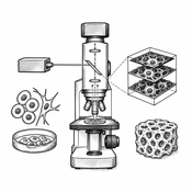
Image Credit:AI-generated illustration
Outline
Confocal Laser Scanning Microscope: A Review of Principles and Applications in Biological Sciences and Regenerative Medicine
Authors

Abstract
Abstract
Context:
Since the commercialization of confocal microscopy in the late 1980s, confocal laser scanning microscopy (CLSM) has become one of the most common fluorescence microscopy techniques for 3D structural studies of biological cells and tissues. The flexibility of this approach has enabled its application in studies ranging from rapid imaging of processes in living cells, to morphological analyses of tissues, and localization of protein expression patterns.
Evidence Acquisition:
The studies published in PubMed, google scholar, Scopus, and Web of Science databases from 2004 to 2025 was used. Selection criteria included studies evaluating confocal microscopy and its advantages in tissue engineering and regenerative medicine.
Results:
CLSM has a major advantage over conventional microscopy because it rejects light that does not come from the focal plane, enabling one to perform optical sectioning and construction of 3D images. Also, its high axial resolution, sharp image quality, and associated quantitative image analysis provide crucial structural information at medium range for full 3D realization of the microstructure. Due to this feature, CLSM is currently finding applications in the study of a variety of processes such as phase separation in polymer blends, fracture toughness in alloys, and in microfabrication. It also has applications in regenerative medicine, such as analyzing cell dynamics, cell interactions, and measuring tissue properties, and can help improve tissue regeneration and repair processes and make progress in this field.
Conclusion:
This article reviews confocal microscopy and its advantages in tissue engineering, regenerative medicine and biomaterials. In this article, we also introduce the principles of confocal microscopy and discuss how this approach has become a mainstay in the life sciences.
Keywords: confocal microscopy, 3D structure, laser scanning microscopy, tissue engineering
References
- 1.Appel AA, Anastasio MA, Larson JC, Brey EM. Imaging challenges in biomaterials and tissue engineering. Biomaterials. 2013;34(28):6615–30. [PubMed ID:23768903]. [PubMed Central ID:PMC3799904]. https://doi.org/10.1016/j.biomaterials.2013.05.033.
- 2.Conchello JA, Lichtman JW. Optical sectioning microscopy. Nat Methods. 2005;2(12):920–31. [PubMed ID:16299477]. https://doi.org/10.1038/nmeth815.
- 3.Minsky M. Memoir on inventing the confocal scanning microscope. Scanning. 2011;10(4):128–38. https://doi.org/10.1002/sca.4950100403.
- 4.Semwogerere D, Weeks ER. Confocal Microscopy. In: Rédei GP, editor. Encyclopedia of Genetics, Genomics, Proteomics and Informatics. Heidelberg, Germany: Springer Science & Business Media; 2005. p. 408–.
- 5.Paddock SW, Eliceiri KW. Laser scanning confocal microscopy: history, applications, and related optical sectioning techniques. Methods Mol Biol. 2014;1075:9–47. [PubMed ID:24052346]. https://doi.org/10.1007/978-1-60761-847-8_2.
- 6.St Croix CM, Shand SH, Watkins SC. Confocal microscopy: comparisons, applications, and problems. Biotechniques. 2005;39(6 Suppl):S2–5. [PubMed ID:20158500]. https://doi.org/10.2144/000112089.
- 7.Elliott AD. Confocal Microscopy: Principles and Modern Practices. Curr Protoc Cytom. 2020;92(1):e68. [PubMed ID:31876974]. [PubMed Central ID:PMC6961134]. https://doi.org/10.1002/cpcy.68.
- 8.Nwaneshiudu A, Kuschal C, Sakamoto FH, Anderson RR, Schwarzenberger K, Young RC. Introduction to confocal microscopy. J Invest Dermatol. 2012;132(12):e3. [PubMed ID:23187113]. https://doi.org/10.1038/jid.2012.429.
- 9.Paganelli A, Tarentini E, Benassi L, Scelfo D, Pisciotta A, Rossi E, et al. Use of confocal microscopy imaging for in vitro assessment of adipose-derived mesenchymal stromal cells seeding on acellular dermal matrices: 3D reconstruction based on collagen autofluorescence. Skin Res Technol. 2022;28(1):133–41. [PubMed ID:34555218]. [PubMed Central ID:PMC9292443]. https://doi.org/10.1111/srt.13103.
- 10.Gigan S, Katz O, de Aguiar HB, Andresen ER, Aubry A, Bertolotti J, et al. Roadmap on wavefront shaping and deep imaging in complex media. Journal of Physics: Photonics. 2022;4(4). https://doi.org/10.1088/2515-7647/ac76f9.
- 11.Guida S, Arginelli F, Farnetani F, Ciardo S, Bertoni L, Manfredini M, et al. Clinical Applications of In Vivo and Ex Vivo Confocal Microscopy. Applied Sciences. 2021;11(5). https://doi.org/10.3390/app11051979.
- 12.Pellacani G, Scope A, Gonzalez S, Guitera P, Farnetani F, Malvehy J, et al. Reflectance confocal microscopy made easy: The 4 must-know key features for the diagnosis of melanoma and nonmelanoma skin cancers. J Am Acad Dermatol. 2019;81(2):520–6. [PubMed ID:30954581]. https://doi.org/10.1016/j.jaad.2019.03.085.
- 13.Yelamos O, Manubens E, Jain M, Chavez-Bourgeois M, Pulijal SV, Dusza SW, et al. Improvement of diagnostic confidence and management of equivocal skin lesions by integration of reflectance confocal microscopy in daily practice: Prospective study in 2 referral skin cancer centers. J Am Acad Dermatol. 2020;83(4):1057–63. [PubMed ID:31202873]. [PubMed Central ID:PMC8285925]. https://doi.org/10.1016/j.jaad.2019.05.101.
- 14.Kiesslich R, Canto MI. Confocal laser endomicroscopy. Gastrointest Endosc Clin N Am. 2009;19(2):261–72. [PubMed ID:19423023]. https://doi.org/10.1016/j.giec.2009.02.007.
- 15.Ragazzi M, Longo C, Piana S. Ex Vivo (Fluorescence) Confocal Microscopy in Surgical Pathology: State of the Art. Adv Anat Pathol. 2016;23(3):159–69. [PubMed ID:27058244]. https://doi.org/10.1097/PAP.0000000000000114.
- 16.Woudberg R. Confocal Microscopy Applications with Integrative Techniques in Cell Mechanics and Tissue Engineering. Bioimaging in Tissue Engineering and Regeneration; 2025. p. 215–56.
- 17.Silvestri L, Mascaro ALA, Lotti J, Sacconi L, Pavone FS. Advanced Optical Techniques to Explore Brain Structure and Function. Journal of Innovative Optical Health Sciences. 2013;06(01). https://doi.org/10.1142/s1793545812300029.
- 18.Nosrati H, Abpeikar Z, Mahmoudian ZG, Zafari M, Majidi J, Alizadeh A, et al. Corneal epithelium tissue engineering: recent advances in regeneration and replacement of corneal surface. Regen Med. 2020;15(8):2029–44. [PubMed ID:33169642]. https://doi.org/10.2217/rme-2019-0055.
- 19.Furrer P, Gurny R. Recent advances in confocal microscopy for studying drug delivery to the eye: concepts and pharmaceutical applications. Eur J Pharm Biopharm. 2010;74(1):33–40. [PubMed ID:19755157]. https://doi.org/10.1016/j.ejpb.2009.09.002.
- 20.Jonkman J, Brown CM, Wright GD, Anderson KI, North AJ. Tutorial: guidance for quantitative confocal microscopy. Nat Protoc. 2020;15(5):1585–611. [PubMed ID:32235926]. https://doi.org/10.1038/s41596-020-0313-9.
- 21.Zafari FSM, Bakhtiyari M. [Teimourian S. Rapid repair of skin ulcers in diabetic rats using polycaproplactone /gelatin nanon-fiber scaffold]. Koomesh. 2019;21(3). Persian.
- 22.Bagherzadeh R, Latifi M, Najar SS, Tehran MA, Kong L. Three-dimensional pore structure analysis of nano/microfibrous scaffolds using confocal laser scanning microscopy. J Biomed Mater Res A. 2013;101(3):765–74. [PubMed ID:22961915]. https://doi.org/10.1002/jbm.a.34379.
- 23.Ilie MA, Caruntu C, Lupu M, Lixandru D, Tampa M, Georgescu SR, et al. Current and future applications of confocal laser scanning microscopy imaging in skin oncology. Oncol Lett. 2019;17(5):4102–11. [PubMed ID:30944603]. [PubMed Central ID:PMC6444326]. https://doi.org/10.3892/ol.2019.10066.
- 24.Seidel T, Edelmann JC, Sachse FB. Analyzing Remodeling of Cardiac Tissue: A Comprehensive Approach Based on Confocal Microscopy and 3D Reconstructions. Ann Biomed Eng. 2016;44(5):1436–48. [PubMed ID:26399990]. [PubMed Central ID:PMC4805509]. https://doi.org/10.1007/s10439-015-1465-6.
- 25.Buttner P, Galli R, Husser D, Bollmann A. Label-free Imaging of Myocardial Remodeling in Atrial Fibrillation Using Nonlinear Optical Microscopy: A Feasibility Study. J Atr Fibrillation. 2018;10(5):1644. [PubMed ID:29988238]. [PubMed Central ID:PMC6006967]. https://doi.org/10.4022/jafib.1644.
- 26.Au M, Almeida-Magana R, Al-Hammouri T, Haider A, Shaw G. Accuracy of Ex-vivo Fluorescence Confocal Microscopy in Margin Assessment of Solid Tumors: A Systematic Review. J Histochem Cytochem. 2023;71(12):661–74. [PubMed ID:37968920]. [PubMed Central ID:PMC10691410]. https://doi.org/10.1369/00221554231212948.
- 27.Marenco J, Calatrava A, Casanova J, Claps F, Mascaros J, Wong A, et al. Evaluation of Fluorescent Confocal Microscopy for Intraoperative Analysis of Prostate Biopsy Cores. Eur Urol Focus. 2021;7(6):1254–9. [PubMed ID:32912840]. https://doi.org/10.1016/j.euf.2020.08.013.
- 28.Blouin S, Roschger A, Varga F, Misof B, Spitzer S, Roschger P, et al. Confocal laser scanning microscopy-a powerful tool in bone research. Wien Med Wochenschr. 2018;168(11-12):314–21. [PubMed ID:29802493]. https://doi.org/10.1007/s10354-018-0639-x.
- 29.Ghanbari E, Naseri L, Khazaei M, Akbari M, Elahian F, Mirzaei SA, et al. [An Introduction to Tissue Engineering and the Most Widely Used Scaffolds]. Journal of Isfahan Medical School. 2018;36(488):823–37. Persian. https://doi.org/10.22122/jims.v36i488.10056.
- 30.Atala A. Tissue engineering and regenerative medicine: concepts for clinical application. Rejuvenation Res. 2004;7(1):15–31. [PubMed ID:15256042]. https://doi.org/10.1089/154916804323105053.
- 31.Golchin A, Ranjbarvan P, Parviz S, Shokati A, Naderi R, Rasmi Y, et al. The role of probiotics in tissue engineering and regenerative medicine. Regen Med. 2023;18(8):635–57. [PubMed ID:37492007]. https://doi.org/10.2217/rme-2022-0209.
- 32.Asgharzade S, Alizadeh A, Arab S. Regenerative Medicine Approaches in COVID-19 Pneumonia. Curr Stem Cell Res Ther. 2021;16(6):647–55. [PubMed ID:33438550]. https://doi.org/10.2174/1574888X16999210112205826.
- 33.Pawley JB. Handbook Of Biological Confocal Microscopy. 2006.
- 34.Tsang VL, Bhatia SN. Three-dimensional tissue fabrication. Adv Drug Deliv Rev. 2004;56(11):1635–47. [PubMed ID:15350293]. https://doi.org/10.1016/j.addr.2004.05.001.
- 35.Porgham Daryasari M, Dusti Telgerd M, Hossein Karami M, Zandi-Karimi A, Akbarijavar H, Khoobi M, et al. Poly-l-lactic acid scaffold incorporated chitosan-coated mesoporous silica nanoparticles as pH-sensitive composite for enhanced osteogenic differentiation of human adipose tissue stem cells by dexamethasone delivery. Artif Cells Nanomed Biotechnol. 2019;47(1):4020–9. [PubMed ID:31595797]. https://doi.org/10.1080/21691401.2019.1658594.
- 36.Cutts LS, Hibberd S, Adler J, Davies MC, Melia CD. Characterising drug release processes within controlled release dosage forms using the confocal laser scanning microscope. Journal of Controlled Release. 1996;42(2):115–24. https://doi.org/10.1016/0168-3659(96)01340-5.
- 37.Lamprecht A, Schafer U, Lehr CM. Structural analysis of microparticles by confocal laser scanning microscopy. AAPS PharmSciTech. 2000;1(3):E17. [PubMed ID:14727903]. [PubMed Central ID:PMC2750345]. https://doi.org/10.1208/pt010317.
- 38.Ilie M, Caruntu C, Lixandru D, Tampa M, Georgescu SR, Constantin MM, et al. In vivo confocal laser scanning microscopy imaging of skin inflammation: Clinical applications and research directions (Review). Experimental and Therapeutic Medicine. 2018. https://doi.org/10.3892/etm.2018.6981.
- 39.Allan IU, Shevchenko RV, Rowshanravan B, Kara B, Jahoda CA, James SE. The Use of Confocal Laser Scanning Microscopy to Assess the Potential Suitability of 3d Scaffolds for Tissue Regeneration, by Monitoring Extra‐Cellular Matrix Deposition and by Quantifying Cellular Infiltration and Proliferation. Soft Materials. 2009;7(4):319–41. https://doi.org/10.1080/15394450903372679.
- 40.Gabor AG, Duma VF, Fabricky MMC, Marsavina L, Tudor A, Vancea C, et al. Ceramic Scaffolds for Bone Augmentation: Design and Characterization with SEM and Confocal Microscopy. Materials (Basel). 2022;15(14). [PubMed ID:35888366]. [PubMed Central ID:PMC9322854]. https://doi.org/10.3390/ma15144899.
- 41.Griffith LG, Swartz MA. Capturing complex 3D tissue physiology in vitro. Nat Rev Mol Cell Biol. 2006;7(3):211–24. [PubMed ID:16496023]. https://doi.org/10.1038/nrm1858.
- 42.Badylak SF, Gilbert TW. Immune response to biologic scaffold materials. Semin Immunol. 2008;20(2):109–16. [PubMed ID:18083531]. [PubMed Central ID:PMC2605275]. https://doi.org/10.1016/j.smim.2007.11.003.
- 43.Leahy M, Thompson K, Zafar H, Alexandrov S, Foley M, O'Flatharta C, et al. Functional imaging for regenerative medicine. Stem Cell Res Ther. 2016;7(1):57. [PubMed ID:27095443]. [PubMed Central ID:PMC4837501]. https://doi.org/10.1186/s13287-016-0315-2.
- 44.Klontzas ME, Protonotarios A. High-Resolution Imaging for the Analysis and Reconstruction of 3D Microenvironments for Regenerative Medicine: An Application-Focused Review. Bioengineering (Basel). 2021;8(11). [PubMed ID:34821748]. [PubMed Central ID:PMC8614770]. https://doi.org/10.3390/bioengineering8110182.
- 45.Lambert TJ, Waters JC. Navigating challenges in the application of superresolution microscopy. J Cell Biol. 2017;216(1):53–63. [PubMed ID:27920217]. [PubMed Central ID:PMC5223610]. https://doi.org/10.1083/jcb.201610011.
- 46.Wan B, Ganier C, Du-Harpur X, Harun N, Watt FM, Patalay R, et al. Applications and future directions for optical coherence tomography in dermatology. Br J Dermatol. 2021;184(6):1014–22. [PubMed ID:32974943]. https://doi.org/10.1111/bjd.19553.
- 47.Takbhate BP. A Narrative Review on Confocal Laser Scanning Microscopy: Principle, Applications, Advancements and Challenges. Journal of Clinical and Diagnostic Research. 2024. https://doi.org/10.7860/jcdr/2024/73869.20245.
Copyright
© 2026, Author(s). This open-access article is available under the Creative Commons Attribution 4.0 (CC BY 4.0) International License (https://creativecommons.org/licenses/by/4.0/), which allows for unrestricted use, distribution, and reproduction in any medium, provided that the original work is properly cited.
Similar Articles
Role of Modern Technologies in Tissue Engineering
Ali Q, Malik S, Malik A, Hafeez MN, Salman S. Role of Modern Technologies in Tissue Engineering. Arch Neurosci. 2020;7(1):e90394. doi: https://doi.org/10.5812/ans.90394

Basic Application of Stereology in Histology and Medical Sciences
Mahmoudzadeh Sagheb HR, Moudi B. Basic Application of Stereology in Histology and Medical Sciences. Gene Cell Tissue. 2014;1(3):e24237. doi: https://doi.org/10.17795/gct-24237
Innovative Platforms in Regenerative Medicine: Bridging Research and Clinical Solutions
Jalali SN, Fathi Z, Mohebbi S. Innovative Platforms in Regenerative Medicine: Bridging Research and Clinical Solutions. Jentashapir J Cell Mol Biol. 2025;16(1):e158285. doi: https://doi.org/10.5812/jjcmb-158285
Advancements in Digital Pathology and Whole Slide Imaging: Technical Challenges and Emerging Technologies
Sharini H, Azarpira N, Masjoodi S. Advancements in Digital Pathology and Whole Slide Imaging: Technical Challenges and Emerging Technologies. J Clin Res Paramed Sci. 2026;15(1):e171493. doi: https://doi.org/10.5812/jcrps-171493
CRISPR System and Its Applications in Medicine and Stem Cell Engineering
Abdolmaleki A, Nahumi A, Basharzad Seddigh H, Yazdannasab N, Panahi A, et al. CRISPR System and Its Applications in Medicine and Stem Cell Engineering. Gene Cell Tissue. 2024;11(4):e150914. doi: https://doi.org/10.5812/gct-150914
- Scopus by DOI: 0
Last Update: 6 days ago
- Scopus by Title: 0
Last Update: 6 days ago
- Scopus by Title (Ref): 0
Last Update: 6 days ago
- CrossRef: 0
Last Update: 7 hours ago
- OpenAlex: 0
Last Update: 6 hours ago
- OpenCitations: 0
Last Update: 5 days ago