
1. Introduction
Percutaneous vertebroplasty and kyphoplasty stabilize vertebral compression fractures by injecting polymethyl methacrylate into the vertebral body (1). Cement escapes into the venous system in approximately one-quarter of patients, and venous cement complications have been reported in 1% to 10% (2). Cement that leaks into the perivertebral veins can travel through the azygos system to the right heart and then to the pulmonary arteries (1, 2). In a single-center series of 1854 vertebroplasty procedures, intracardiac cement was detected after 3.9% of procedures, was accompanied by pulmonary cement in 86% of cases, and caused symptoms after only 0.3% of procedures (3).
The clinical spectrum ranges from incidental findings to cardiac perforation and death (2, 4). Incidental discovery is uncommon; in a systematic review of 71 patients, only 4 were identified incidentally, and 28 presented on the first postoperative day (4). Two features distinguish the present case from most published reports. First, the interval between vertebroplasty and detection was at least 2 years, whereas symptoms usually begin within 48 hours (5). Second, the cement was confined to the heart, without the pulmonary emboli that usually accompany it (3). We describe this patient, whose embolus was identified during the evaluation for chest pain and removed during coronary artery bypass grafting (CABG). This report follows the CARE guideline.
2. Case Presentation
A 72-year-old woman presented to an outpatient clinic with an episode of typical chest pain. Her medical history included long-standing hypertension and hyperlipidemia, both controlled with medication, and a total hip replacement 13 years earlier. She had sustained osteoporotic vertebral compression fractures 2 and 4 years earlier. These fractures were treated with percutaneous cement vertebroplasty followed by spinal fixation. Records of the vertebroplasty procedures were unavailable.
Electrocardiography and echocardiography were performed before coronary angiography. The electrocardiogram showed sinus rhythm with nonspecific changes. Transthoracic echocardiography revealed a highly echo-dense, bone-like mass measuring 17 × 12 mm attached to the right atrial free wall. The ejection fraction was normal, there was no significant valvular abnormality, and the estimated pulmonary artery pressure was 35 mm Hg. Transesophageal echocardiography localized the mass to the right atrial appendage and showed no attachment to the valves or the interatrial septum (Figure 1A).
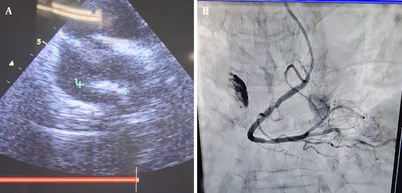
Figure 1.
Detection of the embolus during the cardiac evaluation. A, Transesophageal echocardiography, bicaval view, showing a plate-like, bone-like mass on the right atrial appendage with no attachment to the interatrial septum or tricuspid valve. B, Fluoroscopy during coronary angiography, showing a plate-like, dense mass with no relation to the coronary arteries.
Coronary angiography showed 3-vessel disease with very severe stenosis of the left anterior descending artery. During the same study, cineangiography and fluoroscopy demonstrated a plate-like, semimobile, bone-like density that moved with the heart and had no relation to the coronary arteries (Figure 1B). Chest radiography and noncontrast chest computed tomography (CT) showed a bone-like density on the right side of the heart, corresponding to the right atrial shadow. There was no evidence of cement in the pulmonary arteries (Figure 2).
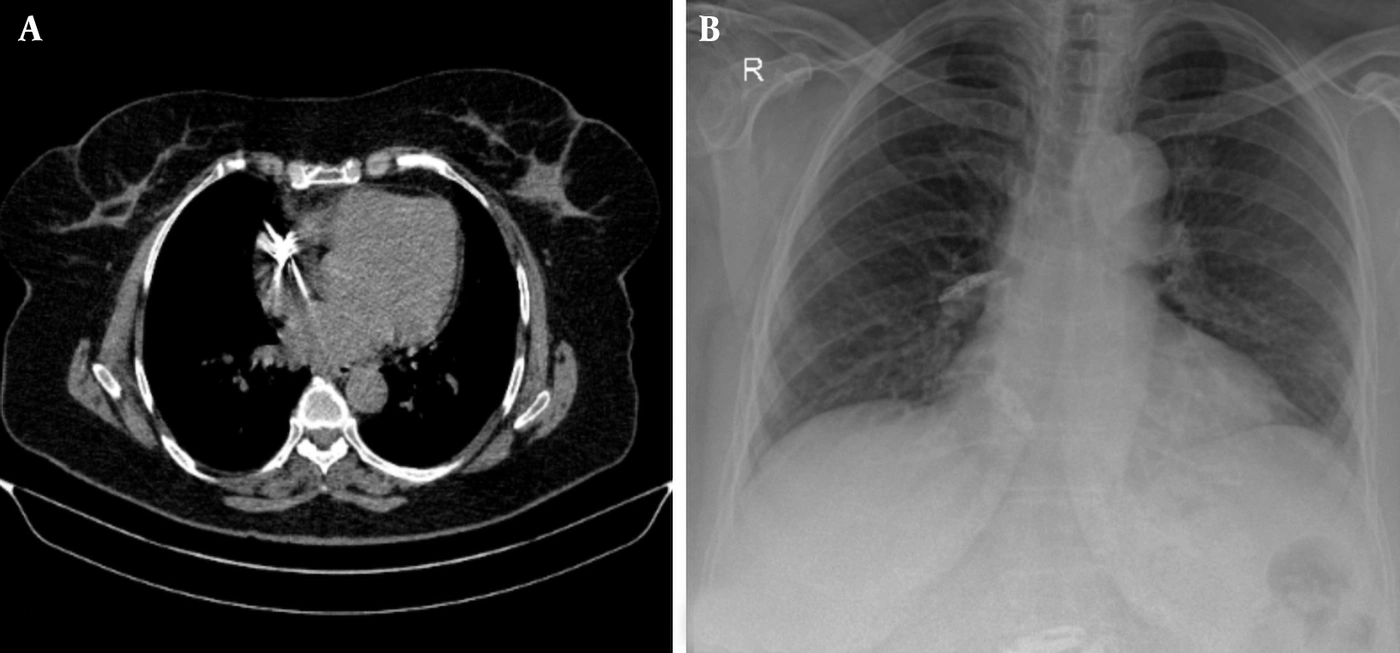
Figure 2.
Radiographic density of the embolus. A, Posteroanterior chest radiograph and B, noncontrast chest computed tomography, each showing a bone-like density on the right atrial contour.
Three findings suggested cement embolism: 1) the bone-like density of the mass on every imaging modality, 2) its attachment to the appendage, and 3) the history of repeated vertebroplasty. The patient was scheduled for CABG with concomitant removal of the right atrial mass.
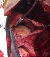
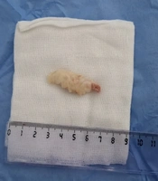
With cardiopulmonary bypass using bicaval cannulation and cardioplegic arrest, the right atrium was opened, exposing a long, bone-like mass attached to the right atrial appendage. The mass was completely excised along with part of the appendage tissue, the right atrium was repaired with sutures, and CABG with 4 grafts was performed. Histopathological examination showed a flat, rectangular, very firm cement fragment measuring 40 × 15 × 2 mm with scant fibrinoid material, consistent with cement embolism (Figure 3).
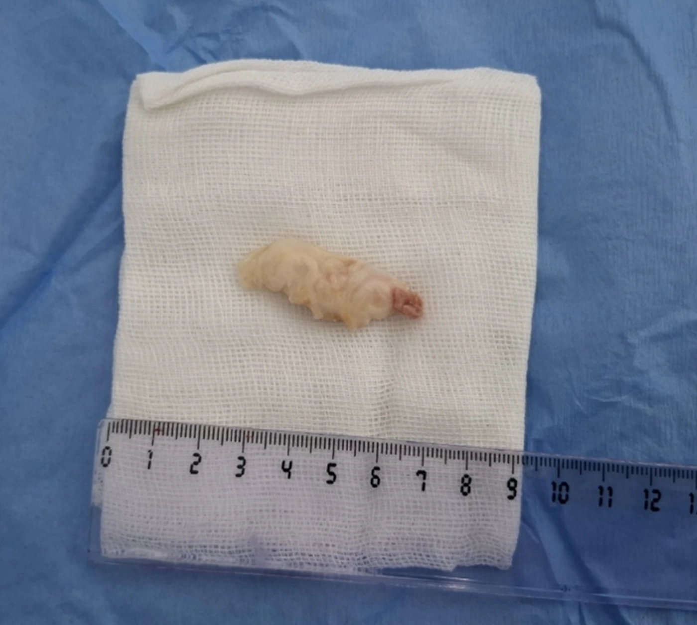
Figure 3.
The excised specimen: a flat, rectangular, firm cement fragment measuring 40 × 15 × 2 mm, removed together with part of the right atrial appendage tissue.
The operation was uneventful, and the patient was discharged 6 days later without complications. Postoperative echocardiography showed a normal ejection fraction, no residual right atrial mass, and no pericardial effusion. Outpatient follow-up is ongoing.
3. Discussion
Three aspects of this case warrant discussion: the interval between vertebroplasty and diagnosis, the absence of pulmonary involvement, and the discrepancy between the echocardiographic and pathological dimensions of the fragment. Each has implications for the recognition and management of these patients.
Symptoms of intracardiac cement embolism usually begin within 48 hours of the procedure, although delays of months to 5 years have been reported (5). In this patient, at least 2 and possibly 4 years elapsed between vertebroplasty and detection, and the embolus was detected only because echocardiography was performed for chest pain. Li et al. described a patient whose symptoms began 2 years after vertebral augmentation (6), and Guo et al. found cement in the right ventricle 2.5 years after vertebroplasty (7). Two mechanisms could explain such delays. Cement may embolize during the procedure and remain quiescent in a low-pressure, trabeculated chamber such as the appendage; alternatively, a fragment retained in the perivertebral veins or inferior vena cava may migrate later. Because the vertebroplasty records were unavailable, we cannot determine which mechanism occurred in this case. In either scenario, an uneventful recovery from vertebroplasty does not exclude a cement embolus, and a history of vertebral augmentation remains relevant when a cardiac mass is identified years later.
In the series reported by Fadili Hassani et al., pulmonary cement accompanied 86% of intracardiac emboli (3), and pulmonary involvement is common in individual reports (8-10). In our patient, CT showed no pulmonary cement; thus, the dense pulmonary arterial opacities that often suggest the diagnosis were absent. A single plate-like fragment of this size may have lodged in the trabeculated appendage on its first passage rather than fragmenting and continuing into the lungs. When cement is confined to the heart, the differential diagnosis includes myxoma, thrombus, and vegetation, and the history of vertebroplasty becomes more important.
The distinguishing imaging feature of cement is its density. In this patient, the mass appeared bone-like on echocardiography, fluoroscopy, chest radiography, and CT. In the context of prior vertebroplasty, this concordance was sufficient for a presumptive diagnosis. Hamade et al. supported the same inference by showing that the intracardiac material had the same density as the cement in the treated vertebrae (11), a comparison worth making whenever CT is available. Without a history of vertebroplasty, an echo-dense right atrial mass may be misinterpreted as a calcified thrombus or tumor. As in our patient, cement emboli have previously been found incidentally during cardiac catheterization (12) or on echocardiography (10).
The most useful practical lesson concerns size. Transthoracic echocardiography measured the mass as 17 × 12 mm, whereas the excised fragment measured 40 × 15 × 2 mm; thus, the longest dimension was underestimated by more than half. Cement is a strong acoustic reflector: its near surface produces intense echoes with dense shadowing behind it, so only part of the fragment is displayed, and a thin plate viewed obliquely is further foreshortened. When removal is planned, particularly percutaneous retrieval, echocardiographic dimensions should be regarded as a lower bound, and multimodality imaging should be used for procedural planning (8).
No comparative data guide management, and the 2 recent systematic reviews reach different conclusions. Tong et al. considered monitoring appropriate for asymptomatic patients and surgery for symptomatic patients (5), whereas Lubbad et al., noting 2 deaths among 14 conservatively managed patients and complete retrieval in all 46 patients treated with open surgery, advised against conservative management because of the risk of delayed complications (4). Conservative management can nonetheless be durable in selected patients: Setty et al. reported an asymptomatic patient who received no anticoagulation and remained well at 24 months (13), and Mills et al. (14) and Shin et al. (9) each reported uneventful 4-year follow-up. Hatzantonis et al. proposed anticoagulation for asymptomatic patients, percutaneous retrieval for symptomatic right atrial emboli, and surgery for right ventricular involvement or perforation (2). Our patient fell outside these categories because she already required cardiopulmonary bypass for coronary revascularization; therefore, removing the fragment added only a right atriotomy to the planned operation. Guo et al. took the same approach in a patient with right ventricular cement and severe left anterior descending artery stenosis (7), and Coso Garrido et al. surgically removed an asymptomatic right atrial embolus because of its size, mobility, and embolic risk (8). In the absence of another indication for cardiac surgery, this case should not be interpreted as supporting surgery for an asymptomatic appendage fragment.
After at least 2 years in the circulation, the fragment carried only scant fibrinoid material. This finding is consistent with the view that cement fragments become endothelialized over time and lose much of their thrombogenic potential, which is the rationale for the 3 to 6 months of anticoagulation often recommended after diagnosis (2, 13, 14).
Prevention remains the most effective strategy. Higher-viscosity cement reduces the risk of venous leakage (1), and in the series reported by Fadili Hassani et al., treating several levels in the same session was associated with more intracardiac emboli, whereas flat-panel fluoroscopy was associated with fewer (3).
This report has limitations. It describes a single patient. The vertebroplasty records were unavailable, so the treated levels, cement volume, and whether leakage was recognized at the time are unknown, and the timing of migration cannot be established. Finally, the available follow-up extends only to hospital discharge, so the durability of the result cannot yet be assessed.
3.1. Conclusions
An intracardiac cement embolus can remain silent in the right atrial appendage for years and be detected incidentally during the evaluation of another cardiac problem. Bone-like density on echocardiography, fluoroscopy, radiography, and CT, together with a history of vertebral augmentation, supports the diagnosis. Echocardiography can substantially underestimate fragment size and should not be the sole basis for procedural planning. When cardiac surgery is indicated for another reason, concomitant removal is a reasonable option; otherwise, the choice between surveillance and removal remains unsettled.