
Panitumumab-Induced Severe Hypomagnesemia and Risk of Atrial Fibrillation and QT Prolongation: An Electrophysiologic Perspective

Author(s):
Azin AlizadehaslAzin AlizadehaslAzin Alizadehasl ORCID1, Haniyeh Faraji AzadHaniyeh Faraji Azad1, Sara AdimiSara AdimiSara Adimi ORCID1, Amirhossein ShirinezhadAmirhossein ShirinezhadAmirhossein Shirinezhad ORCID1,*
1Cardio-Oncology Research Center, Rajaie Cardiovascular Institute, Tehran, Iran
*Corresponding Author: Cardio-Oncology Research Center, Rajaie Cardiovascular Institute, Tehran, Iran. Email: [email protected]

Multidisciplinary Cardiovascular Annals:Vol. 17, issue 1; e172632
Published online:Aug 02, 2026
Article type:Case Report
Received:Jun 07, 2026
Accepted:Jun 30, 2026
How to Cite:Alizadehasl A, Faraji Azad H, Adimi S, Shirinezhad A. Panitumumab-Induced Severe Hypomagnesemia and Risk of Atrial Fibrillation and QT Prolongation: An Electrophysiologic Perspective. Multidiscip Cardio Annal. 2026;17(1):e172632. doi: https://doi.org/10.69107/mca-172632

Abstract

Introduction:

Panitumumab-induced hypomagnesemia disrupts calcium and potassium channel function, prolongs the corrected QT (QTc) interval, and may create a substrate for atrial arrhythmogenesis. We describe a patient with metastatic colon cancer who developed severe hypomagnesemia and marked QTc prolongation during panitumumab therapy; however, atrial fibrillation did not occur. This case demonstrates rapid electrophysiological recovery after magnesium repletion and underscores the importance of cardiac monitoring in patients receiving epidermal growth factor receptor inhibitors.

Case Presentation:

A 68-year-old man with metastatic colon cancer receiving folinic acid, fluorouracil, and irinotecan plus panitumumab developed progressive hypomagnesemia, with the serum magnesium level decreasing from 0.78 mmol/L at baseline to 0.33 mmol/L by cycle 8, accompanied by hypokalemia. He remained asymptomatic. Before cycle 9, electrocardiography showed normal sinus rhythm without atrial fibrillation; however, the QTc interval had increased by 50 ms from baseline, from 410 to 460 ms. Panitumumab was withheld, and magnesium and potassium were repleted. Follow-up electrocardiography showed normalization of the QTc interval to 415 ms without atrial or ventricular arrhythmia.

Conclusions:

Panitumumab can cause clinically silent yet progressive hypomagnesemia, which may disrupt cardiac conduction and increase the risk of arrhythmias. Timely electrolyte repletion may reverse QTc prolongation, prevent arrhythmias, and allow continuation of anticancer therapy. Vigilant monitoring and prompt intervention are essential.

2. Introduction

Advances in anticancer therapy have improved life expectancy among patients with cancer; however, treatment-related adverse effects, particularly arrhythmias, have become increasingly recognized (1). Atrial fibrillation (AF) and corrected QT (QTc) interval prolongation are among the cardiac toxicities associated with cancer therapy (2). The relationship between anticancer medications and arrhythmias is complex and may involve electrolyte imbalances caused by the malignancy and further exacerbated by cancer treatment.
Panitumumab, an epidermal growth factor receptor inhibitor (EGFRI), can cause hypomagnesemia by blocking magnesium reabsorption in the renal tubules (3). Nevertheless, electrolyte disturbances that cause electrophysiologic abnormalities may be overlooked in cardio-oncology. Severe hypomagnesemia directly affects calcium and potassium channels, and dysfunction of these channels can prolong the QTc interval. Evidence also suggests that hypomagnesemia may promote atrial arrhythmogenesis by increasing triggered activity via delayed afterdepolarizations (4, 5).
Here, we present a case of metastatic colon cancer in which severe hypomagnesemia and QTc prolongation developed during panitumumab therapy, increasing the risk of atrial and ventricular arrhythmias, although atrial fibrillation was not documented. This case highlights the rapid response to magnesium repletion and the importance of close monitoring for electrolyte depletion in patients receiving targeted anticancer therapy.

3. Case Presentation

A 68-year-old man with colon cancer and liver metastases initiated folinic acid, fluorouracil, and irinotecan (FOLFIRI) plus panitumumab every 2 weeks as second-line treatment. His medical history included hypertension, type 2 diabetes mellitus, and gastroesophageal reflux disease. His medications included amlodipine 5 mg once daily, metformin 1000 mg twice daily, and omeprazole 20 mg once daily. Before treatment, his serum magnesium level was 0.78 mmol/L (reference range, 0.70 - 0.91 mmol/L), potassium level was 4 mmol/L (reference range, 3.5 - 5.0 mmol/L), and baseline QTc interval was 410 ms.
The patient completed 8 cycles of FOLFIRI plus panitumumab without cardiac symptoms. Serial imaging showed a slight radiographic response. However, periodic laboratory monitoring demonstrated a progressive decline in serum magnesium, from 0.78 mmol/L at baseline to 0.62 mmol/L at cycle 4 and 0.33 mmol/L at cycle 8. The patient remained asymptomatic, with no muscle cramps, weakness, tremor, palpitations, or paresthesia. He had grade 1 diarrhea and a stable grade 2 acneiform rash.
During the pre-cycle 9 evaluation, a 12-lead electrocardiogram was obtained because of progressive hypomagnesemia. The electrocardiogram showed normal sinus rhythm without atrial fibrillation and a QTc interval of 460 ms, representing a 50-ms increase from baseline. Although this value remained below the conventional upper reference limit for men, the marked change from the patient's baseline was clinically important. No atrial fibrillation was documented.
Although no overt arrhythmia occurred, the combination of severe hypomagnesemia, hypokalemia, and a 50-ms increase in QTc placed the patient at increased risk of atrial and ventricular arrhythmias. Panitumumab was therefore withheld before cycle 9, and intravenous magnesium sulfate 2 g and potassium replacement were administered. Serial electrocardiography was performed after electrolyte repletion.
Follow-up electrocardiography showed normal sinus rhythm and a QTc interval of 415 ms. The patient remained asymptomatic, and no atrial or ventricular arrhythmias were documented. Serial electrocardiograms obtained during the subsequent week confirmed persistent normalization of the QT interval.

4. Discussion

This case illustrates severe, progressive hypomagnesemia during panitumumab-containing chemotherapy, associated with substantial QTc prolongation and an increased risk of atrial and ventricular arrhythmias. The patient did not develop AF or a ventricular arrhythmia because the abnormality was identified and treated promptly. The rapid normalization of the QTc interval after electrolyte repletion supports a reversible association between EGFRI-associated electrolyte loss and repolarization abnormalities.
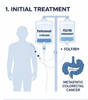
Panitumumab and other EGFRIs cause hypomagnesemia primarily by reducing renal magnesium reabsorption through effects on transient receptor potential melastatin 6 channels in the distal convoluted tubule (6). Progressive magnesium loss may remain clinically silent for prolonged periods because symptoms are nonspecific (7). Magnesium is essential for cardiac electrophysiology because it regulates inward and outward ionic currents, including L-type calcium channels, inward rectifier and delayed rectifier potassium currents, and Na+/K-ATPase activity (8). Severe hypomagnesemia prolongs the QTc interval through direct effects on repolarizing potassium currents, which increase action-potential duration, and through indirect hypokalemic effects caused by increased renal potassium wasting and disrupted cellular potassium handling (4). Magnesium deficiency may also promote afterdepolarizations, particularly delayed afterdepolarizations, by increasing intracellular calcium loading and triggered activity, thereby providing a potential mechanism for atrial ectopy and AF initiation (9). In addition, hypomagnesemia causes refractory electrolyte abnormalities through 2 pathways. First, it activates renal outer medullary K+ channels, increasing renal potassium excretion and worsening hypokalemia (10). Second, it suppresses parathyroid hormone secretion and activity, leading to refractory hypocalcemia (11). Therefore, potassium or calcium repletion alone may be ineffective unless magnesium deficiency is corrected first or concurrently. These mechanisms and the clinical course are summarized in Figure 1.
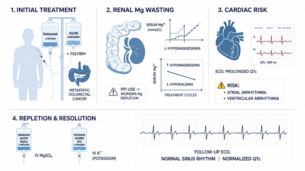
Schematic representation of panitumumab-induced magnesium wasting, QTc prolongation, and recovery after electrolyte repletion. Progressive hypomagnesemia during panitumumab therapy may cause QTc prolongation and predispose patients to arrhythmias, whereas prompt magnesium and potassium replacement can reverse the electrophysiologic abnormality.
Figure 1.

Schematic representation of panitumumab-induced magnesium wasting, QTc prolongation, and recovery after electrolyte repletion. Progressive hypomagnesemia during panitumumab therapy may cause QTc prolongation and predispose patients to arrhythmias, whereas prompt magnesium and potassium replacement can reverse the electrophysiologic abnormality.

This case is notable because it demonstrates overlapping atrial and ventricular electrophysiologic vulnerability caused by an isolated metabolic abnormality associated with targeted therapy. AF is increasingly observed in patients with cancer and may result from inflammation, autonomic imbalance, direct cardiotoxicity, or metabolic disturbances (12). In addition to directly promoting atrial ectopy, hypomagnesemia may indirectly increase AF risk by prolonging the QT interval and promoting heterogeneous refractoriness (4). In the presence of low magnesium and potassium levels, the observed 50-ms increase in QTc from baseline, although still within a borderline range, represented a clinically meaningful change and signaled impending electrical instability. In cardio-oncology practice, relative changes from a patient’s baseline electrocardiogram and electrolyte values may be as important as absolute thresholds.
The patient was also receiving omeprazole for gastroesophageal reflux disease. Proton pump inhibitors can independently increase the risk of severe hypomagnesemia, possibly by reducing intestinal magnesium absorption (13). Concurrent proton pump inhibitor use may therefore have exacerbated panitumumab-associated magnesium wasting. In such cases, switching to an H2-receptor blocker may be considered after appropriate clinical assessment.
Electrolyte-mediated arrhythmias in patients with cancer may be underrecognized for several reasons. First, medication-induced hypomagnesemia may remain asymptomatic unless it becomes severe (6). Second, oncology follow-up often focuses on myelosuppression and gastrointestinal and dermatologic toxicities, whereas electrolyte monitoring may be less frequent. Third, concomitant medications, including proton pump inhibitors, may worsen magnesium and potassium depletion (14). Finally, intervention may be delayed when population-based thresholds are applied without accounting for patient-specific trends.
Prompt magnesium and potassium replacement led to rapid normalization of the QTc interval and reduced concern about imminent arrhythmia. This response supports magnesium repletion as an appropriate preventive strategy when magnesium-related QTc prolongation is suspected. Early intervention is important. Available evidence indicates that initiating magnesium replacement before the concentration decreases below 1.1 mg/dL, or approximately 0.45 mmol/L, can reduce the incidence of grade 3 or higher hypomagnesemia from 30% to 3.3% (P = 0.012) (3, 15). Although this patient had already developed grade 3 hypomagnesemia, earlier recognition of the progressive decline from 0.78 to 0.62 to 0.33 mmol/L might have enabled preventive magnesium supplementation.
Withholding panitumumab while correcting the electrolyte imbalance was reasonable because the abnormality was reversible and the patient was at increased arrhythmic risk. If panitumumab is resumed, the oncologic benefit should be balanced against the risk of recurrent electrolyte abnormalities and arrhythmias. Regular electrolyte monitoring, magnesium supplementation, minimization of interacting medications such as proton pump inhibitors, and electrocardiographic monitoring should be considered in at-risk patients. Electrocardiography should be performed not only at baseline but also urgently in patients with severe hypomagnesemia, palpitations, syncope, or concomitant use of QT-prolonging medications. The transient effect of intravenous magnesium must also be considered; its benefit may last only approximately 48 hours, and magnesium levels may decrease again after initial repletion. In severe cases, daily or alternate-day intravenous magnesium may be required.
Other potential causes include the cardiotoxic effects of chemotherapeutic agents (16). Although ischemia, myocardial dysfunction, and vasospasm were not observed in this case, these complications should be considered in patients receiving anticancer therapy. Autonomic modulation, drug interactions, and occult electrolyte losses from diarrhea might also have contributed. Nevertheless, the progressive decline in magnesium during panitumumab therapy was not explained by the patient’s baseline comorbidities, although these conditions may have increased his arrhythmic risk. In patients with persistent or recurrent hypomagnesemia despite replacement, amiloride, a potassium-sparing diuretic, may reduce renal magnesium wasting. Although amiloride was not used in this patient, it could be considered as an adjunct when panitumumab cannot be discontinued or interrupted (17).
This case has several limitations. Intracellular magnesium and urinary magnesium excretion were not measured; therefore, magnesium handling could not be fully characterized. In addition, continuous cardiac monitoring was not performed during the period of greatest hypomagnesemia. Such data could have provided a more clinically actionable assessment of the electrophysiologic effects.
In conclusion, panitumumab and potentially other targeted anticancer therapies can cause progressive, clinically silent hypomagnesemia that disrupts cardiac conduction and increases arrhythmic risk. Simple and timely interventions may prevent arrhythmias and permit continuation of therapy. Vigilant monitoring and prompt action are essential.
Patient Consent for Publication: Written informed consent was obtained from the patient for publication of this case report and any accompanying anonymized clinical data. The patient was informed that all identifying information would be removed to ensure anonymity, and he provided permission to publish the article based on his clinical course and treatment.

Footnotes

  • AI Use Disclosure:For the purpose of Figure Design, the Perplexity And Chatgpt was used Completely in the Figure 1 section.

  • Authors' Contribution:A. A.: Patient diagnosis and clinical management, project supervision, validation of electrophysiologic interpretation, critical manuscript revision, and final approval; H. F. A.: Data curation, investigation, literature review, writing – original draft, figure/table preparation, and manuscript formatting; S. A.: Writing – review and editing, including assistance with drafting sections of the case report; A. S.: Conceptualization of the electrophysiologic perspective, methodology, formal analysis, supervision of electrolyte and arrhythmia risk assessment, manuscript review and editing, journal submission, peer-review correspondence, data availability, and final approval.

  • Conflict of Interests Statement:The authors declare that they have no known competing financial interests or personal relationships that could have appeared to influence the work reported in this paper.

  • Data Availability:All relevant clinical data supporting the findings of this case report are fully described within the manuscript. No further datasets were generated or analyzed during this study.

  • Ethical Approval:This case report was reviewed and approved by the Institutional Review Board (IRB) / Ethics Committee. The study was conducted in accordance with the principles of the Declaration of Helsinki.

  • Funding/Support:No funding was received for this study.

  • Informed Consent:Written informed consent was obtained from the patient for publication of this case report and any accompanying anonymized clinical data. The patient was informed that all identifying information would be removed to ensure anonymity, and he provided permission to publish the article based on his clinical course and treatment.

References

  • 1.
    Buza V, Rajagopalan B, Curtis AB. Cancer treatment-induced arrhythmias: Focus on chemotherapy and targeted therapies. Circ Arrhythm Electrophysiol. 2017;10(8). e005443. [PubMed ID: 28798022]. https://doi.org/10.1161/CIRCEP.117.005443.
  • 2.
    Link MG, Yan GX, Kowey PR. Evaluation of toxicity for heart failure therapeutics: Studying effects on the QT interval. Circ Heart Fail. 2010;3(4):547-555. [PubMed ID: 20647490]. https://doi.org/10.1161/CIRCHEARTFAILURE.109.917781.
  • 3.
    Kimura M, Usami E, Teramachi H, Yoshimura T. Identifying optimal magnesium replenishment points based on risk of severe hypomagnesemia in colorectal cancer patients treated with cetuximab or panitumumab. Cancer Chemother Pharmacol. 2020;86(3):383-391. [PubMed ID: 32789758]. https://doi.org/10.1007/s00280-020-04126-9.
  • 4.
    Negru AG, Pastorcici A, Crisan S, Cismaru G, Popescu FG, Luca CT. The role of hypomagnesemia in cardiac arrhythmias: A clinical perspective. Biomedicines. 2022;10(10):2356. [PubMed ID: 36289616]. [PubMed Central ID: PMC9598104]. https://doi.org/10.3390/biomedicines10102356.
  • 5.
    Ducceschi V, Di Micco G, Sarubbi B, Russo B, Santangelo L, Iacono A. Ionic mechanisms of ischemia-related ventricular arrhythmias. Clin Cardiol. 1996;19(4):325-331. [PubMed ID: 8706374]. https://doi.org/10.1002/clc.4960190409.
  • 6.
    Cheng H, Gammon D, Dutton TM, Piperdi B. Panitumumab-related hypomagnesemia in patients with colorectal cancer. Hosp Pharm. 2009;44(3):234-238. https://doi.org/10.1310/hpj4403-234.
  • 7.
    Kothari M, Wanjari A, Shaikh SM, Tantia P, Waghmare BV, Parepalli A, et al. A comprehensive review on understanding magnesium disorders: Pathophysiology, clinical manifestations, and management strategies. Cureus. 2024;16(9). e68385. [PubMed ID: 39355467]. [PubMed Central ID: PMC11444808]. https://doi.org/10.7759/cureus.68385.
  • 8.
    White RE, Hartzell HC. Magnesium ions in cardiac function: Regulator of ion channels and second messengers. Biochem Pharmacol. 1989;38(6):859-867. [PubMed ID: 2467677]. https://doi.org/10.1016/0006-2952(89)90272-4.
  • 9.
    Chambers P. Mgwater. Magnesium Online Library; 2003, [cited 2026 Aug 2]. Magnesium and potassium in lone atrial fibrillation. Available from: http://www.mgwater.com/laf.shtml.
  • 10.
    Verzicco I, Regolisti G, Quaini F, Bocchi P, Brusasco I, Ferrari M, et al. Electrolyte disorders induced by antineoplastic drugs. Front Oncol. 2020;10. 779. [PubMed ID: 32509580]. [PubMed Central ID: PMC7248368]. https://doi.org/10.3389/fonc.2020.00779.
  • 11.
    Bobrowicz M, Pachucki J, Popow M. Hypomagnesaemia leading to parathyroid dysfunction, hypocalcaemia, and hypokalaemia as a complication of long-term treatment with a proton pump inhibitor: A literature review. Endokrynol Pol. 2024;75(4):359-365. [PubMed ID: 39279305]. https://doi.org/10.5603/ep.98576.
  • 12.
    Madnick DL, Fradley MG. Atrial fibrillation and cancer patients: Mechanisms and management. Curr Cardiol Rep. 2022;24(10):1517-1527. [PubMed ID: 35976601]. https://doi.org/10.1007/s11886-022-01769-3.
  • 13.
    Park CH, Kim EH, Roh YH, Kim HY, Lee SK. The association between the use of proton pump inhibitors and the risk of hypomagnesemia: A systematic review and meta-analysis. PLoS One. 2014;9(11). e112558. [PubMed ID: 25394217]. [PubMed Central ID: PMC4230950]. https://doi.org/10.1371/journal.pone.0112558.
  • 14.
    William JH, Danziger J. Magnesium deficiency and proton-pump inhibitor use: A clinical review. J Clin Pharmacol. 2016;56(6):660-668. [PubMed ID: 26582556]. https://doi.org/10.1002/jcph.672.
  • 15.
    Ito D, Asano H, Kimura M, Usami E. Evaluation of the usefulness of protocol-based magnesium supplementation for hypomagnesemia in patients with advanced or recurrent colorectal cancer treated with panitumumab. Support Care Cancer. 2025;33(6). 472. [PubMed ID: 40366467]. https://doi.org/10.1007/s00520-025-09531-5.
  • 16.
    Chaulin AM, Abashina OE, Duplyakov DV. Pathophysiological mechanisms of cardiotoxicity in chemotherapeutic agents. Russ Open Med J. 2020;9(3). e0305. https://doi.org/10.15275/rusomj.2020.0305.
  • 17.
    Wazny LD, Brophy DF. Amiloride for the prevention of amphotericin B-induced hypokalemia and hypomagnesemia. Ann Pharmacother. 2000;34(1):94-97. [PubMed ID: 10669191]. https://doi.org/10.1345/aph.19127.

Copyright

Copyright © 2026, Multidisciplinary Cardiovascular Annals. This open-access article is available under the Creative Commons Attribution-NonCommercial 4.0 (CC BY-NC 4.0) International License (https://creativecommons.org/licenses/by-nc/4.0/), which allows for the copying and redistribution of the material only for noncommercial purposes, provided that the original work is properly cited.

Similar Articles

30
Jun
2015

High-Grade Atrioventricular Block and Takotsubo Cardiomyopathy: Case Report and Review of the Literature

Panagiotis Korantzopoulos,
Dimitrios N. Nikas,
Konstantinos Letsas,
Ioannis Gkirdis,
Marios D. Kolios,
Ioannis V. Ntalas
,et al.

Korantzopoulos P, N. Nikas D, Letsas K, Gkirdis I, Kolios MD, et al. High-Grade Atrioventricular Block and Takotsubo Cardiomyopathy: Case Report and Review of the Literature. Int Cardiovasc Res J. 2017;9(2):e11407. doi:

3
Nov
2018
Hyopomagnesemia in Critically Ill Children

Hyopomagnesemia in Critically Ill Children

Seher Erdoğan,
Tuba Seven Menevşe

Erdoğan S, Seven Menevşe T. Hyopomagnesemia in Critically Ill Children. Inn J Pediatr. 2018;28(6):e66444. doi: https://doi.org/10.5812/ijp.66444

14
Jan
2020
International Journal of Endocrinology and Metabolism

Association Between Pre-Transplant Magnesemia and Post-Transplant Dysglycemia in Kidney Transplant Recipients

Azam Alamdari,
Ghazal Asadi,
Farzaneh Sadat Minoo,
Mohammad-Reza Khatami,
Seyed Mansour Gatmiri,
Simin Dashti-Khavidaki
,et al.

Alamdari A, Asadi G, Minoo FS, Khatami M, Gatmiri SM, et al. Association Between Pre-Transplant Magnesemia and Post-Transplant Dysglycemia in Kidney Transplant Recipients. Int J Endocrinol Metab. 2020;18(1):e97292. doi: https://doi.org/10.5812/ijem.97292

30
Apr
2016

Atorvastatin for Prevention of Amikacin-induced Electrolytes Imbalances; a Randomized Clinical Trial

Behrooz Heydari,
Hossein Khalili,
Simin Dashti-Khavidaki,
Mohammad Taghi Beig- Mohammadi,
Mostafa Mohammadi

Heydari B, Khalili H, Dashti-Khavidaki S, Beig- Mohammadi MT, Mohammadi M. Atorvastatin for Prevention of Amikacin-induced Electrolytes Imbalances; a Randomized Clinical Trial. Iran J Pharm Res. 2016;15(2):e125174. doi: https://doi.org/10.22037/ijpr.2016.1796

31
Oct
2014

Elevated Vancomycin Trough Concentration: Increased Efficacy and/or Toxicity?

Sepideh Elyasi,
Hossein Khalili,
Simin Dashti-Khavidaki,
Hamid Emadi-Koochak,
Amirhooshang Mohammadpour,
Alireza Abdollahi

Elyasi S, Khalili H, Dashti-Khavidaki S, Emadi-Koochak H, Mohammadpour A, et al. Elevated Vancomycin Trough Concentration: Increased Efficacy and/or Toxicity?. Iran J Pharm Res. 2014;13(4):e125586. doi: https://doi.org/10.22037/ijpr.2014.1584

Download PDF447.89 KB

Crossmark

Crossmark

Checking

Share on
Cited by
Metrics

Ordering Reprints

Articles are published under the Creative Commons license stated on each article. No permission or royalty fee is required for uses permitted by that license. CCC handles optional bulk and customized reprint orders. Any quotation covers production and delivery services only, not copyright permission. > Request Reprints from CCC 

Search Relations

Author(s):

Related Articles