
1. Background
Painful diabetic peripheral neuropathy (PDPN) is a common and disabling complication of diabetes. Symptoms may include burning, electric, aching, and nocturnal pain, which may interfere with sleep and quality of life. Current guidelines recommend pharmacological treatment with agents such as pregabalin, other gabapentinoids, serotonin-norepinephrine reuptake inhibitors, tricyclic antidepressants, and selected sodium-channel blockers. Importantly, patients should also be assessed for sleep disturbance (1, 2).
Despite these treatments, a proportion of patients remain symptomatic. The concept that metabolically injured nerves may be particularly vulnerable to focal mechanical compression has generated interest in peripheral nerve decompression. Surgical studies have reported improvements in pain; however, the evidence remains heterogeneous, and recent sham-controlled evidence has raised concerns about placebo and procedure-related effects (3-7).
Ultrasound-guided perineural hydrodissection is a minimally invasive technique intended to mechanically separate a peripheral nerve from adjacent tissues. Existing evidence is concentrated mainly in focal entrapment neuropathies, particularly carpal tunnel syndrome, and therefore cannot be directly extrapolated to PDPN (8, 9).
2. Objectives
This study evaluated six-month changes in pain, sleep-related patient-reported outcomes, and electrodiagnostic severity following a single standardized five-nerve ankle block with perineural hydrodissection in patients who remained symptomatic despite treatment with pregabalin and duloxetine.
3. Methods
3.1. Study Design and Participants
This prospective, single-arm longitudinal study included 34 patients with diabetes and painful peripheral neuropathy. All patients had persistent pain despite a standardized pharmacological regimen of pregabalin 75 mg twice daily and duloxetine 30 mg once daily. This medication regimen was continued throughout follow-up. Because there was no untreated, sham, or surgical control group, the study was designed to evaluate within-patient temporal change rather than comparative efficacy.
3.2. Intervention
All patients underwent a standardized ultrasound-guided intervention. Five standard peripheral nerves at the ankle were targeted: the tibial, deep peroneal, superficial peroneal, sural, and saphenous nerves. A total of 10 mL of solution containing 1% lidocaine and 8 mg dexamethasone was prepared. Two milliliters were administered around each nerve under real-time ultrasound guidance. The injection was performed in the perineural plane with the intention of separating the nerve from surrounding tissues and releasing local tissue restriction or adhesions (perineural hydrodissection). No open surgical decompression was performed, and no repeat procedure was scheduled.
3.3. Outcomes
Pain intensity was recorded at baseline and at 1, 3, and 6 months. Sleep quality was recorded at baseline using the categories good, moderate, or bad; at 6 months, overall satisfaction with sleep quality was recorded. Because these sleep measures were not identical instruments, sleep was treated as a descriptive outcome rather than a formal longitudinal scale.
Electrodiagnostic findings were recorded at baseline and at 6 months using the categories mild (mi), mild-to-moderate (mi to m), moderate (m), moderate-to-severe (m to s), and severe (s). For statistical analysis, these categories were assigned ordinal scores of 1, 1.5, 2, 2.5, and 3, respectively. This ordinal transformation was used solely to permit paired nonparametric analysis and should not be interpreted as a quantitative nerve conduction measurement.
3.4. Statistical Analysis
Continuous variables are presented as mean ± standard deviation, and categorical variables as counts and percentages. The Friedman test was used to assess overall changes in pain across four repeated time points. Kendall’s W was calculated to quantify the magnitude of the repeated-measures effect. Pairwise Wilcoxon signed-rank tests were performed as exploratory post hoc comparisons and adjusted using the Holm method. Baseline-to-6-month electrodiagnostic severity was compared using the Wilcoxon signed-rank test. A two-sided P < 0.05 was considered statistically significant. Analyses were performed using the complete dataset of 34 patients.
4. Results
4.1. Baseline Characteristics
Thirty-four patients were included in the analysis. The mean age was 69.85 ± 4.38 years, and 19 (55.9%) were men. The mean duration of diabetes was 20.56 ± 4.02 years, and the mean HbA1c was 7.04 ± 0.18%. All participants were receiving pregabalin 75 mg twice daily and duloxetine 30 mg once daily. Baseline characteristics are presented in Table 1.
Table 1.
Baseline Characteristics of the Study Participants a
| Variables | Value |
|---|---|
| Patients | 34 |
| Age, y | 69.85 ± 4.38 |
| Male | 19 (55.9) |
| Female | 15 (44.1) |
| Diabetes duration, y | 20.56 ± 4.02 |
| HbA1c, % | 7.04 ± 0.18 |
| Pregabalin | 75 mg twice daily |
| Duloxetine | 30 mg once daily |
a Values are expressed as number, mean ± SD or No. (%) unless otherwise indicated.
4.2. Pain Trajectory
Pain scores decreased progressively at each follow-up assessment. Mean pain declined from 7.76 ± 0.96 at baseline to 6.09 ± 1.00 at 1 month, 5.62 ± 0.82 at 3 months, and 5.32 ± 0.81 at 6 months. The Friedman test demonstrated a strong overall time effect (χ2 = 86.27, df = 3, P = 1.38 × 10⁻¹⁸), with Kendall’s W = 0.846. The mean baseline-to-6-month reduction was 2.44 points (95% CI, 2.18 - 2.71), corresponding to a 31.4% reduction from baseline. These values are summarized in Table 2 and illustrated in Figure 1.
Table 2.
Pain Scores at Baseline and Follow-Up Assessments
| Time Point | Mean ± SD | Median | Range |
|---|---|---|---|
| Baseline | 7.76 ± 0.96 | 7 | 7 - 10 |
| 1 mo | 6.09 ± 1.00 | 6 | 5 - 8 |
| 3 mo | 5.62 ± 0.82 | 5 | 5 - 8 |
| 6 mo | 5.32 ± 0.81 | 5 | 4 - 7 |
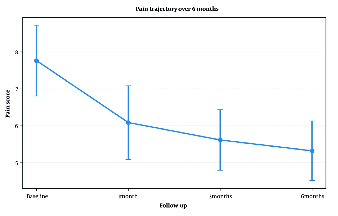
Figure 1.
Mean pain score (± SD) over six months.
4.3. Pairwise Pain Comparisons
Exploratory post hoc pairwise comparisons showed statistically significant differences between all pairs of pain assessments after Holm adjustment. The largest baseline-to-follow-up differences occurred at 3 and 6 months, and the 3-to-6-month comparison also remained statistically significant. The raw and Holm-adjusted P values are presented in Table 3.
Table 3.
Pairwise Comparisons of Pain Scores with Holm Adjustment
| Comparison | Raw P | Holm-adjusted P |
|---|---|---|
| Baseline vs 1 mo | 2.96 × 10⁻⁷ | 1.18 × 10⁻⁶ |
| Baseline vs 3 mo | 1.41 × 10⁻⁷ | 8.49 × 10⁻⁷ |
| Baseline vs 6 mo | 2.23 × 10⁻⁷ | 1.11 × 10⁻⁶ |
| 1 month vs 3 mo | 0.000786 | 0.00157 |
| 1 month vs 6 mo | 5.02 × 10⁻⁶ | 1.51 × 10⁻⁵ |
| 3 months vs 6 mo | 0.00753 | 0.00753 |
4.4. Electrodiagnostic Outcomes
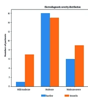
Electrodiagnostic severity shifted toward less severe categories over the six-month follow-up. The number of participants classified as severe decreased from 11 (32.4%) at baseline to 3 (8.8%) at six months, whereas the mild-to-moderate category increased from 1 (2.9%) to 7 (20.6%). Using the prespecified ordinal scoring system, mean severity decreased from 2.40 at baseline to 2.12 at six months. The paired Wilcoxon signed-rank test was statistically significant (W = 0, P = 3.38 × 10⁻⁵), with a mean ordinal reduction of 0.28 points (95% CI, 0.19 - 0.37). The distribution is shown in Table 4 and Figure 2.
Table 4.
Electrodiagnostic Severity Categories at Baseline and Six Months a
| Severity | Baseline | 6 mo |
|---|---|---|
| Mild-to-moderate | 1 (2.9) | 7 (20.6) |
| Moderate | 16 (47.1) | 15 (44.1) |
| Moderate-to-severe | 6 (17.6) | 9 (26.5) |
| Severe | 11 (32.4) | 3 (8.8) |
a Values are expressed as No. (%).
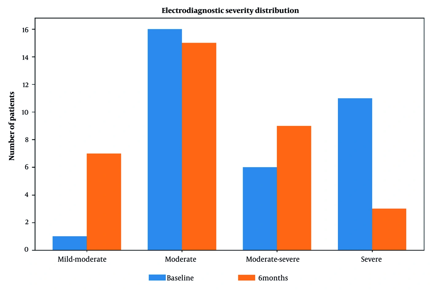
Figure 2.
Distribution of electrodiagnostic severity categories at baseline and six months.
4.5. Sleep-Related Outcomes
Sleep findings were descriptive because the baseline and six-month measures did not assess identical constructs. At baseline, 22 patients (64.7%) reported good sleep, compared with 23 (67.6%) reporting good overall satisfaction with sleep quality at six months. The distributions of the recorded categories are presented in Table 5. Because the response constructs differed, these data do not support a formal paired statistical claim of improvement.
Table 5.
Sleep Category at Baseline and Overall Sleep Satisfaction at Six Months a
| Sleep Categories | Baseline | 6-month Overall Satisfaction |
|---|---|---|
| Good | 22 (64.7) | 23 (67.6) |
| Moderate | 10 (29.4) | 7 (20.6) |
| Moderate-to-severe | 0 (0.0) | 2 (5.9) |
| Bad/poor | 2 (5.9) | 2 (5.9) |
a Values are expressed as No. (%).
5. Discussion
In this prospective single-arm cohort study, a single ultrasound-guided five-nerve ankle block with perineural hydrodissection was followed by a progressive reduction in pain over six months. Mean pain decreased by 2.44 points, from 7.76 at baseline to 5.32 at six months, and the repeated-measures effect size was large (Kendall’s W = 0.846). Electrodiagnostic severity also shifted toward less severe categories. These findings are encouraging but should be interpreted as an association rather than proof of efficacy.
The pharmacological context is important. Pregabalin and duloxetine are established, evidence-based options for painful diabetic neuropathy, and guidelines recommend offering agents from effective medication classes while considering adverse effects and patient factors (1, 10). All participants in this study were already receiving pregabalin 75 mg twice daily and duloxetine 30 mg once daily yet continued to experience clinically relevant pain. Thus, the study population represents patients with persistent symptoms despite standard medical management. Nevertheless, because medication was continued, the design cannot isolate the independent contribution of the procedure.
The biological rationale for hydrodissection is extrapolated from focal entrapment neuropathies. Ultrasound allows visualization of the nerve and surrounding tissue and can guide delivery of injectate into the perineural plane. Systematic reviews of hydrodissection have mainly included carpal tunnel syndrome and, to a lesser extent, ulnar neuropathy; they suggest that ultrasound-guided hydrodissection can be performed safely, but the optimal injectate, volume, and mechanism-specific contribution remain uncertain (8, 9). Therefore, our findings extend an existing interventional concept to a different and more diffuse neuropathic condition, but they should not be interpreted as confirmation that diabetic neuropathy is primarily an entrapment disorder.
Our results are also relevant to the literature on surgical peripheral nerve decompression. A 2019 pragmatic randomized trial reported a significant difference in pain trajectory favoring lower-extremity decompression over observation in patients with painful diabetic neuropathy and superimposed nerve compression (3). A 2014 randomized study similarly reported lower pain after decompression of four sites in one limb compared with the contralateral control limb (5). However, a 2018 structured review highlighted uncertainty related to sham effects and concluded that the evidence was insufficient to recommend decompression routinely (4).
More recently, a double-blind randomized trial published in 2024 found that decompression was associated with lower pain than observation, but decompressed and sham-operated legs improved similarly at the earlier time point, raising the possibility that part of the observed benefit was related to placebo or nonspecific procedural effects (6). A 2026 systematic review of randomized trials likewise concluded that the current surgical evidence is limited by a high risk of bias, substantial publication bias, heterogeneity, and wound-related complications, despite pooled pain reductions at 6 and 12 months (7).
Compared with open surgical decompression, the present technique is less invasive and does not require a surgical incision. This may be attractive in older patients with diabetes; however, our dataset contains no systematic adverse-event assessment and therefore cannot establish a safety advantage. Moreover, the injectate included both lidocaine and dexamethasone, so the observed effect could reflect local anesthetic effects, corticosteroid effects, mechanical hydrodissection, or a combination of these mechanisms.
The electrodiagnostic findings warrant cautious interpretation. The shift from 11 severe cases at baseline to 3 at six months and the significant paired ordinal test are encouraging. However, the categories were not based on quantitative nerve conduction measurements. Future work should record nerve conduction velocity, distal latency, sensory and motor amplitudes, and F-wave parameters, when available. Objective electrophysiological recovery should then be analyzed alongside validated clinical outcomes.
Sleep findings were less conclusive. The proportion classified as “good” was similar at baseline and at six months, and the wording of the follow-up variable differed. Current guidelines specifically recommend assessment for sleep disorders in PDPN, making this an important outcome for future trials (1). A validated instrument such as the Pittsburgh Sleep Quality Index should be administered at baseline and at every follow-up visit.
5.1. Strengths and Limitations
Strengths include a uniform, single-session intervention, ultrasound guidance, standardized concomitant medication, repeated pain assessments through six months, and an objective electrodiagnostic outcome. The principal limitation is the absence of a control or sham group. Additional limitations include the small sample size, lack of blinding, inability to separate the effects of lidocaine/dexamethasone from hydrodissection, the use of ordinal rather than quantitative electrodiagnostic data, and nonstandardized sleep measurement. Regression to the mean, natural symptom fluctuation, expectations, and other time-varying factors may also explain part of the observed improvement.
5.2. Conclusions
Among 34 patients with painful diabetic peripheral neuropathy who remained symptomatic despite pregabalin and duloxetine, a single ultrasound-guided five-nerve ankle block with perineural hydrodissection was associated with a statistically significant reduction in pain over six months and a significant shift toward less severe electrodiagnostic categories. These findings support further investigation but do not establish causality or superiority over medical therapy or surgery. A multicenter randomized trial with a sham-injection control, standardized sleep and quality-of-life measures, quantitative nerve conduction parameters, and prespecified adverse-event reporting is warranted.