
1. Background
Cerebral ischemia is one of the most serious complications of neurosurgical procedures and may result in permanent neurological deficits, prolonged hospitalization, and reduced quality of life (1). Despite improvements in surgical techniques and perioperative care, the incidence of perioperative stroke has increased over the past two decades, mainly because a growing number of older patients with multiple chronic comorbidities are undergoing surgery (2, 3).
Neuroinflammation encompasses inflammatory responses to noxious stimuli affecting the central nervous system. Resident microglial cells play a significant role in neuroinflammatory processes by producing inflammatory mediators (4). Interleukin-6 (IL-6) and tumor necrosis factor-alpha (TNF-α) are important pathophysiological components of a wide range of neurological conditions, including traumatic brain and spinal cord injuries, neurodegenerative disorders such as Alzheimer disease and Parkinson disease, CNS infections, and stroke (5). Neuroinflammatory processes can disrupt the blood-brain barrier, subsequently contributing to cerebral edema, impaired cerebral autoregulation, and microthrombus formation (6).
Recent investigations have demonstrated that general anesthetic techniques can significantly influence systemic inflammatory cytokine profiles, including IL-6, during the perioperative period (7). Furthermore, comparative trials in neurosurgery have reported longer operative and anesthetic durations with general anesthesia than with regional anesthesia (8). Regional anesthesia also significantly reduced the need for analgesic administration. Different anesthetic techniques may differentially affect systemic and local inflammatory responses. Several studies have investigated the neuroprotective and anti-inflammatory effects of propofol and thiopental in neurosurgical procedures. In 2020, Hausburg et al. reported that propofol may attenuate ischemia-related brain injury by reducing inflammatory markers; its use was associated with lower neuroinflammation and improved neurological outcomes compared with other anesthetic agents (9). Owusu et al. (10) further suggested that the anti-inflammatory properties of propofol, particularly its ability to reduce TNF-α and IL-6 levels, may make it a potentially beneficial anesthetic agent for neurosurgical procedures.
Despite experimental and animal evidence supporting the anti-inflammatory and neuroprotective effects of propofol, clinical evidence demonstrating its superiority over thiopental in neurosurgical procedures remains insufficient (11). Like propofol, thiopental can reduce cerebral metabolic activity, but the agents may differ in their effects on inflammation, ischemic injury, and the cellular response to surgical stress.
2. Objectives
This randomized controlled study aimed to determine the effects of propofol and thiopental induction on postoperative inflammatory markers and intraoperative hemodynamic parameters, and to assess whether propofol may be a more favorable option for anesthetic induction in neurosurgical procedures, potentially contributing to improved clinical outcomes.
3. Methods
3.1. Study Design and Ethics
This single-center, two-arm, parallel-group randomized controlled trial used a 1:1 allocation ratio and a superiority framework. It was designed in 2025 to compare the effects of propofol and sodium thiopental on markers of cerebral injury and the inflammatory response in patients undergoing neurosurgical procedures. The study was conducted in accordance with the Declaration of Helsinki. Ethical approval was obtained from the Ethics Committee of Shahid Beheshti University of Medical Sciences (IR.SBMU.RETECH.REC.1404.271), and the trial was registered in the Iranian Registry of Clinical Trials (IRCT20250722066589N2) on August 3, 2025. The registry record is available at https://www.irct.ir/trial/85283. The study was conducted at the Department of Anesthesiology, Shohada Tajrish Hospital, affiliated with Shahid Beheshti University of Medical Sciences, Tehran, Iran, between August 2025 and March 2026. Written informed consent was obtained from all participants after the study procedures and intervention had been explained in detail and their questions and concerns had been addressed.
The primary outcomes were between-group differences in changes in serum IL-6 and TNF-α concentrations from baseline to 6 hours postoperatively.
Inflammatory markers were measured using enzyme-linked immunosorbent assay with commercially available, standardized kits. Systolic blood pressure (SBP), diastolic blood pressure (DBP), and heart rate (HR) were assessed as secondary hemodynamic outcomes. A sample size of 37 patients per group was determined. Patients scheduled for elective neurosurgical procedures who provided written informed consent were eligible if they were aged 18 - 60 years and had an American Society of Anesthesiologists physical status of I or II. Exclusion criteria were severe cardiovascular disease, including heart failure or uncontrolled arrhythmias (affecting hemodynamic parameters); uncontrolled diabetes mellitus, defined as glycated hemoglobin (HbA1c) > 8% (influencing inflammatory and biochemical outcomes); known hypersensitivity to propofol or thiopental; and inability or unwillingness to comply with the study protocol, including refusal to undergo postoperative follow-up.
This randomized controlled trial was designed, conducted, and completed in accordance with the Consolidated Standards of Reporting Trials (CONSORT) guidelines to ensure transparent and comprehensive reporting of the clinical methodology.
3.2. Randomization and Interventions
Each participant was assigned a unique identification number using a computer-generated random number. Randomization was then performed using a random number table, allocating participants to two groups of 37 in a 1:1 ratio. Allocation concealment was achieved using sequentially numbered, opaque, sealed envelopes prepared by an independent researcher who was not involved in recruitment, patient care, or outcome assessment. Because propofol and thiopental differ in appearance, the anesthesiologist administering the induction agent could not be blinded. Participants, laboratory personnel, and statisticians were blinded to group allocation.
All patients underwent a preanesthetic evaluation on the day before surgery. Baseline demographic variables, including age, gender, and body mass index (BMI), were recorded, and blood samples were collected to assess baseline TNF-α and IL-6 levels. Participants were admitted to the operating room after following the standard fasting protocol. After standard anesthetic monitoring was established, the attending investigator administered the induction regimen according to the assigned study group. Both groups received standardized premedication with midazolam and fentanyl. Anesthesia was induced with propofol (1.5 - 2 mg/kg) or thiopental (4 - 5 mg/kg), titrated until loss of the eyelash reflex. After anesthetic induction, an arterial line was established for each patient to allow accurate monitoring of intraoperative hemodynamic variables, and baseline values were recorded. Anesthesia was maintained with a continuous propofol infusion (0.2 mg/kg/min) in both groups, titrated to achieve and maintain a bispectral index of 40 - 60. Hemodynamic variables were recorded every 15 minutes throughout surgery, and an overall intraoperative mean was calculated for each patient across all surgical time points.
Blood samples were centrifuged at 3000 rpm for 10 minutes at 4 °C. Serum IL-6 and TNF-α levels were measured in duplicate using commercially available human ELISA kits (Karmania Pars Gene, Kerman, Iran; Cat. No. KPG-HIL6 and KPG-HTNFA), following the manufacturer’s instructions. The analytical sensitivity was 1.5 pg/mL for IL-6 and 2.0 pg/mL for TNF-α. Intra-assay and interassay coefficients of variation were < 8% and < 10%, respectively.
3.3. Statistical Analysis
The sample size was calculated to compare the effects of propofol and thiopental on postoperative inflammatory markers, including IL-6 and TNF-α, in patients undergoing neurosurgical procedures. Based on the anticipated between-group difference in postoperative inflammatory marker levels and the corresponding variability reported in previous studies (12), the sample size was estimated using the formula for comparing two independent means. Assuming a two-sided significance level of 5% (α = 0.05) and 80% statistical power (1 - β = 0.80), 30 evaluable patients were required in each group. To allow for possible attrition, loss to follow-up, or nonevaluable laboratory samples, the target sample size was increased to 37 patients per group. Accordingly, 74 patients were enrolled, with 37 allocated to each treatment group.
To evaluate changes in inflammatory markers, the difference between postoperative and preoperative values (Δ) was calculated. Data were initially extracted from Excel files and entered into the analysis after cleaning and correction of incomplete or corrupted values. Descriptive statistics were reported as mean ± standard deviation (SD), and the normality of the data distribution was assessed using the Shapiro-Wilk test. Within-group comparisons of preoperative and postoperative values were performed using the paired t-test or Wilcoxon signed-rank test, as appropriate for the data distribution. Between-group comparisons were performed using Welch’s t-test or the Mann-Whitney U test, as appropriate.
In addition to comparing Δ values, analysis of covariance was performed, with adjustment for baseline values, to further evaluate between-group differences. Effect sizes were calculated using Cohen’s d, and 95% confidence intervals (CIs) were reported. A two-sided P < 0.05 was considered statistically significant. Statistical analyses were performed using Python with the SciPy, pandas, and Matplotlib packages. The study protocol and statistical analysis plan are available from the corresponding author upon reasonable request.
4. Results
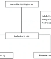
A total of 80 patients were screened for eligibility between August 2025 and March 2026. Before randomization, 6 patients were excluded because of a history of heart failure or poorly controlled diabetes mellitus. Consequently, 74 patients were randomized to two groups (Figure 1): 37 to the propofol group and 37 to the thiopental group. All randomized participants were included in the primary analysis. No participants were lost to follow-up, and no primary outcome data were missing. No serious adverse events related to the study interventions were observed during the study period.
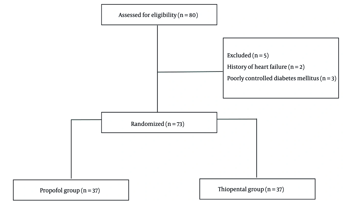
Figure 1.
Flow chart of study
Table 1 summarizes the baseline demographic and clinical characteristics of the study population. The mean age was 40.95 ± 8.55 years in the propofol group and 42.84 ± 8.69 years in the thiopental group, with no statistically significant between-group difference (P = 0.348). The sex distribution was also comparable: 18 males (48.6%) and 19 females (51.4%) in the propofol group versus 20 males (54.1%) and 17 females (45.9%) in the thiopental group (P = 0.816). Body mass index (BMI) was similar between the groups (24.74 ± 2.59 versus 23.29 ± 2.83 kg/m2, respectively; P = 0.479). ASA physical status classifications also did not differ significantly between the groups (P = 0.465). Overall, baseline demographic and clinical variables were well balanced between the treatment groups, confirming successful randomization.
Table 1.
Baseline Demographic and Clinical Characteristics a
| Variables | Propofol (n = 37) | Thiopental (n = 37; pg/mL) | P value |
|---|---|---|---|
| Age (y) | 40.95 ± 8.55 | 42.84 ± 8.69 | 0.348 |
| Gender: Males | 18 (48.6) | 20 (54.1) | 0.816 |
| BMI (kg/m2) | 24.74 ± 2.59 | 23.29 ± 2.83 | 0.479 |
a Values are expressed as mean ± Sd or No. (%).
In the propofol group, the mean TNF-α level increased from 7.09 ± 1.96 before surgery to 7.17 ± 5.67 after surgery; however, this change was not statistically significant (P = 0.815). In the thiopental group, TNF-α increased significantly from 8.16 ± 1.72 to 12.59 ± 5.43 (P < 0.001). Postoperative IL-6 levels also increased significantly in both groups, from 8.74 ± 3.42 to 11.74 ± 6.75 in the propofol group (P = 0.004) and from 7.64 ± 1.67 to 13.85 ± 5.10 in the thiopental group (P < 0.001).
Comparison of changes in inflammatory markers between the groups showed that the increase in TNF-α was significantly smaller in the propofol group than in the thiopental group. The increase in IL-6 was also lower in the propofol group (Table 2).
Table 2.
Changes in TNF-Α and IL-6 Before and After Surgery in the Propofol and Thiopental Groups a
| Groups and Markers | Preoperative (pg/mL) | Postoperative (pg/mL) | Δ (mean ± SD; pg/mL) |
|---|---|---|---|
| Propofol (n = 37) | |||
| TNF-α (pg/mL) | 7.09 ± 1.96 | 7.17 ± 5.67 | 0.08 ± 4.94 |
| IL-6 (pg/mL) | 8.74 ± 3.42 | 11.74 ± 6.75 | 3.00 ± 5.65 |
| Thiopental (n = 37) | |||
| TNF-α (pg/mL) | 8.16 ± 1.72 | 12.59 ± 5.43 | 4.44 ± 5.00 |
| IL-6 (pg/mL) | 7.64 ± 1.67 | 13.85 ± 5.10 | 6.21 ± 5.46 |
a Values are expressed as mean ± SD. IL-6, interleukin-6.
Direct comparison of postoperative values showed a statistically significant between-group difference in TNF-α levels, whereas the difference in postoperative IL-6 levels was not statistically significant. However, comparison of changes from baseline showed that the increases in both TNF-α and IL-6 were significantly smaller in the propofol group than in the thiopental group (Table 3).
Table 3.
Between-Group Comparison of Postoperative TNF-Α and IL-6 Levels and Changes in the Propofol and Thiopental Groups a
| Marker | Propofol (n = 37; pg/mL) | Thiopental (n = 37) | Δ mean (pg/mL) | 95% CI (pg/mL) | Statistical test | P-Value | Cohen’s d |
|---|---|---|---|---|---|---|---|
| Δ TNF-α (pg/mL) | 0.08 ± 4.94 | 4.44 ± 5.00 | -4.36 | [-6.66, -2.05] | Mann–Whitney U | 0.0006 | -0.876 |
| Postoperative TNF-α (pg/mL) | 7.17 ± 5.67 | 12.59 ± 5.43 | -5.42 | [-8.00, -2.85] | Mann–Whitney U | 0.0003 | -0.978 |
| Preoperative TNF-α (pg/mL) | 7.09 ± 1.96 | 8.16 ± 1.72 | -1.07 | [-1.92, -0.21] | Welch t-test | 0.0152 | -0.578 |
| Δ IL-6 (pg/mL) | 3.00 ± 5.65 | 6.21 ± 5.46 | -3.21 | [-5.18, -0.03] | Welch t-test | 0.0472 | -0.469 |
| Postoperative IL-6 (pg/mL) | 11.74 ± 6.75 | 13.85 ± 5.10 | -2.11 | [-4.89, 0.66] | Welch t-test | 0.1335 | -0.353 |
| Preoperative IL-6 (pg/mL) | 8.74 ± 3.42 | 7.64 ± 1.67 | 1.09 | [-0.16, 2.35] | Mann–Whitney U | 0.0443 | 0.407 |
a Values are expressed as mean ± Sd unless otherwise indicated. Abbreviation: IL-6, interleukin-6.
After adjustment for baseline values using ANCOVA, the between-group difference in postoperative TNF-α levels remained statistically significant, with adjusted means of 7.91 in the propofol group and 11.85 in the thiopental group. Although the unadjusted comparison of postoperative IL-6 levels did not show a statistically significant difference, adjustment for baseline values revealed a significant between-group difference, with adjusted means of 11.35 in the propofol group and 14.23 in the thiopental group (Table 4).
Table 4.
Analysis of Covariance of Postoperative TNF-Α and IL-6 Levels Adjusted for Baseline Values
| Marker | Propofol adjusted mean (pg/mL) | Thiopental adjusted mean (pg/mL) | 95% CI (pg/mL) | T | P-Value | η2p |
|---|---|---|---|---|---|---|
| TNF-α (pg/mL) | 7.91 | 11.85 | [-6.33, -1.55] | -3.283 | 0.0016 | 0.132 |
| IL-6 (pg/mL) | 11.35 | 14.23 | [-5.59, -0.17] | -2.122 | 0.0373 | 0.060 |
Abbreviation: IL-6, interleukin-6.
SBP, DBP, and heart rate (HR) values were significantly lower in the propofol group than in the thiopental group (Table 5).
Table 5.
Comparison of Hemodynamic Parameters Between the Propofol and Thiopental Groups a
| Variables | Propofol (mmHg or beats/min) | Thiopental (mmHg or beats/min) | Mean Difference (mmHg or beats/min) | P value |
|---|---|---|---|---|
| SBP (mmHg) | 116.81 ± 8.42 | 122.97 ± 11.49 | − 6.16 (− 10.83 to − 1.49) | 0.010 |
| DBP (mmHg) | 70.03 ± 4.80 | 76.27 ± 7.13 | − 6.24 (− 9.06 to −3.42) | < 0.001 |
| HR (beats/min) | 70.68 ± 6.80 | 79.81 ± 10.14 | − 9.13 (− 13.13 to − 5.13) | < 0.001 |
a Values are expressed as mean ± SD or 95% CI. Abbreviations: SBP, systolic blood pressure; DBP, diastolic blood pressure.
5. Discussion
Surgical injury and the subsequent inflammatory response are influenced by factors that may exacerbate or attenuate tissue injury and thereby affect surgical outcomes. One important factor is the choice of anesthetic agents (13). These agents may modulate the inflammatory response by altering the activity and distribution of immune cell populations, including natural killer cells, B lymphocytes, T lymphocytes, and macrophages. These effects may influence cytokine secretion and inflammatory marker levels, including IL-6, thereby modifying the magnitude of the inflammatory response (14, 15). Because perioperative brain injury may cause substantial morbidity, including new neurological deficits and postoperative cognitive dysfunction, identifying effective strategies to reduce its clinical impact is important (16).
Historically, sodium thiopental and propofol have been widely used for neurosurgical procedures because they reduce cerebral metabolic activity and intracranial pressure. Previous studies have demonstrated the effects of propofol on different immune cells and proposed potential anti-inflammatory mechanisms (17). In a 2020 systematic review, Alhayyan et al. reported reductions in IL-6 and C-reactive protein levels following anesthesia with propofol. However, no specific anesthetic technique was identified as superior for reducing inflammatory markers (18). Early experimental work by Goldstein et al. demonstrated increased tolerance to cerebral anoxia with pentobarbital in dogs (19). Thiopental also exhibits anti-inflammatory effects, and its inhibitory effects on the nonspecific immune system have been documented in several studies (20).
Few studies have directly compared the anti-inflammatory effects of propofol and thiopental. We therefore compared these agents by measuring inflammatory markers following neurosurgical procedures to evaluate their potential to attenuate the postoperative inflammatory response. Our findings suggest that propofol induction was associated with lower postoperative inflammatory marker levels, including IL-6 and TNF-α, than thiopental induction.
Regarding the clinical outcomes of anesthesia, Bilotta et al., in a 2013 qualitative review of randomized clinical trials, found no significant difference between thiopental and propofol in terms of POCD (16). Providing indirect evidence relevant to our study, Safari et al. (21) investigated the effect of anesthetic technique on postoperative inflammation following craniotomy by evaluating inflammatory markers comparable to those assessed here. Their patients received either intravenous propofol or inhaled isoflurane for maintenance of anesthesia. In addition to IL-6 and TNF-α, they evaluated interleukin-1. Inflammatory marker levels were lower in the propofol group. In 2017, Berger et al. compared propofol and isoflurane for maintenance of anesthesia during intracranial surgery. Unlike our study, they examined cerebrospinal fluid samples and measured several inflammatory markers, including IL-6 and TNF-α. They found no between-group differences in cytokine levels at baseline or 24 hours after surgery (12).
In a study similar to ours, Tajadiny et al. compared the effects of thiopental and propofol on inflammatory responses during electroconvulsive therapy in patients with major depressive disorder (22). They assessed IL-23 and IL-17 levels and found no statistically significant between-group differences. A retrospective analysis by Kim et al. (23) reported a lower incidence of neurological complications following thiopental-based general anesthesia in patients undergoing clipping of unruptured intracranial aneurysms.
Accurate monitoring of intraoperative hemodynamic parameters is crucial when evaluating the safety and efficacy of anesthetic interventions, as demonstrated in recent clinical trials examining pharmacological responses during surgery (24). As a secondary outcome, we investigated the effects of the two induction regimens on intraoperative hemodynamic variables. Patients receiving propofol induction had significantly lower intraoperative HR, SBP, and DBP than those receiving thiopental. Our findings are consistent with those of Aslan et al., who compared hemodynamic changes following rapid-sequence induction with propofol and thiopental (25). Kareem Aurooq et al. also reported that propofol produces a more rapid and pronounced hemodynamic effect, characterized by a greater reduction in blood pressure than thiopental (26). The lower hemodynamic values observed with propofol in our study may therefore reflect greater attenuation of sympathetic activation.
5.1. Study Limitations
This study has several limitations. First, it was conducted at a single center and included only patients undergoing intracranial surgery. Different intracranial pathologies may have introduced heterogeneity because the underlying pathology could influence inflammatory marker levels and responses. Larger studies focused on specific neurosurgical procedures and involving more homogeneous populations are therefore warranted. Second, inflammatory markers and changes in vital signs alone are insufficient to establish the superiority of one anesthetic agent over another in terms of clinical outcomes. Future studies should evaluate clinically relevant patient outcomes and compare postoperative outcomes among patients receiving different anesthetic agents. Third, inflammatory assessment was limited to serum IL-6 and TNF-α levels. Assessment of a broader range of inflammatory and other relevant biomarkers may provide a more comprehensive understanding of the effects of these agents and improve the generalizability of the findings. Fourth, hemodynamic adverse events, such as transient hypotension or administration of rescue vasoactive medications (e.g., ephedrine or phenylephrine), were not systematically recorded as predefined trial endpoints because the protocol focused on continuous hemodynamic monitoring. Future trials should include predefined categorical thresholds for hypotension and track vasopressor requirements.
Another limitation is the difficulty of isolating the specific anti-inflammatory effects of the induction agent from those of the broader intraoperative and perioperative anesthetic regimen. Although both groups received standardized maintenance anesthesia, other perioperative variables, such as surgical duration, intraoperative blood loss, opioid consumption, and fluid management, could independently modulate systemic inflammatory cytokine levels. The observed differences in IL-6 and TNF-α should therefore be interpreted cautiously as reflecting the overall clinical impact of the induction-inclusive protocol rather than an exclusive pharmacological property of propofol or thiopental. Both groups received propofol for maintenance; therefore, the comparison concerns the induction agents within a shared maintenance regimen. Accordingly, the conclusions should be interpreted as comparing propofol and thiopental induction rather than entirely separate propofol- and thiopental-based anesthetic regimens. Future studies comparing balanced general anesthesia with total intravenous anesthesia using alternative induction agents are warranted to further delineate these effects.
5.2. Conclusions
Propofol induction in patients undergoing intracranial surgery was associated with lower early postoperative levels of IL-6 and TNF-α than thiopental induction. Patients receiving propofol also had lower intraoperative HR, SBP, and DBP. Because both groups received propofol for maintenance, these findings concern the induction agents within a shared maintenance regimen rather than a comparison of complete anesthetic regimens. Reduced inflammatory marker levels alone should not be interpreted as evidence of the superiority of propofol as an anesthetic agent in neurosurgical patients. Further studies evaluating clinically relevant patient outcomes and comparing different anesthetic regimens are warranted.