
1. Introduction
Influenza is a contagious respiratory disease caused by influenza A, B, C, and D viruses in humans. Symptoms range from mild illness to severe pneumonia (1-4). Avian influenza A virus comprises 16 unique hemagglutinin (HA) and nine unique neuraminidase (NA) serotypes. The most common seasonal influenza A strains in the human population are H1N1 and H3N2 (1, 3). Seasonal influenza spreads primarily through respiratory droplets among people in close contact. Vaccines and antiviral medications are critical for preventing and treating influenza virus infections (1, 4). Influenza vaccination is the main preventive strategy, with an effectiveness of approximately 70 - 90% in people younger than 60 years and 17 - 53% in those aged 60 years or older. Drug-resistant viruses pose a substantial challenge to antiviral treatment. Innovative strategies, including increased support for research on the microbiome’s role in shaping host immunity to protect high-risk populations, could provide substantial public health benefits (5).
Beyond digestion and metabolism, the microbiome plays an important protective role against pathogenic microbes. Understanding microbiome dynamics may provide insights into maintaining homeostasis and preventing or treating viral infections (6). The microbiome has been shown to influence the immune system during viral infections (7). For a long time, the lungs were believed to be free of microbes; however, new diagnostic methods have revealed a microbial community in the lungs. This community consists mainly of bacteria, fungi, and viruses and is primarily derived from the oropharynx, which is part of the upper respiratory tract (8-10). Transient dysbiosis caused by acute infection suggests that, although microbial communities are generally stable, specific ecological niches may lose protective microbiota and become susceptible to bacterial invasion (11).
For example, research suggests that patients with coronavirus disease 2019 (COVID-19) who have a higher lung microbial load are less likely to recover from invasive mechanical ventilation and have an increased risk of mortality (8). The interaction between the microbiome and influenza is complex and bidirectional. Multiple factors, including the immune response and microbiota, influence the severity of influenza-related respiratory illness (2, 12). Although viral components have been extensively studied, recent research highlights the important roles of the microbiota and coinfections in severe and fatal viral infections (2, 13). Microbiome imbalance and altered immune responses can affect susceptibility to and severity of influenza infection (2, 13). One study found that antibiotic-induced disruption of the gut microbiome can alter the immune response to influenza vaccination (12). Conversely, influenza virus infection may alter the structure of the respiratory bacterial community, thereby increasing the risk of secondary bacterial pneumonia and otitis media (2).
Therefore, understanding the relationship between influenza and the respiratory tract microbiome (RTM) is crucial for developing strategies to reduce the effects of influenza on the host. However, the association between the microbiota and viral infections is complex and requires further study.
This systematic review aimed to gather comprehensive scientific evidence on the association between influenza and alterations in the lower respiratory tract and oropharyngeal microbiomes.
2. Methods
2.1. Search Strategy and Eligibility Criteria
We searched PubMed, Web of Science, Scopus, and Embase (Excerpta Medica Database) for studies published from January 2010 to January 2024 that compared the lung or lower respiratory tract (LRT) microbiome of patients with influenza with that of healthy controls. The search was limited to full-text, English-language manuscripts. Initially, only English-language abstracts were considered. Medical Subject Headings (MeSH) search terms included "Microbiota" OR "Microbiotas" OR "Microbiome" AND "lung" AND "Influenza" OR "Flu."
2.2. Inclusion and Exclusion Criteria
Two independent reviewers (E. I. and H. A.) screened titles and abstracts according to the inclusion and exclusion criteria. Eligible studies assessed variations in the lower respiratory tract microbiome in patients with influenza; collected respiratory tract samples from the lung, endotracheal aspirates, bronchoalveolar lavage (BAL), or oropharyngeal swabs; included human or animal studies; and were published in English.
This review also included studies of the oropharyngeal microbiome when BAL and oropharyngeal swabs were collected simultaneously, because the oropharyngeal microbiome has been reported to serve as a reservoir for the lower respiratory tract and lung microbiome. In addition, studies identified from the reference lists of included articles, such as references (14-16), were considered. Studies were excluded if they focused on upper respiratory tract microbiome data or were conference proceedings, letters to the editor, or reviews.
2.3. Data Extraction
Extracted data included descriptive variables such as the study, author, year of publication, study objective, country of enrollment, sequencing method, bacteria identified in the lungs, number of patients with influenza, number of healthy controls, and age of influenza-infected patients. Following full-text review, data extracted from eligible studies were checked for completeness and accuracy.
2.4. Risk of Bias and Quality Assessment
The quality and risk of bias of the included studies were assessed using the Critical Appraisal Skills Programme (CASP) checklist to provide an overall appraisal of methodological rigor across study designs. The CASP checklist includes 10 questions for each case-control study. We developed a quality-assessment chart using a traffic-light system to indicate "good," "adequate," or "poor," as recommended by Cochrane. Disagreements were resolved by consensus among the authors.
3. Results
The search strategy yielded 382 articles, of which 159 were duplicates. We documented the search, extraction, and exclusion decisions (Figure 1). All 227 articles were screened by title and abstract. Seventy-four articles met the inclusion criteria. After abstract review, 63 papers were excluded based on the PICOS criteria. In addition, two articles were excluded during the final screening and full-text review because they addressed only the upper respiratory tract microbiome. Finally, nine articles that examined lower respiratory tract and/or oropharyngeal microbiome changes, their effects on lower respiratory tract infections, or lung infections associated with microbiome changes met the PICOS criteria and were included in this review.
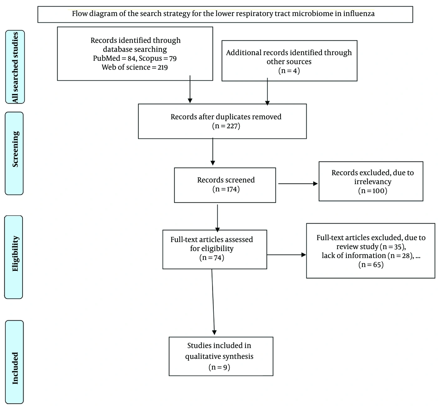
Figure 1.
Flow diagram of the search strategy for the lower respiratory tract microbiome in influenza.
3.1. Main Study Characteristics
Table 1 summarizes changes in the relative abundance of lower respiratory tract (LRT) bacterial communities in patients with influenza across multiple studies. Two clinical studies conducted in Mexico City and China in 2023 analyzed the BAL microbiome (17, 18). One study examined patients hospitalized in Mexico City with influenza A H1N1pdm09 (17). The study conducted in China included patients aged 18 years or older with severe pneumonia and compared those who tested positive for influenza virus (influenza virus-positive [IFVP]) with those who tested negative (influenza virus-negative [IFVN]) (18). A case report described a 61-year-old woman with H7N9 infection who received treatment; only the lung microbiome data were included in this review (19). The clinical studies collected BAL samples for analysis of the lung and lower respiratory tract microbiomes (17-19). DNA was extracted using the QIAmp Cador Pathogen Mini Kit (Qiagen N.V., Hilden, Germany) and the DNeasy PowerSoil Kit (Qiagen, Hilden, Germany) (17, 18).
Table 1.
Some Characteristics of the Analyzed Studies a
| Author (Year) and Reference | Study Title (Study Design) | Country | Age Category | Sex | Participants/Antibiotic Use | Methods |
|---|---|---|---|---|---|---|
| Hu et al. (2016) (19) | A case report demonstrating the utility of next-generation sequencing in analysing serial samples from the lung following an infection with influenza A (H7N9) virus | China | 61 years | Female | H7N9 infection; treated with extended-spectrum antibiotic therapy | Next-generation sequencing |
| Yildiz et al. (2018) (11) | Influenza A virus infection impacts systemic microbiota dynamics and causes quantitative enteric dysbiosis (experimental animal study, in vivo) | Switzerland | 8 - 9 weeks | Female | Mice; data not shown | 16S rRNA gene-specific NGS, qPCR, and bacterial culture |
| Zhou et al. (2023) (18) | Impact of influenza virus infection (IFV) on lung microbiome in adults with severe pneumonia (experimental human study, in vivo) | China | IFVP and IFVN; data not shown | Nucleic acid extraction and PCR | ||
| Ramos-Sevillano et al. (2019) (24) | The effect of the influenza virus on the human oropharyngeal microbiome (randomized controlled human study) | United Kingdom | 52 with influenza A/Wisconsin/67/2005 (H3N2); healthy controls; data not shown | 16S rRNA sequencing | ||
| Chen et al. (2023) (21) | Topography of the respiratory tract and gut microbiota in mice with influenza A virus (IAV) infection (controlled experimental animal study, in vivo) | China | 6 weeks | C57BL/6 female mice | (n = 10): PBS mock-infected (Mock) and IAV groups | |
| Gu et al. (2019) (22) | Dynamic Changes in the Microbiome and Mucosal Immune Microenvironment of the Lower Respiratory Tract by Influenza Virus Infection (controlled experimental animal study, in vivo) | China | 8 weeks | Female | C57BL/6 mice, SPF; data not shown | Next-generation sequencing |
| Hernández-Terán et al. (2023) (17) | Microbiota composition in the lower respiratory tract is associated with severity in patients with acute respiratory distress caused by influenza (observational hospital-based cohort study) | Mexico City | 45.5 (36.5 - 51) years | 63.3% male; 36.6% female | Deceased and recovered influenza A patients; 76.6% used antibiotics | |
| Li-juan et al. (2022) (16) | Klebsiella pneumoniae infection following H9N2 influenza A virus infection contributes to pneumonia development in mice (experimental animal study, in vivo) | China | 6 weeks | Kunming (KM) mice; data not shown | ||
| Lu et al. (2017) (15) | Disordered oropharyngeal microbial communities in H7N9 patients with or without secondary bacterial lung infection (observational case-control study) | China | 60.5 years | Male = 12; female = 9 | 51 H7N9 patients b and 30 healthy controls; no antibiotic use |
a Abbreviations: SPF, specific-pathogen-free; NGS, next-generation sequencing; IFVP, influenza virus-positive group; IFVN, influenza virus-negative group; IAV, influenza A virus-infected.
b Patients: n = 21 with secondary bacterial lung infections and n = 30 without secondary bacterial lung infections.
Specific microbiota were detected using 16S rRNA gene-specific next-generation sequencing, qPCR, and bacterial culture of organ contents (11, 19). The V3-V4 region of 16S rRNA was amplified by PCR using specific primers, and raw Illumina sequences were analyzed using QIIME2 (Quantitative Insights into Microbial Ecology, version 2020.8). Bronchoalveolar lavage fluid was used for 16S rDNA sequencing. Contaminating reads in raw paired-end sequence data were removed using Trimmomatic (version 0.35).
In a case-report study, total RNA was extracted from BAL samples using the Qiagen RNeasy Mini Kit. Double-stranded cDNA was generated using the SuperScript Double-Stranded cDNA Synthesis Kit (Invitrogen) for library preparation and analyzed using next-generation sequencing and bioinformatics (19). 16S rRNA gene sequencing and next-generation sequencing (NGS) were used in animal-model experiments (11, 14, 16, 21, 22). Four studies used specific-pathogen-free (SPF) C57BL/6J mouse models with animals aged 6 - 9 weeks (11, 14, 16, 21, 22).
3.2. Microbiome Characteristics
The review identified six phyla, 11 genera, and five bacterial species associated with respiratory tract infections in patients with influenza. Six microbiome datasets from humans and mice were used for extraction and analysis. Another study identified diverse microbial communities in the lower respiratory tract of healthy individuals. The dominant phyla were Firmicutes, Proteobacteria, Actinobacteria, and Bacteroidetes, and the identified genera included Prevotella, Veillonella, Streptococcus, Fusobacterium, and Haemophilus, as determined by NGS (23).
Table 2 summarizes differences in microbial composition at the phylum, family, genus, and species levels. In the study by Hernández-Terán et al. (17), which compared recovered and deceased patients with influenza A, the respiratory microbiota of deceased patients were composed primarily of Proteobacteria, Bacteroidetes, and Firmicutes, with Firmicutes as the dominant phylum and Staphylococcus and Granulicatella as the dominant genera. Recovered patients had a distinct community composition, with Veillonella, Streptococcus, and Rothia as the dominant genera. Overall, microbial diversity was lower in deceased patients than in recovered patients (17).
Table 2.
Phylum-, Family-, Genus-, and Species-Level Differences in Microbial Composition of the Lower Respiratory Tract in Influenza-Positive Versus Influenza-Negative Subjects in Human and Animal Studies a
| First Author (Year) and Reference | Variable Abundance | Phylum/Class | Family | Genus or Species |
|---|---|---|---|---|
| Zhou et al. (2023) (18) | Dominant microbiome in the IFVP group | Gaiella; Rhodoplanes; Staphylococcus; Haemophilus; Pseudomonas; Acinetobacter (LEfSe analysis) | ||
| Zhou et al. (2023) (18) | Dominant microbiome in the IFVN group | Streptococcus; Klebsiella; Escherichia-Shigella; Corynebacterium-1; Burkholderia-Caballeronia-Paraburkholderia | ||
| Lu et al. (2017) (15) | Increased in H7N9 vs healthy groups | Fusobacteria (P < 0.005) | Bifidobacteriaceae; Pseudomonadaceae; Bacteroidaceae (P = 0.003) | |
| Lu et al. (2017) (15) | Decreased in H7N9 vs healthy groups | Gammaproteobacteria, except Pseudomonas (P < 0.005) | ||
| Yildiz et al. (2018) (11) | Decreased | Alpha- and beta-proteobacteria | Clostridia | |
| Chen et al. (2023) (21) | Increased in lung | Proteobacteria | Enterococcus faecalis; Streptococcus danieliae; Streptococcus respiraculi; Streptococcus suis | |
| Chen et al. (2023) (21) | Decreased | Firmicutes | Lactobacillus animalis; Lactobacillus johnsonii; Lactobacillus murinus | |
| Gu et al. (2019) (22) | Increased in the acute phase | Gammaproteobacteria (ANOSIM, R > 0, P < 0.05) | Streptococcus; Staphylococcus; Pseudomonas; Acinetobacter (ANOSIM, R > 0, P < 0.05) | |
| Gu et al. (2019) (22) | Decreased in the acute phase | Alphaproteobacteria (ANOSIM, R > 0, P < 0.05) | Sphingomonas (ANOSIM, R > 0, P < 0.05) |
a Abbreviations: IFVP, influenza virus-positive patients; IFVN, influenza virus-negative patients; ANOSIM, analysis of similarities.
3.3. Influenza
The included studies examined confirmed cases in patients or animals with influenza subtypes H1N1 and H7N9 and compared them with healthy controls (11, 15-18, 21, 22, 24). Influenza A virus (IAV) can be identified by viral culture in embryonated eggs or mammalian cells followed by hemagglutination (HA) testing (11, 16). Real-time RT-PCR was used to detect laboratory-confirmed influenza in most studies, and one study defined infection as a ≥ 4-fold rise in hemagglutination inhibition (HAI) or microneutralization titers between day −2 and day 28 (15, 18, 19, 21, 22, 24).
The studies compared the relative abundance of each phylum in patients with influenza with that in healthy controls. Studies reported decreased proportions of Firmicutes and increased proportions of Proteobacteria and other bacterial phyla in patients with influenza compared with healthy controls (21). In human studies, patients with influenza showed increased levels of opportunistic pathogens such as Acinetobacter and Staphylococcus and decreased levels of Veillonella, Streptococcus, Prevotella, and Rothia compared with healthy individuals (18). In mouse studies, influenza infection was associated with increased levels of Enterococcus, Staphylococcus, Acinetobacter, Delftia, and Rubrobacter (21, 22).
Microbial community diversity was assessed using the Shannon Diversity Index and Chao1, and the data were analyzed using principal component analysis (PCA) and principal coordinate analysis (PCoA) to assess similarities between groups. One human study found that recovered patients had significantly greater microbial richness (Chao1 index, P = 0.01) and diversity (Shannon index, P = 0.004) than deceased patients (17). Conversely, another study reported slightly higher Shannon and Simpson indexes in influenza-infected patients, although the differences were not statistically significant. The PCA plot showed distinct clustering that differentiated healthy and H1N1-infected animals (18).
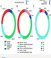
Because of the difficulty of sampling the lower respiratory tract, most studies have focused on the upper respiratory tract. In this review, two original studies and one case report were used to evaluate changes in the lower respiratory tract microbiome in patients with influenza (17-19). The case report involved serial collection of BAL fluid samples from a 61-year-old woman with H7N9 after illness onset. The microbiome was analyzed using next-generation sequencing and microbiological methods in China (19). Bacteria were substantially more abundant than viruses and fungi, accounting, on average, for 85% of detected organisms compared with 0.6% viruses and 0.1% fungi. Proteobacteria dominated the microbiome, representing at least 94% of the total bacterial community. After H7N9 was suppressed and testing became negative, A. baumannii, a gram-negative bacterium, was detected in the lungs. Its relative abundance increased as H7N9 declined, suggesting a potential secondary bacterial infection after viral invasion (19). Four animal studies were also included.
3.4. Handling of Antibiotic Exposure
Antibiotic exposure was handled differently across studies. Some studies excluded participants with recent antibiotic use, others evaluated the effects of prior antibiotic treatment, one case study followed microbiome alterations during consecutive antibiotic regimens, and experimental models largely avoided antibiotic administration except as a control intervention. Sampling strategies also varied and included longitudinal sampling across the disease course, early-hospitalization sampling, and fixed postinfection time points, allowing researchers to examine microbiome changes associated with influenza infection (11, 15, 17-19). No information on antibiotic use was provided in the other studies (16, 21, 22, 24).
4. Discussion
This systematic review analyzed nine clinical and animal studies examining changes in the lower respiratory tract (LRT) microbiome during influenza infection. Mouse model studies provide both a foundation for and a complement to human research by enabling direct access to the LRT microbiota. The findings highlight the important role of the LRT microbiome in lung immunity and morphology and its potential influence on the progression of respiratory infections.
Mouse and human microbiomes differ substantially with respect to host physiology and immune function, diet and environment, microbial composition, and relative abundance. Comparative analyses of human and animal studies can provide mechanistic insights, but their limitations should be carefully considered. The reviewed studies used full-length 16S rRNA gene sequencing and next-generation sequencing to characterize microbial communities in healthy or recovered individuals and in those infected with influenza A virus (IAV) (11, 14, 15-19, 21, 22, 24-26). These analyses indicate that IAV infection can substantially alter the composition and diversity of the LRT microbiome (15, 16, 17, 19, 22, 26), although some studies reported transient or nonsignificant changes (11, 18, 24), possibly because of differences in virus subtype and in methods of sample collection and microbiome detection.
In healthy human subjects, the LRT harbors a diverse microbial community predominantly composed of Prevotella, Veillonella, Streptococcus, Fusobacterium, and Haemophilus (18). In influenza, human studies have reported specific changes, including increased diversity within Fusobacteria, Bifidobacteriaceae, Pseudomonadaceae, and Bacteroidaceae in infected patients (15). Similarly, the oropharyngeal microbiomes of H7N9 patients, especially those with secondary bacterial infections, showed higher abundances of bacterial families, including Bacteroidaceae, Fusobacteriaceae, Leptotrichiaceae, and Pseudomonadaceae, than those of healthy controls. These findings support the hypothesis that the oropharynx acts as a reservoir for lung pathogens in humans (15).
In addition, avian influenza H7N9 has been reported to increase bacterial adherence to oropharyngeal epithelial cells, potentially increasing the risk of secondary bacterial lung infections. Previous research has also linked greater oropharyngeal microbiome diversity with infection (15). Across several studies, infected patients consistently showed increased relative abundance of potentially pathogenic bacteria. In particular, Acinetobacter baumannii and Staphylococcus aureus were more abundant in these patients (17-19), and patients with severe influenza-associated pneumonia often showed this enrichment.
The relationship between the LRT microbiome and influenza virus in humans is also influenced by host immunity and antibiotic use. Other studies have reported lower microbiome diversity and distinct changes after antibiotic treatment, as well as frequent coinfections with Staphylococcus aureus, Pseudomonas aeruginosa, and Acinetobacter baumannii in influenza-infected patients. Antibiotic pretreatment before hospitalization reduced microbial richness and altered respiratory microbiome composition, increasing Actinobacteria and pathogenic genera such as Rothia and Enterococcus and producing substantial structural differences between treated and untreated groups (17, 18).
Mouse models provide detailed insights into LRT-specific changes and mechanistic pathways. The healthy murine LRT microbiome differs from that of humans. In one study, 9-week-old mice showed dominance by Sphingomonas, Halomonas, Shewanella, and Acinetobacter, with Sphingomonas comprising up to 80% of the population (22). Following IAV infection, mouse studies reported a higher prevalence of classes such as Gammaproteobacteria, including the families Enterobacteriaceae, Vibrionaceae, and Pseudomonadaceae, and genera such as Halomonas and Shewanella (22).
Infection also depletes immune cells, such as alveolar macrophages, thereby impairing microbial regulation in the LRT (25). Thus, disruption of the LRT microbiome may be attributable to several factors. Influenza virus infection depletes the mouse host immune system (25). For example, decreased alveolar macrophage numbers weaken the host’s ability to regulate the LRT microbiome in mouse models. In addition, metabolomic analysis in a mouse model showed that the LRT microenvironment changed 28 days after infection, contributing to a substantial imbalance in the LRT microbiome (22). A human study found that avian influenza H7N9 could increase bacterial adherence to oropharyngeal epithelial cells, thereby increasing susceptibility to secondary bacterial lung infections (15). However, some studies have shown no significant changes in microbial diversity or richness in the upper respiratory tract or LRT of mice infected with IAV (11, 21).
Immune responses mediated by lipopolysaccharide activation of Toll-like receptor 4 in innate immune cells can inhibit influenza virus infection, whereas signaling from commensal bacteria also facilitates an effective innate immune response to viral invasion (15, 26, 27). Previous studies have shown that higher levels of Th1 cytokines, such as IP-10, IL-6, IL-8, and MCP-1, are associated with disease severity in human H7N9 infection (15, 26, 27). Other studies found that higher cytokine levels in recovered patients correlated with different stages of infection. The initial cytokine storm during the acute phase, involving IP-10, MIG, MCP-1, and MCP-2, subsided as viral load declined. However, SDF-1, BCA-1, and RANTES increased substantially because of secondary bacterial infections (19).
Several studies reported changes in microbial composition and diversity during influenza A virus infection. However, the directionality of this relationship remains unclear. Most human studies are observational and cross-sectional, making it difficult to determine whether preexisting microbiome dysbiosis increases susceptibility to influenza or whether viral infection drives microbial changes. The relationship may be bidirectional. Influenza infection can disrupt epithelial barriers, impair innate immune defenses, and alter the lung microenvironment, thereby promoting microbial imbalance. Baseline microbial composition may influence antiviral responses and susceptibility to secondary bacterial infections. Human studies with preinfection sampling are needed to clarify the temporal relationship.
A major limitation of this review is the limited understanding of the human lower respiratory tract microbiome. Mouse and human microbiomes are biologically distinct and cannot be directly assessed together without risking overinterpretation. Although there was considerable heterogeneity in specimen types, patient characteristics, sequencing methods, and sampling timelines across the included studies, we addressed this by conducting a qualitative narrative synthesis with subgroup analyses rather than a formal meta-analysis to avoid overinterpreting pooled estimates.
4.1. Conclusions
Primary evidence from a limited number of human observational studies and supporting animal models suggests that influenza A infection may promote lower respiratory tract dysbiosis, with reduced diversity and increased opportunistic pathogens correlating with severe disease and secondary infections. Antibiotics and host immunity may further affect these microbial dynamics, but the observational evidence and methodological diversity across studies preclude strong mechanistic or biomarker claims. These exploratory findings require prospective, controlled validation to determine their clinical relevance.