
1. Background
Cervical cancer is the fourth most common cancer and the fourth leading cause of cancer-related death among women worldwide, accounting for approximately 7 - 8% of all female cancer deaths. More than 85% of these deaths occur in low-income and middle-income countries, whereas developed countries have screening programs that detect most precancerous lesions at various stages (1). The primary known environmental cause of this cancer is human papillomavirus (HPV), identified by Zur Hausen in 1976 as the principal cause of cervical cancer (2). Several studies have demonstrated a significant association between HPV infection and the development of precancerous and cancerous changes in epithelial cells (2). HPV is the most prevalent viral infection of the genital tract, affecting young and sexually active individuals equally (3, 4). Women with this infection and a normal immune system typically take 15 to 20 years to develop cervical cancer, whereas those with weakened immune systems, such as those with untreated HIV infection, may develop it in only 5 to 10 years (5). Additionally, environmental risk factors for HPV persistence and cervical cancer development include early sexual activity, frequent intercourse, multiple sexual partners, oral contraceptive use, smoking, and immunosuppressive medications (1).
The HPV family comprises more than 200 types of non-enveloped viruses with double-stranded DNA that infect cells of the skin, upper respiratory tract, and genital tract (6). Approximately 40 HPV genotypes are present in the genital tract and fall under the alpha papillomavirus group (7). The most prevalent high-risk HPV genotypes globally are HPV16 and HPV18 (8). Genital warts commonly occur on the penis, scrotum, vulva, perineum, and perineal skin. In men, HPV-16 is primarily linked to penile cancer, whereas HPV-6 and HPV-11 are commonly associated with genital warts (9).
Cervical cancer is increasing in developing nations because of inadequate screening and higher HPV prevalence (42.4% vs. 22.6% in developed countries) (10). Studies have reported high HPV infection rates in Iranian women with cervical cancer (77.4%) (11, 12). Although it is the 12th most common cancer among Iranian women, it remains a concern (13). Iranian men show 55.7% HPV prevalence (14). Screening and HPV genotyping in low-income countries without vaccination can improve public health and reduce spread (15).
Existing HPV diagnostic assays, such as PCR, immunohistochemistry (IHC), and Pap smears, have substantial limitations, particularly in resource-limited areas (16). PCR is costly, requires specialized equipment and personnel, and has long turnaround times, making it inaccessible in low-income countries where most cervical cancer deaths occur. Cytology and IHC are less sensitive and reproducible, are prone to subjective interpretation, and perform poorly with some samples, often missing low viral loads or leading to unnecessary follow-up. These limitations contribute to insufficient screening and delayed detection of high-risk HPV16 and HPV18, which cause most cervical cancers (16).
To improve the early detection and prevention of cervical cancer, there is a critical need for rapid, sensitive, specific, and portable diagnostic tools suitable for point-of-care use. Loop-mediated isothermal amplification combined with lateral flow dipstick (LAMP-LFD) assays offer a solution by providing isothermal amplification, high analytical sensitivity (comparable to PCR), and rapid visual results within 30 - 60 minutes (17). The dipstick format, which uses gold nanoparticles, ensures portability and user-friendly detection, even in low-resource settings. A recent review comprehensively discussed the pathogenesis, vaccination, and various detection methods for HPV16 and HPV18, with special emphasis on the advantages of LAMP-LFD assays (18).
2. Objectives
This study developed and evaluated a novel LAMP-LFD assay for HPV16 and HPV18, aiming to provide a cost-effective, sensitive, and specific tool to overcome current diagnostic limitations and support widespread screening (19). The assay was validated using clinical cervical specimens, including both HPV-positive and HPV-negative samples, as determined by real-time PCR, which served as the reference standard. This approach addresses key limitations of conventional molecular diagnostics, including the need for thermocyclers, long turnaround times, and extensive infrastructure, thereby making it suitable for resource-limited settings.
3. Methods
3.1. Patients and Sampling Method
Vaginal and cervical swab specimens were collected from women attending gynecology clinics who were suspected of HPV-16 and/or HPV-18 infection based on clinical signs, symptoms, or risk factors. Real-time PCR was used as the reference standard method for HPV detection and genotyping. Samples with adequate volume and acceptable DNA purity (A260/280 ratio between 1.8 - 2.0) were included in the study. All participants provided written informed consent for the use of their clinical specimens. The study was approved by the Ethics Committees of Shiraz University of Medical Sciences and Tabriz University of Medical Sciences (IR.TBZMED.REC.1401.022). Polyurethane or sterile nylon swabs with plastic shafts were used to minimize contamination risk and PCR inhibition. Samples were collected in sterile, clearly labeled screw-cap tubes at Sina Clinical Laboratory in the Alborz Central Laboratory. Cervical epithelial cells were obtained by inserting the swab into the cervical canal, and samples were immediately placed in appropriate transport containers. Inclusion in the study was based on clinical suspicion of HPV infection and adequate sample purity, not on previous PCR results. Exclusion criteria included sample contamination, insufficient volume, low-purity DNA (A260/280 ratio outside the acceptable range), lack of signed informed consent, or failure to meet quality control standards. These criteria ensured sample integrity and the reliability of subsequent molecular analyses.
3.2. HPV DNA Samples and Positive Control Plasmids
The study assessed the sensitivity, specificity, and efficiency of HPV genotype detection using positive controls (plasmids, qPCR-verified DNA). The synthetic plasmid was cloned and cultured. HPV positivity was defined by E6/E7 amplification (visible gel bands or positive qPCR), confirmed by size markers. The absence of amplification, with successful internal controls, indicated HPV negativity. Recombinant plasmid transfer involved centrifugation and incubation on LB plates. Colony screening on antibiotic media confirmed transformation, and results were evaluated by gel analysis.
3.3. HPV LAMP Test and Product Detection
LAMP reaction efficiency depends on the specificity of internal (FIP/BIP) and external (F3/B3) primers. Reactions were performed in a 1X final volume with 10X isothermal amplification Buffer II, MgSO4, dNTP mix, FIP/BIP and LF/LB primers, Bacillus stearothermophilus (BST) 2.0 DNA polymerase, 2 µL Calcein, 0.8 M DNA samples, 7.5% Betaine, 2.75 µL Dimethyl sulfoxide (DMSO), and nuclease-free water, using distilled water as a negative control. LAMP reactions were conducted at 60, 63, and 65 °C for 60 minutes in a buffer containing 10 mmol Tris-HCl, KCl, RP/FP HPV16, RP/RF HPV18, dNTP, MgCl2, and 1 µL DNA with Taq DNA polymerase. In positive reactions, LF and LB primers (conjugated to Fluorescein isothiocyanate (FITC) and biotin) incorporate both tags into amplicons, generating a visible test line. The lateral flow device (LFD) detection system includes a control line to validate functionality. Molecular LFDs use capillary action through a series of pads (sample, conjugate, detection, and absorbent) to provide visible results. The sample wicks to the conjugate pad, where antibody-labeled nanoparticles bind the target analyte (if present). This complex migrates to the detection pad, forming a colored test line (T-line) at immobilized antibodies. A control line (C-line) confirms sufficient sample flow, and the absorbent pad collects excess fluid. LFDs are rapid, inexpensive, and portable, with widespread use in medical diagnostics, food safety, and environmental monitoring. However, their lower sensitivity limits broader application. This study employed LAMP with HPV16 and HPV18 genomic DNA and specific primers to evaluate DNA purity, which was initially confirmed by gel electrophoresis.
3.4. Specificity and Sensitivity Testing
Sample authenticity was verified by sequencing at Macrogen Korea and comparing sequences using NCBI MEGABLAST. Plasmids were excluded from sequencing confirmation because their unique genotypes had been previously validated. Statistical analyses were performed using SPSS version 16. Diagnostic performance was evaluated by calculating sensitivity, specificity, positive predictive value (PPV), negative predictive value (NPV), overall accuracy, likelihood ratios (LR+ and LR−), and Cohen's kappa coefficient with corresponding 95% confidence intervals. Analytical sensitivity was evaluated using serial dilutions of plasmid and DNA samples, with a positive control in each assay to determine the detection limit. Test results were validated against PCR and gel electrophoresis (20, 21). Test accuracy, defined as the ratio of true positives and true negatives, reflects its ability to differentiate between patients and negative specimens (22).
3.5. Synthesis of Gold Nanoparticles and pH Determination
To synthesize gold nanoparticles (AuNPs), an aqueous chloroauric acid (HAuCl4) solution was heated to near boiling. Sodium citrate was vigorously stirred in to reduce and cap the nanoparticles, and the color change was observed (typically to ruby red). The pH was adjusted to 7.0 - 9.0 with 0.1 M NaOH or HCl and continuously monitored to control particle size and stability. After synthesis, the AuNPs were cooled and characterized. Consistent pH monitoring and documentation are crucial for reproducibility, quality control, and scalability. Optimal pH determination involves pH titrations and particle stability assessment via dynamic light scattering (DLS) or zeta potential measurements (23), aiming for enhanced stability and reduced size. This includes creating a pure antibody concentration gradient in ELISA and adjusting the pH along it with potassium carbonate.
3.6. Conjugation of Anti-FITC Antibodies with Gold Nanoparticles
Gold nanoparticles are suitable for biosensors because of their electronic, magneto-optical, and catalytic properties (24). Antibodies and gold nanoparticles were combined in solution for 20 minutes at room temperature and then centrifuged. The supernatant was replaced with PBS buffer, centrifuged again, and the resulting solution was dissolved in 2 mL of PBS buffer to prepare strips.
3.7. Removal of Unbound Antibodies from the Medium
Unbound antibodies were separated from the sediment by centrifugation. Alternatively, a molecular lateral flow dipstick (LFD) may remove unbound antibodies (25). In this method, gold nanoparticles bind to specific labeled probes, and unbound antibodies are absorbed by the pad, preventing interference with lateral flow. Conjugating anti-FITC antibodies to gold nanoparticles enhanced efficiency and sensitivity. Transmission electron microscopy (TEM), UV, and zeta potential analyses confirmed antibody binding to the gold nanoparticles.
3.8. Lateral Flow Dipstick Construction
To construct lateral flow dipsticks, several components were prepared: the sample pad, absorbent pad, conjugate pad, bulk pad, and test pad. All components except the absorbent pad were buffered and incubated overnight. The absorbent pad was then placed on the detection pad for the test and control lines (26). To improve sensitivity in molecular lateral flow tests, a mass pad is integrated between the conjugate and test pads. This enhances signal detection through various methods such as membrane dialysis, 2D/3D paper membranes, and fluid flow management shapes, although these can complicate fabrication and sample delivery (26). This study proposes using a mass pad in a gold nanoparticle-based molecular lateral flow assay to capture antibodies and labeled probes, thereby increasing detection sensitivity. Molecular lateral flow dipsticks for HPV16 and HPV18 detection were created using nitrocellulose paper, with gold nanoparticle-conjugated antibodies on both test and control strips. Twenty microliters of LAMP samples with varying concentrations of HPV16 and HPV18 DNA were applied. Ethical considerations regarding patient demographics or laboratory animals were not necessary because plasmid or laboratory samples were used.
3.9. Data Analysis and Descriptive Information
Statistical analyses were performed using SPSS version 16. Agreement between the LAMP-LFD assay and real-time PCR was assessed using Cohen's kappa coefficient, and diagnostic performance indices were calculated with 95% confidence intervals. Participant characteristics, including age, height, weight, and BMI, are presented in Table 1.
Table 1.
The Demographic Information
| Variables | Woman (n = 29) | Man (n = 21) | Total (n = 50) |
|---|---|---|---|
| Age (y) | 39.59 ± 5.38 | 37.62 ± 4.54 | 38.66 ± 5.07 |
| Height (cm) | 162.31 ± 3.55 | 174.36 ± 5.54 | 167.62 ± 7.71 |
| Weight (kg) | 58.17 ± 8.36 | 68.90 ± 9.22 | 62.68 ± 10.17 |
| BMI | 22.09 ± 3.19 | 22.46 ± 2.31 | 22.24 ± 2.84 |
The performance of the developed LAMP-LFD assay was evaluated using 50 clinical cervical specimens collected from women suspected of HPV16/18 infection. Real-time PCR was used as the reference (gold standard) method for HPV detection and genotyping. As shown in Table 2, among the 30 specimens tested for HPV16, real-time PCR identified 17 positive and 13 negative cases. The LAMP-LFD assay detected 19 positives, resulting in two false-positive results and no false negatives in this group. For the 20 specimens tested for HPV18, both methods showed complete agreement, identifying 3 positive and 17 negative cases with no false-positive or false-negative results. Overall, real-time PCR identified 20 positive specimens, while the LAMP-LFD assay identified 22 positives across all 50 specimens. The demographic characteristics of the study participants are summarized in Table 1. The purity of extracted DNA (A260/280 ratio) was consistently within the acceptable range of 1.8 - 2.0 for all analyzed samples.
Table 2.
Descriptive Information Related to Variables Including False Negatives, False Positives, Number of Negatives by LAMP Assay, Number of Negatives by RT-PCR Assay, Number of Positives by LAMP Assay, Number of Positives by RT-PCR Assay
| Variables | False negatives | False positives | LAMP-LFD Negative | RT-PCR negatives | LAMP-LFD positives | RT-PCR positives |
|---|---|---|---|---|---|---|
| Genotype 16 (30 people) | 0 | 2 | 11 | 13 | 19 | 17 |
| Genotype 18 (20 people) | 0 | 0 | 17 | 17 | 3 | 3 |
| Total (50 people) | 0 | 2 | 28 | 30 | 22 | 20 |
4. Results
4.1. Genomic DNA and Plasmid-Positive Samples
All extracted DNA samples met the predefined purity criteria and were included in subsequent analyses. Viral DNA purity (A260/280 ratio) consistently fell within the acceptable range of 1.8 - 2. Positive control plasmids containing the complete HPV16 and HPV18 genomes were validated by PCR using specific and universal (MY09/11) primers targeting the E7 gene regions (Figures 1-3 in Supplementary File). Separate primers for the HPV16 and HPV18 E6 and E7 gene regions were designed using Primer Explorer software (Table 3) and confirmed by 1% agarose gel electrophoresis to produce the expected band sizes. After verifying primer accuracy and adhering to safety protocols, optimal consumable volumes for the LAMP technique were determined. Temperature and time gradients were applied to optimize LAMP performance, thereby establishing the optimal conditions (Table 3).
Table 3.
Specific Primer Sequences for the HPV LAMP Test
| Primers and Fragment Lengths (bp) | Sequence | Target area |
|---|---|---|
| HPV16 | E7 | |
| 21 (F3) | GGAACAACAATTAGAACAGCA | |
| 18 (B3) | CGTGTTCTGATATCTGCA | |
| 50 (FIP) | ACACAGTGGCTTTTGACAGTTATACAACAAACCGTTGTGTGACTGGAACA | |
| 38 (BIP) | CTGAAGAAGCAAAGACATCACAAGACATACATCGACCG | |
| 20 (Probe) | TTGTTTAAAGTGTGATCTAC | |
| HPV18 | E7 | |
| 20 (F3) | CATAGAACTGTGTATATTGC | |
| 20 (B3) | AGTTTTTCCAATGTGTCTCC | |
| 42 (FIP) | CGGTATACTTCTCTATACACCAAACATATTAACTTACAGAGG | |
| 41 (BIP) | CATGCTGCATAAATATAGATACACAGAGTCTGAATAATGTC | |
| 20 (Probe) | CTCAGCACTCGACCTTCGAG |
4.2. DMSO Optimization
Dimethyl sulfoxide inhibits the formation of secondary DNA structures and was added to the reaction components to minimize interference. DMSO is also beneficial in the LAMP technique for primers with long lengths and high GC content, helping to reduce secondary structure formation. A major drawback of the LAMP technique is the risk of false-positive results. To prevent this, DMSO was incorporated at optimal concentrations of 5%, 7.5%, 10%, and 7.5% (Figure 1A).
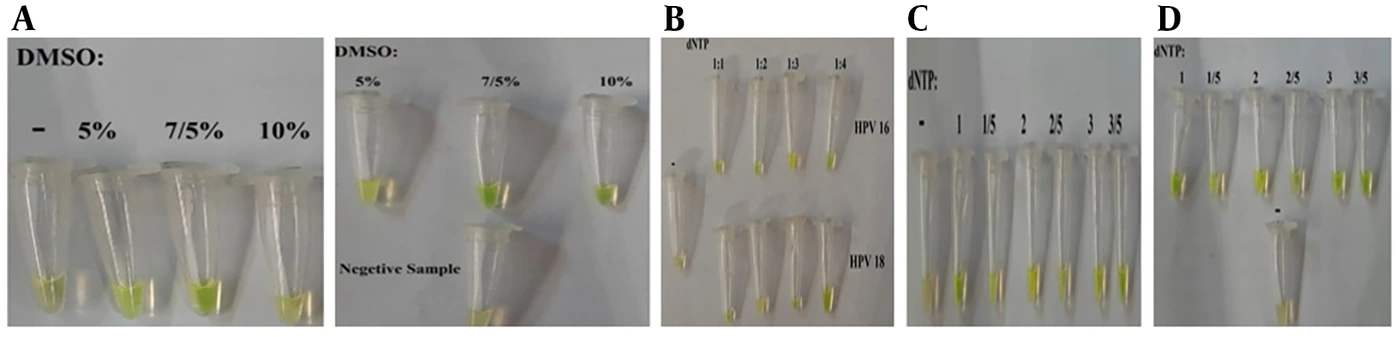
Figure 1.
A, DMSO Optimization in HPV16 and HPV18; B, Optimization of primers concentration in HPV16 and HPV18 samples; C, dNTP optimization in HPV16 and HPV18 sample; D, Optimal result of LAMP technique.
4.3. dNTP and Primer Optimization
For a final volume of 25 µL, B3/F3 to BIP/FIP primer ratios of 1:1, 1:2, 1:3, and 1:4 were tested, with 1:4 identified as the optimal ratio (Figure 1B).
In a final volume of 25 µL, various dNTP volumes were tested: 1 µL, 15 µL, 12 µL, 2.5 µL, 3 µL, and 3.5 µL, with 3.5 µL ultimately selected (Figure 1C).
The results of the LAMP technique for HPV16 and HPV18 samples, based on temperature and time gradients and the optimization of primers, DMSO, and dNTP, are shown in Figure 1D.
4.4. Comparison of LAMP Technique With PCR
Conventional PCR was performed to co-amplify HPV16 and HPV18 in a 50 µL reaction, as detailed in Table 4, for comparison with the LAMP-LFD assay. Deionized water served as the negative control. PCR products were verified by loading 5 µL onto a 2% agarose gel in 1X TBE buffer and running the gel at 120 V for 60 minutes (Table 4).
Table 4.
Composition of the PCR Reaction Mixture Used for Comparison with the LAMP-LFD Assay
| Component | Final Concentration | Volume (in 50 µL Reaction) |
|---|---|---|
| 10X PCR Buffer (Tris-HCl) | 1X | 5 µL |
| KCl | 50 mM | - |
| Forward/Reverse primers HPV16 | 160 nM each | - |
| Forward/Reverse primers HPV18 | 80 nM each | - |
| dNTP mix | 0.2 mM each | - |
| MgCl2 | 2 mM | - |
| Taq DNA polymerase | 1.5 U | - |
| DNA template | - | 1 µL |
| Nuclease-free water | - | Up to 50 µL |
| Total reaction volume | - | 50 µL |
4.5. Validation of Real-Time PCR
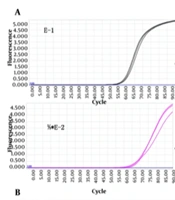
The detection limit of the real-time PCR method was determined using 100-fold dilutions of input DNA. Amplification plots for each HPV type included E2 and E6/7 plasmid DNA. The crossing points for real-time PCR varied from 1010 to 1 copy for HPV-16 E6/E7 DNA. The standard curve demonstrated a dynamic linear range for quantification across the dilution concentrations (Figure 2).
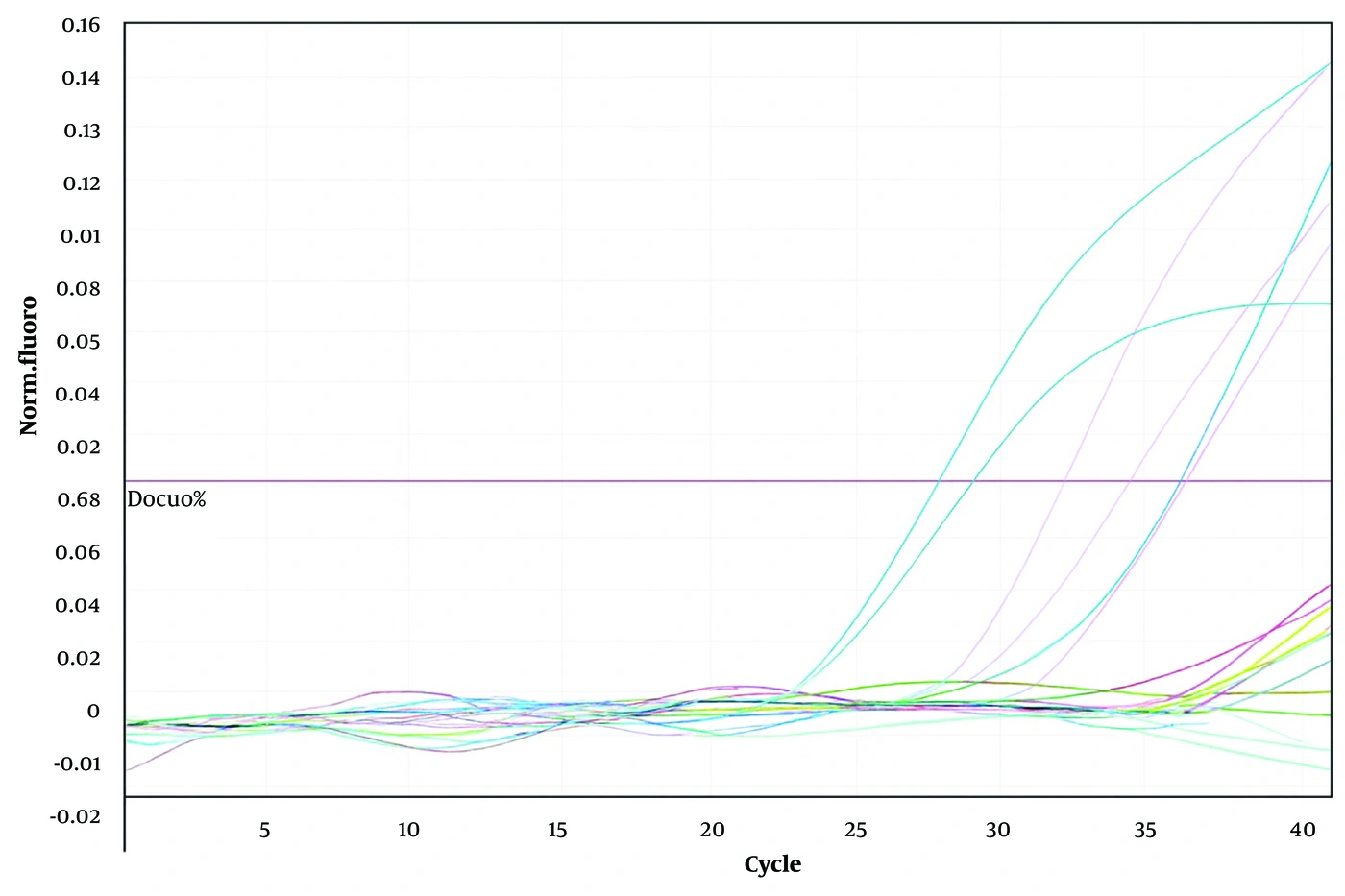
Figure 2.
real-time PCR for human papillomavirus (HPV) DNA. Typical amplification plots obtained by real-time PCR for the 7-point series concentration of HPV-16 E6/7. The x-axis denotes the cycle number and the y-axis the fluorescence intensity over the background.
4.6. Synthesis of Gold Nanoparticles
Citrate functions as both a stabilizing and reducing agent. In 1951, Turkevich pioneered the in-situ synthesis of gold nanoparticles (AuNPs) by reducing HAuCl4 with citrate, a method still widely used today. Slot later introduced a technique using a tannic acid/citrate solution, in which tannic acid acts as the reducing agent, producing AuNPs at 60 °C. Nathan's team developed a method that used citrate as a stabilizer and NaBH4 as the reducer, yielding AuNPs of 6 nm, significantly smaller than the over 20 nm particles produced by the traditional Turkevich method. To synthesize gold nanoparticles, a one-millimolar solution of HAuCl4 was prepared, initially appearing yellow. Upon the addition of sodium citrate and boiling, the solution turned dark red, indicating the formation of gold nanoparticles. The process yielded spherical nanoparticles with diameters of 10 - 30 nm. Higher citrate concentrations promoted nucleation, resulting in smaller, more monodisperse particles, whereas lower concentrations slowed nucleation, leading to larger, more nonuniform gold nanoparticles. For the optimal synthesis of 30 - 15 nm gold nanoparticles, 200 - 400 µL of the gold salt solution was used at pH 10 and 50°C for 4 - 24 hours. These conditions are vital for preparing samples for Zeta potential, TEM, DLS, and UV analyses.
4.7. UV and FTIR Analysis Results
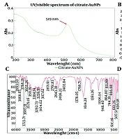
A spectrophotometer was used to confirm the presence of gold nanoparticles by measuring their absorption at different wavelengths. A 1 mL solution of 0.1 mg/mL gold nanoparticles was centrifuged, the supernatant was discarded, and 100 µL of K-PBS solution was added. Optical absorption was then measured, showing an absorption band at 519 nm, which confirms the presence of gold nanoparticles (Figure 3A).
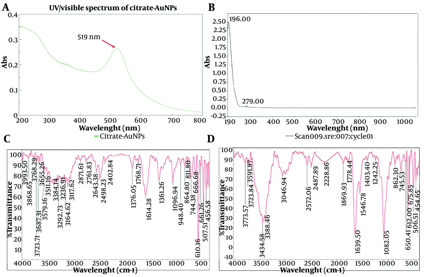
Figure 3.
A, Absorption spectrum of gold nanoparticles at 519 nm wavelength (a) and after binding of anti-FITC antibody (b); B, The FT-IR of nanoparticle sample (a), nanoparticle, and antibody (b).
Infrared spectroscopy analysis of the protein coating on gold showed increased peaks for amide groups I, II, and III compared with the bare gold sample, confirming the presence of protein. The peak at 1639 cm corresponds to amide I (primary amide group of proteins (-CO-NH2)). Additionally, peaks at 1546 cm-1 (N-H bending vibration/C-N stretching vibration) and 1240 cm-1 (C-N stretching vibration/N-H bending vibration) are attributed to the amide II band (secondary amide group of proteins (-CO-NH)) and the amide III bond, respectively. Peaks around 3300 cm-1 (3388 and 3343) are associated with N-H bending vibration (Figure 3B).
4.8. DLS Analysis
The gold nanoparticles were analyzed using DLS with a Zetasizer, showing an approximate diameter of 100 nm (Figure 4 in Supplementary File). Diagram b illustrates that after binding with anti-FITC antibodies, the distribution of the synthesized gold nanoparticles widened, with an average size of 2000 nm. The nonparametric and paired-samples test results for HPV16 and HPV18 are shown in Figure 4A-D.
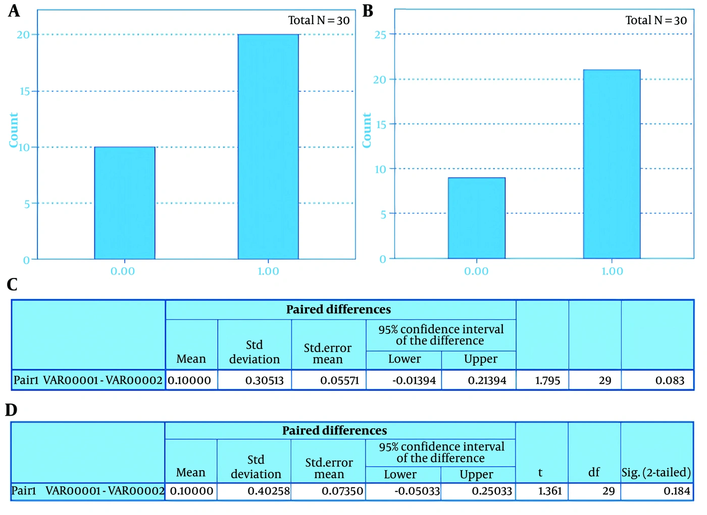
Figure 4.
Nonparametric Tests - Categorical Field Information VAR00002 (A HPV16 and B HPV18). t-Test - Paired Samples Test (C HPV16 and D HPV18).
4.9. Zeta Potential and TEM Analysis
The synthesized gold nanoparticles exhibited an electric charge of -34.8 mV, indicating repulsion that prevents aggregation and promotes stability. TEM analysis showed that the nanoparticles ranged in size from 2 to 10 nm and had spherical and polyhedral shapes. Citrate reduction of HAuCl4 produced gold particles with a diameter of 15 nm. Scanning electron microscopy confirmed their size and dispersion, with an average diameter of approximately 20 nm (Figure 5 in Supplementary File).
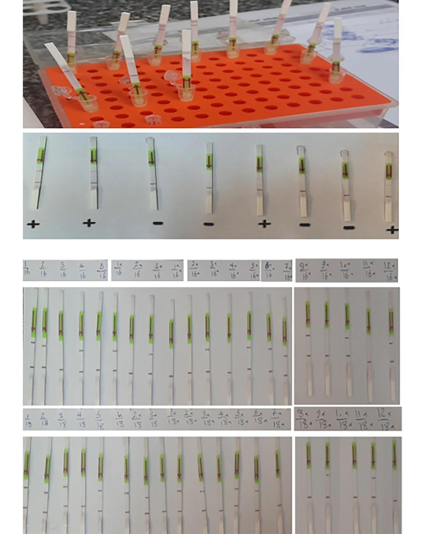
Figure 5.
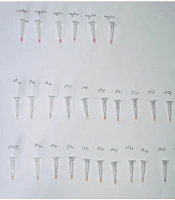
Developing a test strip with nitrocellulose paper coated with anti-FITC antibody linked to gold nanoparticles for LAMP sample identification
4.10. Statistical Analysis
Statistical analyses were performed using SPSS version 16 (IBM Corp., Armonk, NY, USA). The diagnostic performance of the LAMP-LFD assay was evaluated using real-time PCR as the reference standard. Sensitivity, specificity, positive predictive value (PPV), negative predictive value (NPV), overall accuracy, positive and negative likelihood ratios (LR+ and LR−), and Cohen's kappa coefficient were calculated together with their corresponding 95% confidence intervals.
4.11. Molecular Lateral Flow Dipstick Design for Detection of HPV16 and HPV18
To conjugate anti-FITC antibody with gold nanoparticles, pH and antibody concentration gradients were established in a 96-well ELISA plate. Each well received 100 µL of the gold solution, which was incubated for 10 minutes at room temperature, followed by the addition of 100 µL of sodium chloride M1. Wells that remained red indicated successful conjugation. The sample pad, made of cellulose, was cut to 4 mm x 15 mm. The conjugate pad, consisting of synthetic polyolefin or Glass Fiber Membrane, was sized at 4 mm x 13 mm (27). After cutting the Glass Fiber Membrane, the conjugate pad was soaked in a special buffer at room temperature for 24 hours before applying a mixture of gold nanoparticle solution and anti-FITC antibody. A bulk pad enhances antibody-probe interaction, with materials such as polyester, cellulose, and glass fiber available in 4 mm x 4 mm size. The detection pad, made of nitrocellulose, contains test and control lines created by applying avidin and goat anti-mouse IgG monoclonal antibodies. The absorbent pad, resembling flat paper, is made from cellulose fiber, measuring 4 mm x 18 mm and approximately 0.5 mm thick, and is designed to absorb sample residue in the lateral flow device. At this stage, the test strip was constructed using anti-FITC-coated nitrocellulose paper linked with gold nanoparticles. Individual DNA concentrations (1010, 109, 108, 107, 106, 105, 104, 103, 102, 101, and 100) from HPV16 and HPV18 were then prepared, and 120 µL of each concentration was added to the strip. The assay cut-off was established to effectively detect HPV16 and HPV18 (Figure 5).
4.12. Sensitivity Test of the LAMP Method
The sensitivity test showed that the LAMP method performed very well for detecting HPV types 16 and 18. The results were the same as those of the standard real-time PCR test. As shown in Table 5, the sensitivity was 100% (1.0) for type 16, type 18, and for both types together. This indicates that the test correctly identified all positive cases and that no false negatives occurred. The confidence intervals also support this strong performance. Although the range for type 18 was wider (because the sample size was small), the overall results still indicate high accuracy. These findings suggest that the LAMP-LFD assay has high sensitivity for detecting HPV16 and HPV18 in the present study and may represent a promising screening approach. Further validation in larger populations is warranted.
Table 5.
Diagnostic Sensitivity of the LAMP-LFD Assay for HPV16 and HPV18 Detection
| HPV Genotype | True Positives | False Negatives | Sensitivity (%) | 95% CI |
|---|---|---|---|---|
| HPV16 | 17 | 0 | 100.0 | 80.5 - 100.0 |
| HPV18 | 3 | 0 | 100.0 | 29.2 - 100.0 |
| Overall | 20 | 0 | 100.0 | 83.2 - 100.0 |
4.13. LAMP Specificity Test
The specificity test results showed that the LAMP method performed well in detecting HPV genotypes 16 and 18 compared with the standard real-time PCR method. As shown in Table 6, the specificity for genotype 18 was 100% (1.00), indicating that the method correctly identified all negative specimens and produced no false‑positive results in this group. For genotype 16, the specificity was 84.6% (0.846), which was slightly lower. This was due to two false‑positive cases in which the standard method showed negative results but the LAMP test reported positive. However, the 95% confidence intervals indicate that the specificity remained acceptable. For genotype 16, the range was 54.5 - 98.1%, and for both genotypes combined, it was 77.9 - 99.2%. Overall, the total specificity of the LAMP method was 93.3% (0.933), which is high. The lower limit of the confidence interval in the total population (77.9%) also suggests that the true specificity of the test is likely above 77%. These findings indicate good specificity of the LAMP-LFD assay in the study population, although additional validation in larger cohorts is required.
Table 6.
Diagnostic Specificity of the LAMP-LFD Assay for HPV16 and HPV18 Detection
| HPV Genotype | True Negatives | False Positives | Specificity (%) | 95% CI |
|---|---|---|---|---|
| HPV16 | 11 | 2 | 84.6 | 54.5 - 98.1 |
| HPV18 | 17 | 0 | 100.0 | 81.5 - 100.0 |
| Overall | 28 | 2 | 93.3 | 77.9 - 99.2 |
4.14. Accuracy Test of the LAMP Method
As shown in Table 7, the accuracy of the LAMP method was 100% (1.00) for genotype 18 and 93.3% (0.933) for genotype 16. The 100% accuracy for genotype 18 indicates that the results of all 20 samples completely matched those of the real-time PCR method. The accuracy of 93.3% for genotype 16 was due to two false-positive (FP) results among 30 samples. The 95% confidence interval also supports the high accuracy of the method: for genotype 16, it ranged from 77.9% to 99.2%, and for genotype 18, from 88.1% to 100%. Overall, the total accuracy of the LAMP method was 96% (0.960), which is high. The lower bound of the overall confidence interval (86.3%) indicates that, with high confidence, the true accuracy of the kit in the population is above 86%. Therefore, the research hypothesis regarding the acceptable accuracy of the LAMP method is confirmed, indicating that this kit has a strong ability to correctly identify both positive and negative cases.
Table 7.
Diagnostic Accuracy of the LAMP-LFD Assay
| HPV Genotype | Correct Classifications | Total Samples | Accuracy (%) | 95% CI |
|---|---|---|---|---|
| HPV16 | 28 | 30 | 93.3 | 77.9 - 99.2 |
| HPV18 | 20 | 20 | 100.0 | 88.1 - 100.0 |
| Overall | 48 | 50 | 96.0 | 86.3 - 99.5 |
4.15. Predictive Results of the LAMP Method
The results showed that the LAMP test performed well for detecting HPV types 16 and 18. As shown in Table 8, the negative predictive value (NPV) was 100% for HPV16, HPV18, and the overall dataset. In this study population, no false-negative results were observed. However, these findings should be interpreted in the context of the study sample size and HPV infection prevalence. The positive predictive value (PPV) also indicated strong performance: for type 18, the PPV was 100%, with all positive results being correct and no false positives. For type 16, the PPV was 89.5%; this lower value reflected two false‑positive results, indicating that some positive results might not be true positives. When all 50 samples were considered together, the PPV was 90.9%, which remains high. The 95% CI values also support the reliability of these results, although the range for type 16 is wider because the sample size was small.
Table 8.
Predictive Values of the LAMP-LFD Assay
| HPV Genotype | PPV (%) | 95% CI | NPV (%) | 95% CI |
|---|---|---|---|---|
| HPV16 | 89.5 | 66.9 - 98.7 | 100.0 | 71.5 - 100.0 |
| HPV18 | 100.0 | 29.2 - 100.0 | 100.0 | 81.5 - 100.0 |
| Overall | 90.9 | 70.8 - 98.9 | 100.0 | 87.8 - 100.0 |
4.16. Positive and Negative Test Accuracy
In Table 9, the negative test value (LR–) was 0 for both types and overall. The negative likelihood ratio (LR−) was 0 for both HPV16 and HPV18 because no false-negative results were observed in this study. This finding indicates excellent rule-out performance within the studied sample, although the estimate should be interpreted cautiously given the limited sample size. For positive results (LR+), the LR+ for type 18 was infinity (∞) because there were no false positives. For HPV18, the positive likelihood ratio (LR+) approached infinity because no false-positive results were observed in this dataset. This finding indicates very strong evidence supporting HPV18 infection following a positive test result within the study population. For type 16, the LR+ was 6.5, indicating good, although not perfect, performance; it increases the likelihood of true HPV infection, but two false positives occurred. Overall, the LR+ (for all samples) was 15, indicating that the test performs very well in identifying truly infected individuals.
Table 9.
Likelihood Ratios of the LAMP-LFD Assay
| HPV Genotype | Positive Likelihood Ratio (LR+) | Negative Likelihood Ratio (LR−) |
|---|---|---|
| HPV16 | 6.50 | 0.00 |
| HPV18 | ∞ | 0.00 |
| Overall | 15.00 | 0.00 |
4.17. Kappa Coefficient
The results showed that the two tests agreed very well for detecting HPV types 16 and 18. In Table 10, the kappa value was 0.862 for type 16 and 1.000 for type 18. Both values are very high, indicating that the two tests yielded nearly identical results. When all samples were assessed together, the total kappa was 0.918, which is also very strong. Cohen's kappa coefficients demonstrated excellent agreement between the LAMP-LFD assay and real-time PCR for HPV16 (κ = 0.862), HPV18 (κ = 1.000), and the overall dataset (κ = 0.918). These high Cohen's kappa values indicate excellent agreement between the LAMP-LFD assay and real-time PCR in this study population.
Table 10.
Agreement Between the LAMP-LFD Assay and Real-Time PCR
| HPV Genotype | Cohen's κ | Interpretation | P-Value * |
|---|---|---|---|
| HPV16 | 0.862 | Almost perfect agreement | < 0.001 |
| HPV18 | 1.000 | Perfect agreement | < 0.001 |
| Overall | 0.918 | Almost perfect agreement | < 0.001 |
*P-values correspond to the significance of the z-test for Cohen's kappa, testing whether the agreement between the LAMP-LFD assay and real-time PCR was significantly above chance level (one-tailed test).
5. Discussion
Genital wart treatments address cosmetic concerns or remove precancerous lesions, as there is currently no cure for HPV infection itself (28). Therefore, increased screening is essential to reduce cervical cancer, particularly among underscreened women. Most vaccine side effects are mild and localized; however, vaccination should be deferred during an acute febrile illness. Mild respiratory or GI infections are not contraindications. A severe allergic reaction to the first dose precludes additional doses. HPV vaccination during pregnancy is not recommended because of limited data, although no adverse effects have been observed (29). If pregnancy occurs during the vaccination series, remaining doses should be postponed until after delivery. We used real-time PCR, a precise and sensitive method that detects as few as 50 copies of integrated HPV DNA, to measure HPV-16 and -18 viral loads (30). In this diagnostic accuracy study, real-time PCR was used as the predefined reference standard, and the performance of the LAMP-LFD assay was evaluated against PCR results obtained from the same clinical specimens.
Gold nanoparticles are an attractive labeling agent in biotechnology because they are readily produced and can bind to a variety of biomolecules (31). They can be linked to antibodies in three ways (32). First, nanoparticles are coated with citrate, a stabilizing agent that gives them a negative charge and prevents aggregation in solution (33). The first binding mechanism involves hydrophobic interactions between regions of the antibody and the nanoparticle surface, creating a non-fusion bond. The second mechanism relies on electrostatic interactions between positively charged amino acids on the antibody and the negatively charged citrate layer (34). The third mechanism forms a fusion bond with the free sulfhydryl groups of antibodies. This study specifically used the sulfhydryl-group conjugation method, successfully linking citrate-coated gold nanoparticles to anti-FITC antibodies.
Our LAMP-LFD assay for HPV16 and HPV18 demonstrated high sensitivity and rapid turnaround, consistent with previous studies reporting that LAMP-LFD can outperform conventional nested PCR in sensitivity and speed (35). The effectiveness of LAMP in rapidly and accurately identifying clinical isolates has also been established for other bacterial pathogens, such as Nocardia species, underscoring its adaptability and reliability across infectious diseases (36). Similar LAMP-based methods have been used for rapid detection of other viruses, such as Crimean Congo Hemorrhagic Fever Virus, further supporting the strength and versatility of isothermal amplification techniques in point-of-care diagnostics (37). However, as observed in other reports, specificity was somewhat lower, with some HPV-negative samples yielding positive LAMP-LFD results. Further qPCR analysis of discordant samples indicated that most were true positives, likely reflecting the higher analytical sensitivity of LAMP-LFD compared with conventional PCR (35). This finding suggests that LAMP-LFD may be particularly valuable for detecting low-copy HPV infections that might be missed by other methods. Compared with other rapid detection platforms, such as electrochemical LAMP and nanoparticle-based colorimetric assays (38), our LAMP-LFD approach offers a balance of simplicity, speed, and sensitivity, making it well suited for use in low-resource or field settings. Nevertheless, the risk of cross-contamination inherent to isothermal amplification methods should be addressed, for example, by incorporating uracil DNA glycosylase (35).
In the present study, the LAMP-LFD assay demonstrated an overall sensitivity of 100% and an overall specificity of 93.3% compared with real-time PCR. Sensitivity reached 100% for both HPV16 and HPV18, whereas specificity was 84.6% for HPV16 and 100% for HPV18. The two false-positive HPV16 results reduced overall specificity but did not affect sensitivity because no false-negative results were observed. These findings are consistent with previously published LAMP-based assays, which have reported sensitivities ranging from 95% to 100% and specificities between approximately 70% and 100%, depending on primer design, specimen type, and reference standard (35, 38). The nanoparticle-based assays presented intermediate specificity, ranging from 78% to 87%. The LAMP-LFD assays, both in this study and in previously published reports, provided results within 30 to 45 minutes, whereas PCR methods required approximately 2 to 3 hours to complete. The nanoparticle-based tests were the fastest overall, producing results in less than one hour. Regarding equipment requirements, both this study’s LAMP-LFD and the published LAMP-LFD platforms required only minimal laboratory instrumentation, whereas PCR analysis remained dependent on laboratory-based equipment (35, 38). The nanoparticle-based method also required minimal equipment, making it comparable in simplicity to the LAMP-LFD approaches. Finally, regarding field applicability, both this study and the previously reported LAMP-LFD systems demonstrated suitability for on-site or low-resource settings, similar to the nanoparticle-based assays. In contrast, PCR-based detection required complex equipment and controlled conditions, limiting its suitability outside laboratory environments.
Molecular lateral flow is a straightforward diagnostic device that provides observable results by facilitating the movement of a labeled analyte through membranes. In this test, the sample travels from the sample pad to the conjugate pad via capillary action. This method employs a conical network of fiber pads to generate visible results. Molecular lateral flow is simple, rapid, inexpensive, and portable, making it suitable for medical experiments, food safety, and environmental analysis. However, its low sensitivity limits its application.
This study successfully designed a cost-effective, rapid, and highly sensitive LAMP-LFD assay for detecting HPV16 and HPV18, suitable for field or low-resource settings because it does not require a thermocycler and provides easily interpretable colorimetric results. The 30 - 45 min reaction time represents a substantial improvement over conventional PCR assays. The assay demonstrated diagnostic performance comparable to real-time PCR under the conditions evaluated in the present study. Gold nanoparticles enhanced signal visualization and reliability, while optimized pH, DMSO, and primer ratios ensured high amplification efficiency and stability. The system is user-friendly, portable, and well suited for rapid preliminary screening in settings with limited laboratory infrastructure. Ethical approval and standardized sampling strengthened the validity of the clinical results. Internal controls, sequencing, and real-time PCR confirmed result consistency and reproducibility. Limitations include moderately reduced specificity compared with PCR, likely due to LAMP’s higher analytical sensitivity and contamination potential. The absence of uracil-DNA glycosylase increased the risk of false positives. Limited funding restricted the molecular lateral flow dipstick (LFD) to a single test line. The relatively small sample size may limit generalizability. Some plasmid-positive controls lacked independent verification, potentially introducing bias. Extensive manual calibration during optimization reduces scalability. Reagent procurement challenges delayed experimental replication. Nanoparticle synthesis requires controlled pH and temperature, potentially hindering reproducibility in resource-limited laboratories. Finally, although efficient for HPV16/18, the assay’s applicability to other high-risk HPV genotypes requires validation through larger studies. Due to Iran’s economic conditions, a modest budget was allocated for this study. However, additional financial backing was essential for production work. Currency fluctuations, budget constraints, and the need for multiple repetitions made procurement of the required method comparison kits time-consuming. Economic challenges and limited funding also restricted the molecular lateral flow dipstick (LFD) design to a single test line for HPV16 and HPV18 detection, rather than the originally intended two test lines plus a control line.
5.1. Conclusions
Existing treatments for genital warts and precancerous lesions remove lesions but do not eliminate HPV, underscoring the need for improved antiviral therapies. Enhanced screening, particularly in underserved communities, is crucial for reducing cervical cancer rates. Although HPV vaccination is safe, contraindications and special conditions, such as pregnancy, require careful consideration. Innovative technologies, such as gold nanoparticles, offer promising advances in HPV diagnostics. Although molecular lateral flow tests are simple and affordable, their sensitivity requires improvement. Continued research and public health initiatives are vital to improve HPV management and decrease cervical cancer incidence. Our findings support LAMP-LFD as a rapid, cost-effective, and sensitive tool for HPV16/18 detection, with the potential to improve cervical cancer screening, particularly in resource-limited settings. Future large-scale studies comparing it with other rapid assays will clarify its clinical utility.