
1. Background
Urinary tract infection (UTI) is a common bacterial infection in childhood. Early diagnosis and management of acute febrile UTI can alleviate symptoms and potentially prevent long-term complications. However, the prolonged turnaround time for urine culture results, together with the low sensitivity and specificity of urinary symptoms, pyuria, nitrite testing, leukocyte esterase, white blood cell (WBC) count, erythrocyte sedimentation rate (ESR), and C-reactive protein (CRP), highlight the need for a sensitive, rapid, and noninvasive test for the early diagnosis of UTI, particularly in suspected cases (1).
Ultrasonography is a safe, rapid, and inexpensive imaging modality for evaluating the upper and lower urinary tract. It is useful for detecting different types of UTI and for assessing treatment response. Several renal-bladder ultrasound (RBU) findings suggestive of lower UTI have been reported, including bladder wall thickening, air bubbles, fine echoes within the bladder, blood clots in the lumen, and incomplete bladder emptying with urinary retention. Upper UTI may present with focal or global kidney enlargement, increased echogenicity, or mild hydronephrosis (2, 3).
Bladder contents typically appear uniformly anechoic on ultrasonography. However, echogenic material within the bladder lumen, whether floating or layering, may represent technical artifacts or pathological conditions, including dehydration, hematuria, pyuria, fat droplets, inflammation, urothelial sloughing associated with acute tubular necrosis or nephrotic or nephritic syndrome, neurogenic bladder, crystalluria, and urinary stasis (2-5).
Previous studies have reported conflicting findings regarding bladder debris. Some have identified it as a positive indicator of UTI and suggested that bladder debris on RBU warrants UTI screening (3, 4, 6). Conversely, other studies have found bladder debris to be an unreliable indicator of UTI, with no significant correlation with urinalysis or urine culture results across different age groups (7-9). Most previous studies were conducted in adults; therefore, further investigation in pediatric populations is needed.
2. Objectives
This study aimed to evaluate the reliability and predictive value of bladder debris for predicting UTI in children.
3. Methods
3.1. Study Design and Data Collection
This cross-sectional study included 381 children admitted to the emergency department of a tertiary children’s hospital between 2023 and 2024. The children presented with symptoms including unspecified fever, abdominal pain, gastroenteritis, and urinary tract symptoms and were evaluated for suspected UTI.
The inclusion criteria were age < 14 years, suspected UTI requiring RBU evaluation at admission, and urinalysis and urine culture performed on the same day. Participants were classified into 4 groups according to the presence of bladder debris and urine culture results: group A (UTI positive, debris positive), group B (UTI positive, debris negative), group C (UTI negative, debris positive), and group D (UTI negative, debris negative).
Children were excluded if they had moderate-to-severe dehydration, recent antibiotic treatment, a suspected contaminated sample, a neurogenic bladder, known urinary tract abnormalities, previous genitourinary surgery, urine osmolality outside the range of 175 - 1400 mOsm/kg (indicating dilute or concentrated urine), or renal dysfunction.
A total of 381 children met the inclusion and exclusion criteria and were evaluated consecutively. Urine samples were collected aseptically by suprapubic aspiration or urethral catheterization in young, incontinent patients and by the midstream technique in continent children.
UTI was defined as a positive urine culture, characterized by any growth in a sample obtained by suprapubic aspiration, growth of > 105 colony-forming units (CFU)/mL of a single pathogen in a midstream urine sample, growth of > 104 CFU/mL in bladder urine obtained by catheterization, or growth of > 105 CFU/mL in a urine bag sample (1). To exclude contaminated urine bag samples, pyuria and bacteriuria were also considered in the definition of UTI.
Bladder debris was defined as discrete, mobile, hyperechoic, gravity-dependent foci within the bladder lumen that were distinct from the bladder wall and included both floating echogenic foci and layering sediment (4, 5).
A rigorous 2-step verification process was used. First, a dual-probe assessment was performed using both high-frequency linear probes for high-resolution imaging of the anterior wall and low-frequency curved probes to evaluate the entire bladder lumen. This approach reduced the risk of misinterpreting gain-related artifacts or reverberation noise as debris. The echogenicity of potential bladder debris was compared with that of the surrounding anechoic urine and was considered true debris if it remained visible after the gain was reduced to a level at which artifactual noise was eliminated.
Second, dynamic positioning was used. Patients with documented layering debris were repositioned into the lateral decubitus position to assess gravity-dependent particle movement, or the snow-globe effect, thereby distinguishing debris from fixed bladder wall abnormalities or stationary artifacts. To distinguish bladder debris from section-thickness artifacts that may mimic debris on transverse views, sagittal images were acquired, and the transducer was moved away from the center of the bladder.
Ultrasonography was performed by a single expert pediatric radiologist who was blinded to the clinical and laboratory data, using a Philips Affiniti 50 machine equipped with a 6 - 12 MHz linear transducer and a 1 - 6 MHz curved transducer. The examination included measurements of bladder volume and wall thickness and assessment for urolithiasis, urinary crystals, and space-occupying lesions. Color Doppler was used in patients with nonmobile bladder debris attached to the wall to distinguish debris from bladder masses.
3.2. Statistical Analysis
All statistical analyses were performed using SPSS for Windows, version 27.0 (SPSS Inc, Chicago, IL, USA), and MedCalc, version 15.4. Normally distributed continuous variables were reported as mean ± SD and compared between groups using the independent-samples t-test. Non-normally distributed variables were compared using the Kruskal-Wallis test. Categorical variables were reported as absolute frequencies and percentages and compared using the χ2 test or Fisher exact test, as appropriate. The Shapiro-Wilk test was used to assess the normality of continuous variables, and the test statistic and P value were reported for each variable. Logistic regression analysis was used to calculate odds ratios (ORs) and 95% CIs for the potential association between bladder debris and a positive urine culture. Sensitivity and specificity were calculated to assess the ability of bladder debris on RBU to predict a positive urine culture.
3.3. Ethical Approval
The study protocol was approved by the institutional ethics committee (ethical code: IR.SBMU.RICH.REC.1403.019). Informed consent was obtained from the legal guardians before patient enrollment.
4. Results
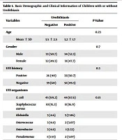
Of the 381 children evaluated, 192 had a confirmed UTI. The mean age was 33 ± 33 months. Females were more prevalent than males among children with UTI, with a female-to-male ratio of 3.8:1. Children with UTI had significantly more pyuria and bacteriuria than those without UTI. Escherichia coli was the most common causative organism, accounting for 70% of infections, followed by Klebsiella species and other organisms. Table 1 presents the demographic and clinical characteristics of the patients.
Table 1.
Comparison of Variables According to Urine Culture and Bladder Debris a
| Variables | Group A (n = 102) | Group B (n = 90) | Group C (n = 95) | Group D (n = 94) | P-Value |
|---|---|---|---|---|---|
| Age (mo) | 35 ± 34 | 30 ± 32 | 56 ± 43 | 60 ± 49 | < 0.001 |
| Gender | < 0.001 | ||||
| Male (n = 118) | 29 (28.4) | 11 (12.2) | 59 (62.1) | 19 (20.2) | |
| Female (n = 263) | 73 (71.6) | 79 (87.8) | 36 (37.9) | 75 (79.8) | |
| WBC | 13752.22 ± 5273 | 13257.77 ± 6307 | 10573.23 ± 4166 | 11015.96 ± 6037 | < 0.001 |
| PLT | 367121.20 ± 138270 | 361655.55 ± 127883 | 330485.15 ± 115957 | 295223.40 ± 134310 | < 0.001 |
| ESR | 42.37 ± 29.29 | 41.52 ± 28.04 | 26.48 ± 20.61 | 30.21 ± 26.88 | < 0.001 |
| CRP | 48.73 ± 36.87 | 51.64 ± 35.87 | 29.56 ± 29.84 | 27.80 ± 33.47 | < 0.001 |
| Hgb | 10.49 ± 1.47 | 10.41 ± 1.40 | 11.02 ± 1.54 | 10.87 ± 1.85 | 0.022 |
| Pyuria | 102 (100) | 90 (100) | 24 (25.3) | N/A | < 0.001 |
| Bacteriuria | 56 (54.9) | 79 (87.8) | 2 (2.1) | N/A | < 0.001 |
| Nitrite | 21 (20.8) | 13 (14.8) | N/A | N/A | 0.283 |
| UC | 0.077 | ||||
| E coli | 78 | 58 | N/A | N/A | |
| Kleb | 19 | 20 | N/A | N/A | |
| others | 5 | 12 | N/A | N/A |
a Values are expressed as mean ± SD, No. (%) or number. Group A, UTI positive/debris positive; group B, UTI positive/debris negative; group C, UTI negative/debris positive; group D, UTI negative/debris negative. Abbreviations: CRP, C-reactive protein; E coli, Escherichia coli; ESR, erythrocyte sedimentation rate; F, female; Hgb, hemoglobin; Kleb, Klebsiella; M, male; N/A, not applicable; PLT, platelet count; UC, urine culture; UTI, urinary tract infection; WBC, white blood cell count.
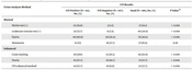
Bladder debris was identified in 102 children with UTI and 95 children without UTI. Conversely, bladder debris was absent in 90 children with UTI and 94 children without UTI. Univariate logistic regression showed no significant association between bladder debris and UTI (OR = 1.121; 95% CI, 0.750 - 1.676; P = 0.576). After adjustment for age and sex, bladder debris was significantly associated with UTI (OR = 1.937; 95% CI, 1.123 - 3.339; P = 0.017) (Table 2). Thus, the adjusted analysis revealed an association that was not apparent in the univariate analysis.
Table 2.
Binary Logistic Regression Analysis of Variables a
| Variables | Univariate OR | Univariate P Value | Multivariate OR | Multivariate P Value |
|---|---|---|---|---|
| Age (mo) | 0.985 (0.980 - 0.990) | < 0.001 | 0.982 (0.975 - 0.990) | < 0.001 |
| Gender | 0.374 (0.238 - 0.589) | < 0.001 | 0.266 (0.144 - 0.491) | < 0.001 |
| Bladder debris | 1.121 (0.750 - 1.676) | 0.576 | 1.937 (1.123 - 3.339) | 0.017 |
a Values are expressed as 95% CI. Abbreviations: CI, confidence interval; OR, odds ratio.
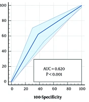
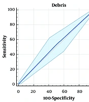
The area under the ROC curve for bladder debris in predicting UTI was 0.514, indicating poor discriminatory ability. At the optimal cutoff, bladder debris had a sensitivity of 53.13% and a specificity of 49.74% for predicting UTI (Table 3 and Figure 1). Specifically, 53.13% of patients with UTI had particulate urine on ultrasound, whereas 49.74% of patients without UTI did not have particulate urine on RBU (P = 0.577).
Table 3.
Area Under the Receiver Operating Characteristic Curve for Bladder Debris in Predicting Urinary Tract Infection a
| Variables | Cutoff | Sensitivity | Specificity | AUC | 95% CI | Standard Error | P Value |
|---|---|---|---|---|---|---|---|
| Bladder debris | > 0 | 53.13% | 49.74% | 0.514 | 0.464 - 0.565 | 0.026 | 0.577 |
a Abbreviations: AUC, area under the curve; CI, confidence interval.
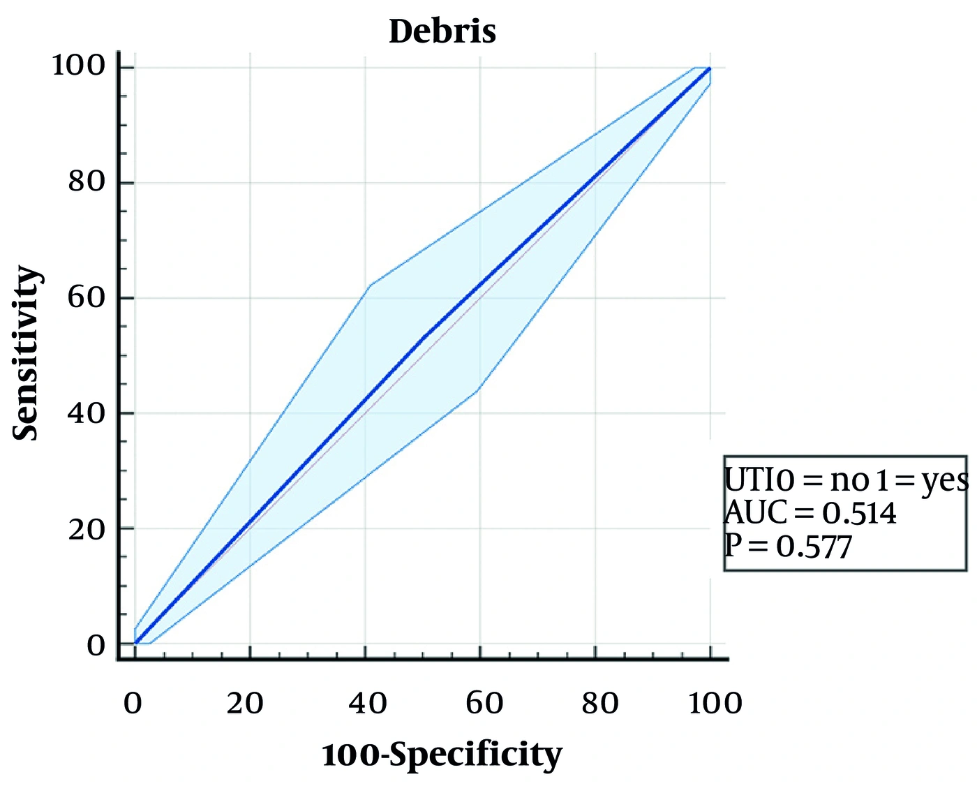
Figure 1.
Receiver operating characteristic curve of bladder debris for predicting urinary tract infection.
The positive predictive value (PPV) of bladder debris was 51.78%, indicating that approximately 52% of children with bladder debris had a positive urine culture. The negative predictive value (NPV) was 51.09%, indicating that approximately 51% of children without bladder debris did not have UTI. The positive likelihood ratio (PLR) was 1.06, indicating that bladder debris was only 1.06 times more likely to be observed in patients with UTI than in those without UTI. The negative likelihood ratio (NLR) was 0.94, indicating that the absence of bladder debris provided little evidence against UTI (Table 4). Figures 2 and 3 show ultrasound images without and with bladder debris, respectively.
Table 4.
Diagnostic Value of Bladder Debris for Predicting Urinary Tract Infection a
| Variables | PPV | NPV | PLR | NLR |
|---|---|---|---|---|
| Bladder debris | 51.78% | 51.09% | 1.06 | 0.94 |
a Abbreviations: NLR, negative likelihood ratio; NPV, negative predictive value; PLR, positive likelihood ratio; PPV, positive predictive value.
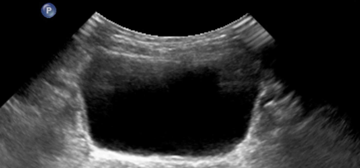
Figure 2.
Normal bladder ultrasound.
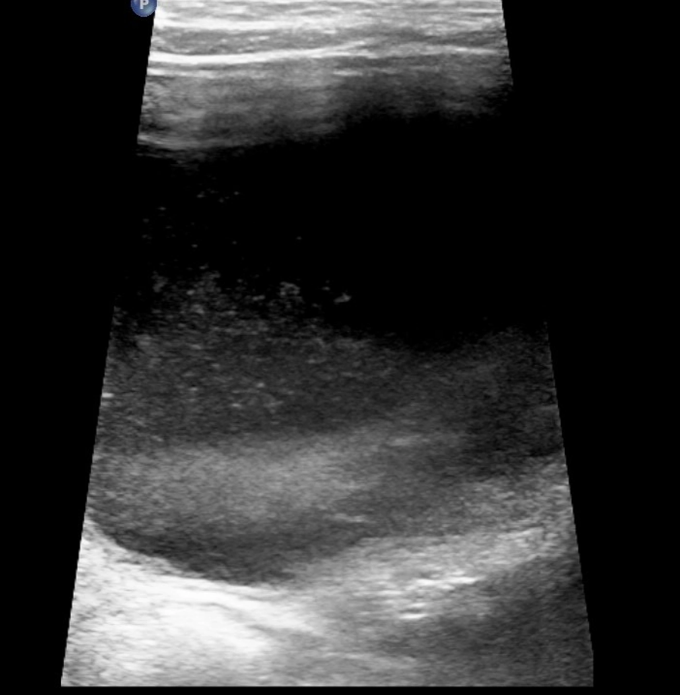
Figure 3.
Bladder ultrasound showing bladder debris.
5. Discussion
Despite the frequent observation of bladder debris in clinical practice, its relationship with UTI remains unclear. Given the limited evidence suggesting that bladder debris may be a warning sign for UTI in children, this study evaluated its significance in symptomatic pediatric patients undergoing assessment for suspected UTI.
The mean age of children diagnosed with UTI was 33 months. Consistent with previous research in this age group, UTI was more prevalent in females. Escherichia coli was the most common causative organism, followed by other enteric gram-negative organisms (1, 3, 6).
In the univariate analysis, bladder debris was not significantly associated with UTI. However, after adjustment for age and sex, bladder debris was significantly associated with UTI, indicating that age and sex influenced the observed relationship between bladder debris and UTI. Nevertheless, this association should be interpreted separately from diagnostic performance: bladder debris had an AUC of 0.514 and low sensitivity, specificity, PPV, and NPV, indicating that it was not an accurate stand-alone marker for identifying UTI.
The poor diagnostic performance of bladder debris is consistent with some previous studies. You et al. reported bladder debris in 18.8% of children younger than 2 years during their first febrile UTI. Although bladder debris was positively correlated with renal pelvic debris, renal parenchymal changes, collecting-system wall thickening, CRP level, and hematuria, it was not correlated with urine culture results. The authors therefore concluded that bladder debris was not an important indicator of UTI (7).
Cheng and Phelps found no correlation between bladder debris and specific abnormalities on urine examination. They recommended that urinalysis be performed within 1 week before or after bladder ultrasound. In their study, urine specimens were collected asynchronously with RBU, whereas urine testing and RBU were performed on the same day in the present study (8). Wachsberg et al. similarly concluded that bladder debris was not a reliable indicator of UTI and could represent a normal finding (9).
In the present study, bladder debris had low sensitivity (53.13%) and specificity (49.74%) for predicting UTI and, therefore, was not an accurate screening marker in children (P = 0.577). Similarly, Wilches et al. reported that bladder debris was common on sonographic examinations in both children and adults. Their reported sensitivity of 72% and specificity of 48% relative to urinalysis also indicated that bladder debris was not a reliable index for UTI screening (2).
Bladder debris also had low PPV and NPV in the present study. This finding is consistent with the study by Sanchez et al., who investigated the association between the ultrasonographic appearance of urine and urinalysis findings in animals. They reported a low PPV of urine echogenicity for detecting active urine sediment and suggested prioritizing urine examination in patients with other markers suggestive of UTI (10).
Although the present findings, together with those of Wilches et al. (2) and Sanchez et al. (10), suggest that bladder debris has limited diagnostic value for UTI, other studies have reached different conclusions. Stevenson et al. detected bladder debris in 22% of patients with UTI and reported a positive urine culture in 47% of patients with debris compared with 12% of those without debris. Although the reported sensitivity was low at 52%, consistent with the present findings, the authors recommended UTI screening in patients with bladder debris on ultrasound (6).
McQuaid et al. reported a 7-fold increase in positive urine cultures among children with bladder debris. They suggested that children younger than 60 months and those with a history of UTI or prenatal genitourinary abnormalities were at increased risk of UTI when bladder debris was present (3). However, patients with preexisting anatomical or functional urinary tract abnormalities were excluded from the present study.
Fananapazir et al. reported an association between bladder debris and urinalysis markers, including nitrite and WBCs, in adults. They recommended urine culture for emergency department patients with bladder debris. However, they also found that the absence of bladder debris did not rule out UTI on urine examination (4).
Moosavian et al. observed significant associations of pyuria, bacteriuria, and positive urine culture with echogenic foci on ultrasound images in dogs. However, they found no association between bladder debris and hematuria, lipiduria, or crystalluria. They therefore recommended posttreatment ultrasound to determine the clinical implications of echogenic urine debris in each patient (11). These varied findings, including evidence from different species, underscore the difficulty of establishing bladder debris as a consistent or sensitive marker of UTI, particularly in the absence of predisposing urinary tract abnormalities.
In the present study, both floating and layering particulate matter were defined as bladder debris. The findings indicate that, in a clinical screening setting, the presence of any mobile echogenic material should be interpreted cautiously. Both linear and curved transducers were used to distinguish true debris from artifactual echoes. Dynamic positioning with decubitus views was also used to confirm sediment mobility and distinguish layering debris from stationary intraluminal findings.
5.1. Study Limitations
This study had a single-center, single-reader design, highlighting the need for multicenter studies involving multiple radiologists to validate the findings. The study included symptomatic children evaluated for suspected UTI; therefore, the results may not be generalizable to asymptomatic or incidentally evaluated children. Future research should compare symptomatic and asymptomatic populations and evaluate RBU findings in both groups.
5.2. Conclusions
Bladder debris was significantly associated with UTI after adjustment for age and sex, but it showed poor discriminatory ability and low sensitivity, specificity, PPV, and NPV. Accordingly, bladder debris has limited predictive value and should not be used as the sole indication for obtaining a urine culture in symptomatic children evaluated for suspected UTI. Further multicenter studies should assess bladder debris before and after treatment in children with UTI to clarify its clinical significance.